Randomized Controlled Trial
. 2024 Mar;271(3):1256-1266.
doi: 10.1007/s00415-023-12060-y. Epub 2023 Nov 10.

Immersive virtual reality-based rehabilitation for subacute stroke: a randomized controlled trial

Affiliations
Randomized Controlled Trial

Immersive virtual reality-based rehabilitation for subacute stroke: a randomized controlled trial

Qianqian Huang et al. J Neurol. 2024 Mar.

Abstract

Objective: Few effective treatments improve upper extremity (UE) function after stroke. Immersive virtual reality (imVR) is a novel and promising strategy for stroke UE recovery. We assessed the extent to which imVR-based UE rehabilitation can augment conventional treatment and explored changes in brain functional connectivity (FC) that were related to the rehabilitation.

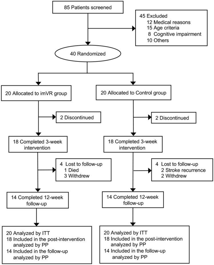
Methods: An assessor-blinded, parallel-group randomized controlled trial was performed with 40 subjects randomly assigned to either imVR or Control group (1:1 allocation), each receiving rehabilitation 5 times per week for 3 weeks. Subjects in the imVR received both imVR and conventional rehabilitation, while those in the Control received conventional rehabilitation only. Our primary and secondary outcomes were the Fugl-Meyer assessment's upper extremity subscale (FMA-UE) and the Barthel Index (BI), respectively. Both intention-to-treat (ITT) and per-protocol (PP) analyses were performed to assess the effectiveness of the trial. For both the FMA-UE/BI, a one-way analysis of covariance (ANCOVA) model was used, with the FMA-UE/BI at post-intervention or at follow-up, respectively, as the dependent variable, the two groups as the independent variable, baseline FMA-UE/BI, age, sex, site, time since onset, hypertension and diabetes as covariates.

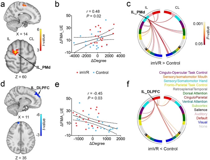
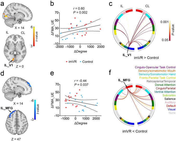
Results: Both ITT and PP analyses demonstrated the effectiveness of imVR-based rehabilitation. The FMA-UE score was greater in the imVR compared with the Control at the post-intervention (mean difference: 9.1 (95% CI 1.6, 16.6); P = 0.019) and follow-up (mean difference:11.5 (95% CI 1.9, 21.0); P = 0.020). The results were consistent for BI scores. Moreover, brain FC analysis found that the motor function improvements were associated with a change in degree in ipsilesional premotor cortex and ipsilesional dorsolateral prefrontal cortex immediately following the intervention and in ipsilesional visual region and ipsilesional middle frontal gyrus after the 12-week follow-up.

Conclusions: ImVR-based rehabilitation is an effective tool that can improve the recovery of UE functional capabilities of subacute stroke patients when added to standard care. These improvements were associated with distinctive brain changes at two post-stroke timepoints. The study results will benefit future patients with stroke and provide evidence for a promising new method of stroke rehabilitation.

Trial registration: ClinicalTrials.gov identifier: NCT03086889.

Keywords: Brain functional connectivity; Functional magnetic resonance imaging; Immersive virtual reality rehabilitation; Stroke.


Conflict of interest statement

The authors report no competing interests.

Figures

Fig. 1
Fig. 1
CONSORT diagram of study enrollment. imVR immersive virtual reality, ITT intention to treat, PP per-protocol
Fig. 2
Fig. 2
Changes in degree in IL_PMd and DLPFC were Significantly Associated with Recovery of Motor Performance at the Post-intervention. a The imVR had a greater degree of IL_PMd at the end of the intervention (week 3) compared with the Control. b A positive correlation between the change in degree in IL_PMd and the change of FMA-UE from baseline to post-intervention. c The circular plot shows the difference in functional connections to IL_PMd between the imVR and Control groups (P < 0.05) in the network space. The IL_PMd region is assigned to the sensory/somatomotor hand network. d The imVR presented a lower degree in IL_ DLPFC at the end of the intervention. e Change in mean degree in IL_DLPFC correlated with change of FMA-UE from baseline to post-intervention. f The circular plot shows the difference in functional connections to IL_DLPFC, which is assigned to the default-mode network. Cluster correction was performed with t > 3.5, P < 0.01. ΔDegree and ΔFMA-UE are defined as the difference of degree and of FMA-UE, respectively, between post-intervention and baseline; IL ipsilesional, CL contralesional, PMd Dorsal Premotor Cortex, DLPFC Dorsolateral Prefrontal Cortex
Fig. 3
Fig. 3
Changes in degree in IL_V1 and _MFG were Significantly Associated with Recovery of Motor Performance at the Fellow-up. a The imVR group had a greater IL_V1 degree at the end of the follow-up (week 15). b Changes in the mean degree of IL_V1 correlated with changes in FMA-UE from baseline to the end of the follow-up. c The circular plot shows the difference in functional connections to IL_V1 between the imVR and Control groups (P < 0.05) in the network space. The IL_V1 region is assigned to the Visual network. d imVR had a lower degree in the IL_MFG region at the end of the follow-up. e Mean IL_MFG degree negatively correlated between a change of mean degree in IL_MFG and the change of FMA-UE at the end of the follow-up. f The circular plot showed the difference in functional connections to IL_MFG between the imVR and Control groups (P < 0.05) in the network space. The IL_MFG region is assigned to the DMN network. cluster-correction was performed with t > 3.5, P < 0.01. ΔDegree and ΔFMA-UE are defined as the difference of degree and FMA-UE, respectively, between follow-up and baseline; IL Ipsilesional, CL contralesional, V1 primary visual cortex, MFG middle frontal gyrus

References

    1. Kelly-Hayes M, et al. The influence of gender and age on disability following ischemic stroke: the Framingham study. J Stroke Cerebrovasc Dis. 2003;12(3):119–126. doi: 10.1016/S1052-3057(03)00042-9. - DOI - PubMed
    1. Wade DT, et al. The hemiplegic arm after stroke: measurement and recovery. J Neurol Neurosurg Psychiatry. 1983;46(6):521–524. doi: 10.1136/jnnp.46.6.521. - DOI - PMC - PubMed
    1. Langhammer B, Stanghelle JK. Bobath or motor relearning programme? A comparison of two different approaches of physiotherapy in st roke rehabilitation: a randomized controlled study. Clin Rehabil. 2000;14(4):361–369. doi: 10.1191/0269215500cr338oa. - DOI - PubMed
    1. Dimyan MA, Cohen LG. Neuroplasticity in the context of motor rehabilitation after stroke. Nat Rev Neurol. 2011;7(2):76–85. doi: 10.1038/nrneurol.2010.200. - DOI - PMC - PubMed
    1. Teasell R, et al. Stroke rehabilitation: an international perspective. Top Stroke Rehabil. 2009;16(1):44–56. doi: 10.1310/tsr1601-44. - DOI - PubMed

Publication types

Associated data