Review
. 2023 Dec 4;222(12):e202309021.
doi: 10.1083/jcb.202309021. Epub 2023 Nov 10.

Mechanisms of actin disassembly and turnover

Affiliations
Review

Mechanisms of actin disassembly and turnover

Bruce L Goode et al. J Cell Biol. .

Abstract

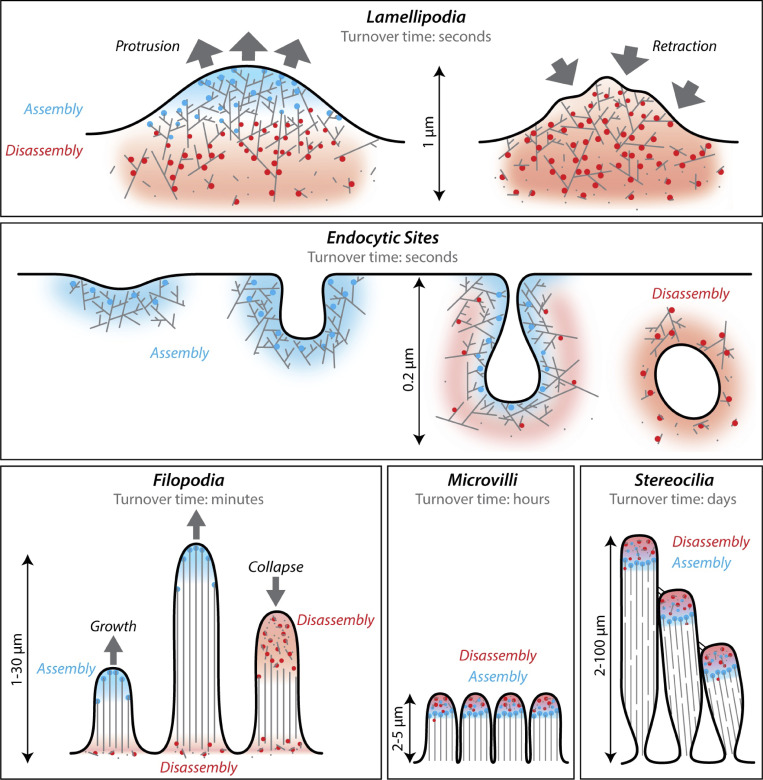
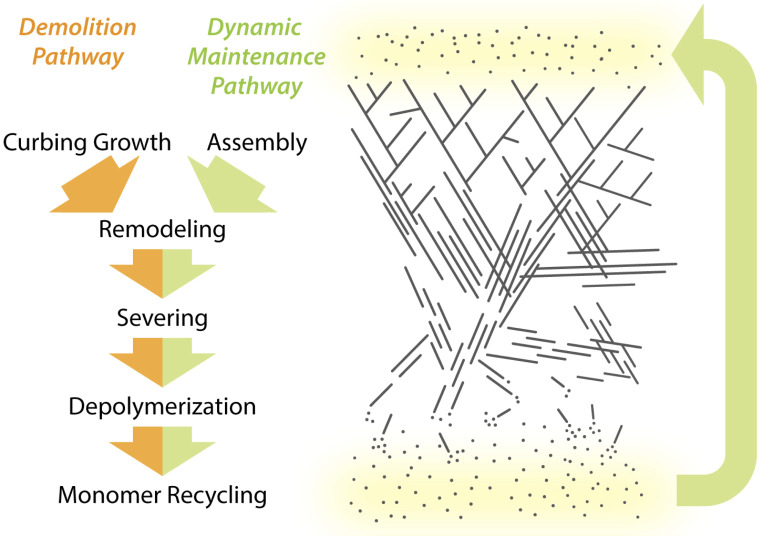
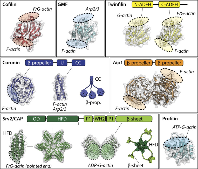
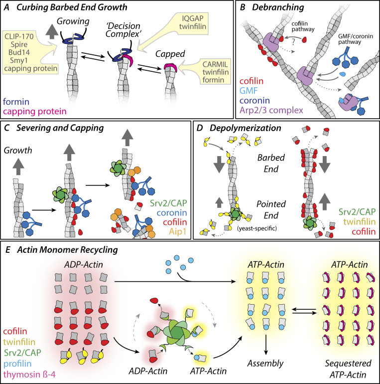
Cellular actin networks exhibit a wide range of sizes, shapes, and architectures tailored to their biological roles. Once assembled, these filamentous networks are either maintained in a state of polarized turnover or induced to undergo net disassembly. Further, the rates at which the networks are turned over and/or dismantled can vary greatly, from seconds to minutes to hours or even days. Here, we review the molecular machinery and mechanisms employed in cells to drive the disassembly and turnover of actin networks. In particular, we highlight recent discoveries showing that specific combinations of conserved actin disassembly-promoting proteins (cofilin, GMF, twinfilin, Srv2/CAP, coronin, AIP1, capping protein, and profilin) work in concert to debranch, sever, cap, and depolymerize actin filaments, and to recharge actin monomers for new rounds of assembly.


Conflict of interest statement

Disclosures: The authors declare no competing interests exist.

Figures

Figure 1.
Figure 1.
Cellular actin structures with different filamentous architectures and turnover dynamics. Panels show different types of in vivo actin networks and highlight the variation in their rates of turnover and filamentous architectures, as well as where new actin assembly occurs (blue) and where actin disassembly (red) occurs within each network. Blue dots, assembly-promoting factors. Red dots, disassembly-promoting factors. The upper row highlights lamellipodial protrusion and retraction. Protrusion (left) is driven by assembly of branched filaments, with disassembly occurring at the rear of the network. Retraction (right) is driven by attenuation of assembly coupled with network disassembly. At endocytic sites (middle row), branched filament assembly drives initial membrane invagination (left) and then a combination of assembly and disassembly drives vesiculation and scission (right). The lower row highlights formation and turnover of several different actin structures composed of unbranched filaments (filopodia, microvilli, and stereocilia). Filopodia are turned over by the demolition pathway, and thus grow until they abruptly collapse. In contrast, microvilli and stereocilia are regulated via the dynamic maintenance pathway, thus persisting as stable structures while their constituent actin filaments are continuously turned over.
Figure 2.
Figure 2.
Two pathways for cellular actin network turnover. Actin networks in cells are either maintained in a state of polarized flux, where filaments undergo dynamic turnover (dynamic maintenance pathway), or they are targeted for net disassembly (demolition pathway). Both pathways involve actin filament remodeling, severing, depolymerization, and monomer recycling. In the dynamic maintenance pathway, assembly and disassembly are balanced. In the demolition pathway, network growth is severely curbed, which promotes network collapse due to filament disassembly mechanisms.
Figure 3.
Figure 3.
Structures of proteins that promote actin turnover. Each protein (or its domains) is shown as surface-rendered views with embedded cartoons of the secondary structural elements. Shaded areas indicate approximate positions of the binding sites for G-actin, F-actin, and Arp2/3 complex (color-coded). Cofilin and GMF share the ADF-H domain fold. Twinfilin consists of two ADF-H domains separated by a short linker and flanked by a C-terminal tail that binds CP. Coronins oligomerize via their coiled-coil (CC) domains, and use their β-propeller and CC domains to bind F-actin, and their unique (U) and CC domains to interact with Arp2/3 complex. EM reconstructions have shown that the N-terminal half of Srv2/CAP, consisting of the oligomerization domain (OD) and HFD, assembles into hexameric shurikens that bind to the sides and pointed ends of actin filaments. The C-terminal half of Srv2/CAP further consists of an actin-binding WH2 domain flanked by two proline-rich domains (P1 and P2) and an actin-binding β-sheet/CARP domain. P1 mediates interactions with profilin, while P2 binds to SH3 domain–containing proteins. PDB ID of structures: 4BEX (human cofilin-1), 1VKK (mouse GMF-γ), 1M4J (N-ADFH mouse TWF1), 3DAW (C-ADFH mouse TWF1), 1PGU (yeast AIP1), 2AQ5 (β-propeller domain of mouse coronin-1A), 2AKF (coiled-coil domain of mouse coronin-1A), 1S0P (HFD of Dictyostelium Srv2/CAP), 1K4Z (β-sheet/CARP domain of yeast Srv2/CAP), and 2PAV (human profilin-1, extracted). N-ADFH, N-terminal actin depolymerizing factor homology; C-ADHF, C-terminal actin depolymerizing factor homology.
Figure 4.
Figure 4.
Molecular mechanisms driving F-actin and G-actin turnover. Each panel highlights a distinct step in actin network turnover. Proteins are color-coded. (A) Formins and CP join each other at the barbed ends of filaments to form decision complexes, and catalyze each other’s displacement. These transitions can be further accelerated by specific ligands of CP (e.g., twinfilin) and formins (e.g., IQGAP1), leading to rapid changes between states of filament growth and capping. Other ligands of CP and formins may influence their lifetimes at barbed ends. (B) Filament debranching mechanisms. The branch junctions nucleated by Arp2/3 complex are inherently stable, yet turn over rapidly in vivo. This is achieved by: (1) a mechanism involving cofilin binding to F-actin and/or Arp2/3 complex, and (2) mechanisms involving GMF and coronin, and their interactions with Arp2/3 complex. Debranching releases Arp2/3 complex, which can be strongly inhibited from nucleating actin assembly by GMF and coronin. (C) Filament severing and capping mechanisms. In the CCA mechanism, coronin binds to filaments first and recruits cofilin to these sites, increasing the efficiency of cofilin binding. Cofilin then recruits AIP1, which induces rapid severing. Severing produces new barbed ends, which are blocked from growth by CCA proteins. Filament severing can also be enhanced (4–10-fold) by a complementary mechanism in which Srv2/CAP and cofilin each bind independently to filament sides, and together accelerate severing. (D) Filament depolymerization mechanisms. At barbed ends, depolymerization can be accelerated by interactions with twinfilin or cofilin. At pointed ends, depolymerization can be accelerated by cofilin decoration of filament sides combined with processive association of Srv2/CAP with the pointed ends of filaments. Pointed end depolymerization also can be accelerated by Srv2/CAP and twinfilin, although the magnitude of the effects is species specific. (E) Regulation of the actin monomer pool. Filament disassembly releases ADP-actin monomers, which must be recycled for new rounds of assembly. Free ADP-actin monomers can bind profilin and rapidly exchange nucleotide (ATP for ADP). However, a large fraction of released ADP-actin monomers are bound to cofilin or twinfilin, which block nucleotide exchange and profilin binding. Srv2/CAP catalyzes the displacement of cofilin and twinfilin from the ADP-actin monomers, accelerates nucleotide exchange on G-actin, and hands off ATP-actin monomers to profilin. These activities stem from Srv2/CAP’s 100-fold higher affinity for ADP-actin (Kd = 18 nM) compared with ATP-actin (Kd = 1.8 µM). In vertebrate cells, high concentrations of ATP-actin monomers are maintained in a dynamic equilibrium between transient binding to thymosin-β4 (which keeps monomers in a sequestered state) and transient binding to profilin (which makes monomers available for assembly). Consumption of ATP-actin monomers by rapid filament assembly releases free profilin, which then rapidly replenishes the assembly-competent pool of profilin-bound ATP-actin monomers via dynamic competition with thymosin-β4.

References

    1. Akin, O., and Mullins R.D.. 2008. Capping protein increases the rate of actin-based motility by promoting filament nucleation by the Arp2/3 complex. Cell. 133:841–851. 10.1016/j.cell.2008.04.011 - DOI - PMC - PubMed
    1. Aktories, K., Lang A.E., Schwan C., and Mannherz H.G.. 2011. Actin as target for modification by bacterial protein toxins. FEBS J. 278:4526–4543. 10.1111/j.1742-4658.2011.08113.x - DOI - PubMed
    1. Almenar-Queralt, A., Lee A., Conley C.A., Ribas de Pouplana L., and Fowler V.M.. 1999. Identification of a novel tropomodulin isoform, skeletal tropomodulin, that caps actin filament pointed ends in fast skeletal muscle. J. Biol. Chem. 274:28466–28475. 10.1074/jbc.274.40.28466 - DOI - PubMed
    1. Andrianantoandro, E., and Pollard T.D.. 2006. Mechanism of actin filament turnover by severing and nucleation at different concentrations of ADF/cofilin. Mol. Cell. 24:13–23. 10.1016/j.molcel.2006.08.006 - DOI - PubMed
    1. Balcer, H.I., Goodman A.L., Rodal A.A., Smith E., Kugler J., Heuser J.E., and Goode B.L.. 2003. Coordinated regulation of actin filament turnover by a high-molecular-weight Srv2/CAP complex, cofilin, profilin, and Aip1. Curr. Biol. 13:2159–2169. 10.1016/j.cub.2003.11.051 - DOI - PubMed

Publication types