. 2023 Nov 9;13(1):19588.
doi: 10.1038/s41598-023-46467-4.

Drug screen identifies verteporfin as a regulator of lipid metabolism in macrophage foam cells

Affiliations

Drug screen identifies verteporfin as a regulator of lipid metabolism in macrophage foam cells

Nicholas Hoeffner et al. Sci Rep. .

Abstract

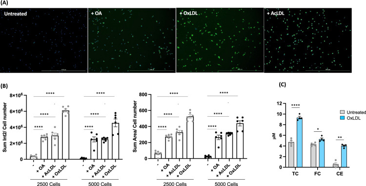
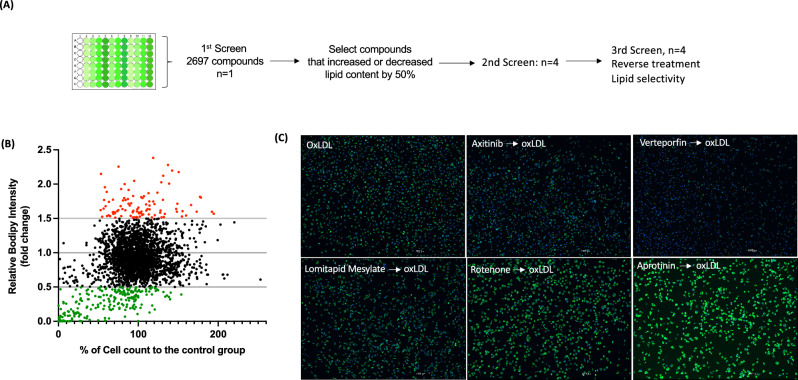
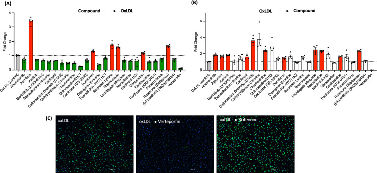
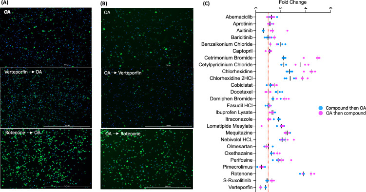
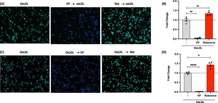
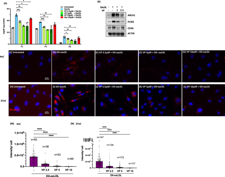
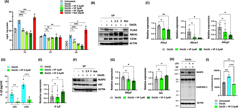
Arterial macrophage foam cells are filled with cholesterol ester (CE) stored in cytosolic lipid droplets (LDs). Foam cells are central players in progression of atherosclerosis as regulators of lipid metabolism and inflammation, two major driving forces of atherosclerosis development. Thus, foam cells are considered plausible targets for intervention in atherosclerosis. However, a compound that directly regulates the lipid metabolism of LDs in the arterial foam cells has not yet been identified. In this study, we screened compounds that inhibit macrophage foam cell formation using a library of 2697 FDA-approved drugs. From the foam cells generated via loading of human oxidized low-density lipoprotein (oxLDL), we found 21 and 6 compounds that reduced and enhanced accumulations of lipids respectively. Among them, verteporfin most significantly reduced oxLDL-induced foam cell formation whereas it did not display a significant impact on foam cell formation induced by fatty acid. Mechanistically our data demonstrate that verteporfin acts via inhibition of oxLDL association with macrophages, reducing accumulation of CE. Interestingly, while other drugs that reduced foam cell formation did not have impact on pre-existing foam cells, verteporfin treatment significantly reduced their total lipids, CE, and pro-inflammatory gene expression. Together, our study identifies verteporfin as a novel regulator of foam cell lipid metabolism and inflammation and a potential compound for intervention in atherosclerosis.


Conflict of interest statement

The authors declare no competing interests.

Figures

Figure 1
Figure 1
Establishment of foam cell detection (A) Images of foam cells treated with oleic acid (OA), oxidized LDL(OxLDL) and acetylated LDL(AcLDL) and stained by BODIPY 493/503, scale bar 1000μm. (B) Sum intensity normalized by cell number (left) and sum area normalized by cell number (right). Six areas containing an average 140 cells of each treatment were analyzed. (C) Total, free, and esterified cholesterols normalized by cell numbers. TC-total cholesterol, FC-free cholesterol, and CE-cholesterol ester. P values are by Student’s t test for paired samples (treated vs. untreated), ****P < 0.00005, **p < 0.005, and *p < 0.05.
Figure 2
Figure 2
Identification of drugs that impact foam cell formation (A) Experimental strategy. (B) Distribution of BODIPY value of all compounds (2697). Relative intensity of a compound treated to oxLDL-treated group. Red dot indicates 50% higher and green dot indicates 50% lower than oxLDL-treated group. (C) Images of cells treated with oxLDL (control) and pretreated with indicated drugs followed by oxLDL loading. Scale bar 1000μm.
Figure 3
Figure 3
Second screening was performed with the 100 compounds selected from 1st screen. Fold change of lipid contents to control (oxLDL treated) group. (A) Compounds were treated prior to oxLDL loading. Twenty-six drugs that significantly decreased (green bar) and increased (red bar) foam cell formation are presented. P values of all colored groups (red and green bars) are < 0.05 by Student’s t test for compared samples (control vs compound treated). (B) The effect of compounds on lipid contents of 26 compounds from (A) were measured after oxLDL loaded for 24 h. P values of all colored groups (red and green bars) are below 0.05. N = 4 per group. (C) The representative images of post drug treatment. Scale bar 1000μm.
Figure 4
Figure 4
Verteporfin reduces the formation and removal of OA treated foam cells but less impactable than oxLDL treated foam cells (A) Representative images of foam cells pretreated with drugs and then stimulated with OA. (B) Representative images of foam cells that treated with OA first then with drugs. Scale bar; 1000μM. (C) Comparison of the impact of 26 drugs treatments before or after OA loading, n = 4 per group. Fold change is relative BODIPY intensity of OA treated group.
Figure 5
Figure 5
Verteporfin decreases lipid contents in human macrophage THP-1 cells. (A) Images and (B) relative BODIPY intensity of VP (10 μM) and Rotenone (10 μM) pre-treated foam cells. (C) Images and (D) relative BODIPY intensity of foam cells treated with VP and Rotenone. P values are by Student’s t test for paired samples. *p < 0.05, **p < 0.005, ****p < 0.0005.
Figure 6
Figure 6
Verteporfin (VP) interferes with LDL association with macrophages. (A) BMMs were pretreated with drugs for 30 min before treating with oxLDL for 24 h. Total cholesterol (TC) and free cholesterol (FC) were measured as described in methods. Cholesterol ester (CE) was obtained by subtracting FC values from TC. N = 4 per group. P vales are by Student’s t test for compared groups (oxLDL treated vs. VP treated with oxLDL) (B) Expression levels of CD36, PLIN2 and ABCA1 in VP pretreated (1 and 2.5μM) foam cells (immunoblotting). (CG) VP was pretreated at 0, 2.5, 5, and 10μM followed by Dil-oxLDL (10 μg/ml) for 2 h at 4 oC. (HL) Decreased internalized oxLDL by VP preincubation. VP was pretreated at 0, 2.5, 5, and 10μM followed by Dil-oxLDL (10 μg/ml) for 3 h at 37 °C. (M) and (N) Quantification of fluorescence intensity on images of EI and GN, respectively. P vales are by Student’s t test for compared groups (with and without VP treatment) (****P < 0.00005, ***P < 0.0005, **p < 0.005 and *p < 0.05).
Figure 7
Figure 7
Treatment of verteporfin to pre-existing foam cells reduces CE contents and inflammation. (A) Foam cells were induced with oxLDL then washed and treated with drugs (μM) as indicated. Total cholesterol (TC) and free cholesterol (FC) were measured as described in methods. Cholesterol ester (CE) was obtained by subtracting FC values from TC. N = 4 per group. (B) Protein levels of CD36, and PLIN2 in VP pretreated (μM) foam cells (immunoblotting) and (C) mRNA expression of PLIN2, ABCA1, and ABCG1 relative to cyclophilin (qPCR). (D) IL-1β secretion and (E) expression (qPCR), N = 4 per group. (F) Protein levels of NLRP3 and ASC in VP pretreated (μM) foam cells (immunoblotting) and (G) mRNA expression of NLRP3 and ASC relative to cyclophilin (qPCR). (H) Immunoblot of cleaved capase-1 and (I) detection of caspase-1 activity in cultured supernatant. P vales are by Student’s t test for compared groups (with and without VP treatment) *0.005 < p ≤ 0.05, **0.0005 < p ≤ 0.005, ***0.00005 < p ≤ 0.0005, ****P < 0.00005.

Similar articles

Cited by

  • Lipid-Laden Macrophages in Pulmonary Diseases.
    Zhu Y, Choi D, Somanath PR, Zhang D. Zhu Y, et al. Cells. 2024 May 22;13(11):889. doi: 10.3390/cells13110889. Cells. 2024. PMID: 38891022 Free PMC article. Review.
  • Efferocytosis in atherosclerosis.
    Adkar SS, Leeper NJ. Adkar SS, et al. Nat Rev Cardiol. 2024 Nov;21(11):762-779. doi: 10.1038/s41569-024-01037-7. Epub 2024 May 15. Nat Rev Cardiol. 2024. PMID: 38750215 Review.

References

    1. Boren J, Williams KJ. The central role of arterial retention of cholesterol-rich apolipoprotein-B-containing lipoproteins in the pathogenesis of atherosclerosis: A triumph of simplicity. Curr. Opin. Lipidol. 2016;27:473–483. doi: 10.1097/MOL.0000000000000330. - DOI - PubMed
    1. Ference BA, et al. Low-density lipoproteins cause atherosclerotic cardiovascular disease. 1. Evidence from genetic, epidemiologic, and clinical studies. A consensus statement from the European Atherosclerosis Society Consensus Panel. Eur. Heart J. 2017;38:2459–2472. doi: 10.1093/eurheartj/ehx144. - DOI - PMC - PubMed
    1. Andersen M, Lenhard B, Whatling C, Eriksson P, Odeberg J. Alternative promoter usage of the membrane glycoprotein CD36. BMC Mol. Biol. 2006;7:8. doi: 10.1186/1471-2199-7-8. - DOI - PMC - PubMed
    1. Dubland JA, Francis GA. Lysosomal acid lipase: At the crossroads of normal and atherogenic cholesterol metabolism. Front. Cell Dev. Biol. 2015;3:3. doi: 10.3389/fcell.2015.00003. - DOI - PMC - PubMed
    1. Nagy L, Tontonoz P, Alvarez JG, Chen H, Evans RM. Oxidized LDL regulates macrophage gene expression through ligand activation of PPARgamma. Cell. 1998;93:229–240. doi: 10.1016/s0092-8674(00)81574-3. - DOI - PubMed

Publication types