. 2023 Nov 10;13(1):19570.
doi: 10.1038/s41598-023-47021-y.

Toward MR protocol-agnostic, unbiased brain age predicted from clinical-grade MRIs

Affiliations

Toward MR protocol-agnostic, unbiased brain age predicted from clinical-grade MRIs

Pedro A Valdes-Hernandez et al. Sci Rep. .

Abstract

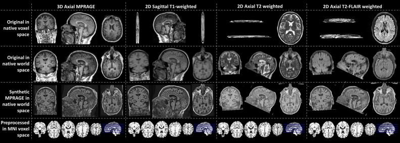
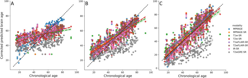
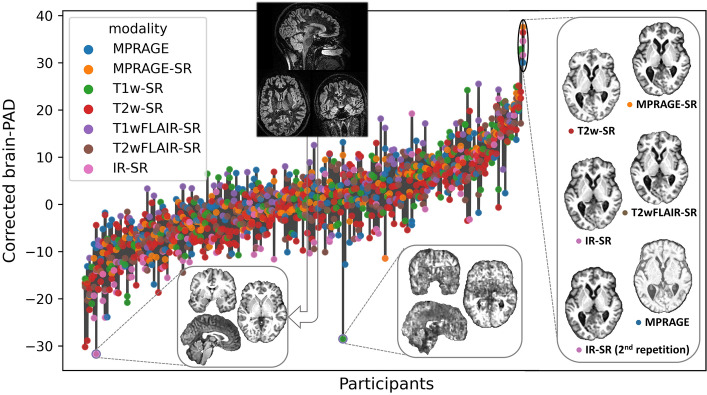
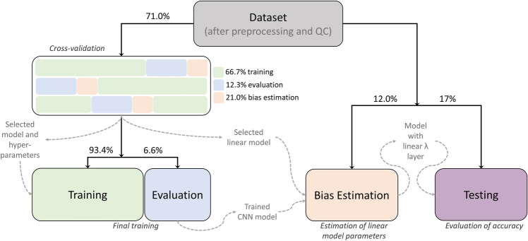
The difference between the estimated brain age and the chronological age ('brain-PAD') could become a clinical biomarker. However, most brain age models were developed for research-grade high-resolution T1-weighted MRIs, limiting their applicability to clinical-grade MRIs from various protocols. We adopted a dual-transfer learning strategy to develop a model agnostic to modality, resolution, or slice orientation. We retrained a convolutional neural network (CNN) using 6281 clinical MRIs from 1559 patients, among 7 modalities and 8 scanner models. The CNN was trained to estimate brain age from synthetic research-grade magnetization-prepared rapid gradient-echo MRIs (MPRAGEs) generated by a 'super-resolution' method. The model failed with T2-weighted Gradient-Echo MRIs. The mean absolute error (MAE) was 5.86-8.59 years across the other modalities, still higher than for research-grade MRIs, but comparable between actual and synthetic MPRAGEs for some modalities. We modeled the "regression bias" in brain age, for its correction is crucial for providing unbiased summary statistics of brain age or for personalized brain age-based biomarkers. The bias model was generalizable as its correction eliminated any correlation between brain-PAD and chronological age in new samples. Brain-PAD was reliable across modalities. We demonstrate the feasibility of brain age predictions from arbitrary clinical-grade MRIs, thereby contributing to personalized medicine.


Conflict of interest statement

The authors declare no competing interests.

Figures

Figure 1
Figure 1
Predicted MPRAGEs for three of the five modalities included in this study. The data belongs to a single participant. The top row shows selected slices in the slice, phase-encoding and readout directions (left, right top and right bottom, respectively). The bottom row shows the bottommost (z =  − 27 mm, slice 46), topmost (z = 53 mm, slice 126), and three intermediate slices (z =  − 7, 13, and 33 mm, slices 66, 86 and 106, respectively) of the 80 slices used for brain age prediction. To show images in world space, trilinear interpolation was used. FLAIR fluid attenuated inversion recovery.
Figure 2
Figure 2
Brain age prediction using the selected CNN (VGG16-based DeepBrainNet) for the MRIs of the testing held-out sample. (A) Predictions using the original (un-retrained) model, (B) predictions using the retrained model and (C) bias-corrected predictions using the retrained model. The colored lines represent the slope of the linear relation between the chronological and the predicted brain age for each modality. MPRAGE magnetization-prepared rapid gradient-echo, T1w T1-weighted, T2w T2-weighted, FLAIR fluid attenuated inversion recovery, GRE gradient echo, IR inversion recovery, [Modality]-SR super-resolution synthetic MPRAGE version of [modality].
Figure 3
Figure 3
Variability of the bias-corrected predicted brain-PADs for each participant (ordered from the lowest to the highest brain-PAD). The bars represent the range of PAD values within each subject. To illustrate the possible causes of some of the outliers that affect the intra-subject reliability, we show the synthetic MPRAGEs (below the scatter plots) of the two images with bias-corrected brain-PAD farthest from the mean in their own within-subject group. The final image used to estimate brain age, to the left, had no apparent issues. However, it can be appreciated that the corresponding actual MRI (above the scatter plots) is highly noisy. This is because SynthSR is sometimes able to reconstruct any noisy MRI to a synthetic MPRAGE with a relatively good SNR. Thus, QC has to be also done on the actual MRIs. The case to the right had simply such a very poor quality that even SynthSR was not able to recover a meaningful synthetic MPRAGE. This image was not detected by the QC described in this study. Moreover, the inset to the right also shows the actual MPRAGE and synthetic MPRAGEs for the subject having the highest average bias-corrected brain-PAD (26.7 years) in the test set (this participant had no T1wFLAIR or T1w but had two IRs).
Figure 4
Figure 4
Flowchart of the definition of the data domains and the different stages of training and evaluation. After selecting the whole brain MRIs, preprocessing, and performing QC, the final dataset was split for training the CNN model (training set), estimating the parameters of the bias model in a different independent sample (the bias estimation set) and testing accuracy (external validation in the held-out testing set) and reliability. In more detail, three-fold cross-validation was used on the training set to determine the optimal combination of CNN model, tune hyperparameters and select a bias model. The final training was performed on the training set using the optimal configuration. The trained CNN model was then used to predict the bias-uncorrected brain ages that were used to estimate the parameters of the bias model in the independent bias estimation set. Finally, the bias model was added on top of the CNN for deployment and tested in the held-out testing set.

Update of

References

    1. Cole JH, Franke K. Predicting age using neuroimaging: Innovative brain ageing biomarkers. Trends Neurosci. 2017;40:681–690. doi: 10.1016/j.tins.2017.10.001. - DOI - PubMed
    1. Bashyam VM, et al. MRI signatures of brain age and disease over the lifespan based on a deep brain network and 14,468 individuals worldwide. Brain. 2020;143:2312–2324. doi: 10.1093/brain/awaa160. - DOI - PMC - PubMed
    1. Yin C, et al. Anatomically interpretable deep learning of brain age captures domain-specific cognitive impairment. Proc. Natl. Acad. Sci. U.S.A. 2023;120:1–11. doi: 10.1073/pnas.2214634120. - DOI - PMC - PubMed
    1. Cole JH, et al. Brain age predicts mortality. Mol. Psychiatry. 2018;23:1385–1392. doi: 10.1038/mp.2017.62. - DOI - PMC - PubMed
    1. Montesino-Goicolea S, Valdes-Hernandez PA, Cruz-Almeida Y. Chronic musculoskeletal pain moderates the association between sleep quality and dorsostriatal-sensorimotor resting state functional connectivity in community-dwelling older adults. Pain Res. Manag. 2022;2022:1–12. doi: 10.1155/2022/4347759. - DOI - PMC - PubMed

Publication types

MeSH terms