. 2023 Nov 10;6(1):1145.
doi: 10.1038/s42003-023-05506-6.

Repeated multi-domain cognitive training prevents cognitive decline, anxiety and amyloid pathology found in a mouse model of Alzheimer disease

Affiliations

Repeated multi-domain cognitive training prevents cognitive decline, anxiety and amyloid pathology found in a mouse model of Alzheimer disease

Jogender Mehla et al. Commun Biol. .

Abstract

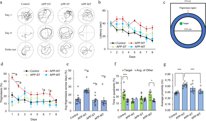
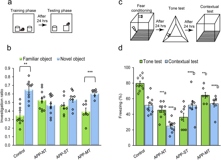
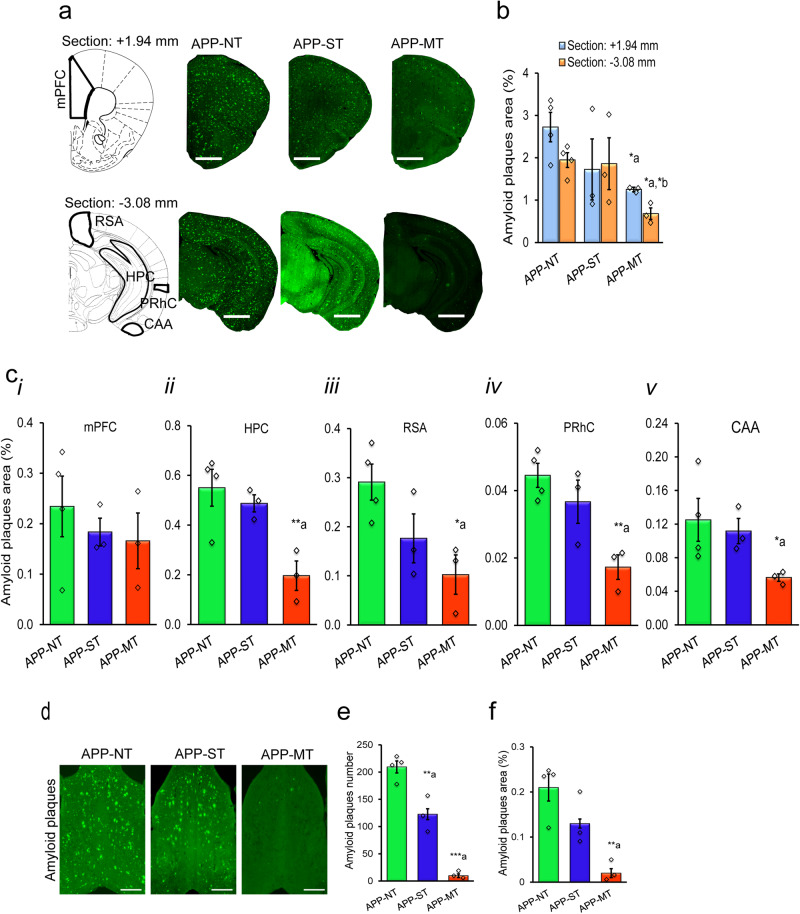
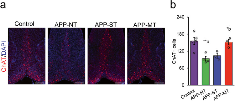
Education, occupation, and an active lifestyle, comprising enhanced social, physical, and mental components are associated with improved cognitive functions in aged people and may delay the progression of various neurodegenerative diseases including Alzheimer's disease. To investigate this protective effect, 3-month-old APPNL-G-F/NL-G-F mice were exposed to repeated single- or multi-domain cognitive training. Cognitive training was given at the age of 3, 6, & 9 months. Single-domain cognitive training was limited to a spatial navigation task. Multi-domain cognitive training consisted of a spatial navigation task, object recognition, and fear conditioning. At the age of 12 months, behavioral tests were completed for all groups. Then, mice were sacrificed, and their brains were assessed for pathology. APPNL-G-F/NL-G-F mice given multi-domain cognitive training compared to APPNL-G-F/NL-G-F control group showed an improvement in cognitive functions, reductions in amyloid load and microgliosis, and a preservation of cholinergic function. Additionally, multi-domain cognitive training improved anxiety in APPNL-G-F/NL-G-F mice as evidenced by measuring thigmotaxis behavior in the Morris water maze. There were mild reductions in microgliosis in the brain of APPNL-G-F/NL-G-F mice with single-domain cognitive training. These findings provide causal evidence for the potential of certain forms of cognitive training to mitigate the cognitive deficits in Alzheimer disease.


Conflict of interest statement

The authors declare no competing interests.

Figures

Fig. 1
Fig. 1. Experimental design for the study.
The study was conducted for 12 months. For multi-domain cognitive training (MT), we performed the spatial version of the Morris water task (MWM), novel object recognition (NOR) and fear conditioning (tone and context) tests. We used only MWM for single-domain cognitive training (ST).
Fig. 2
Fig. 2. Effects of cognitive training on spatial learning and memory functions of 12 months old control and APPNL-G-F/NL-G-F mice in the MWM.
a Representative swim path on Day 1, Day 8, and probe trial for various experimental groups. Overall comparison of experimental groups considering mean latency to find the hidden escape platform during the acquisition phase. b Mean latency to find the hidden escape platform during the acquisition phase for various groups. c Representative pool schematic showing the size of the outer and inner regions of the pool. Any swimming in the blue region (thigmotaxis area) was counted as time in thigmotaxis. Adopted from the ReadMe in the wtr2100 documentation. d Percent thigmotaxis showed by mice on each day during the acquisition trials. e Average thigmotaxis (%) across the days shown by mice during the acquisition trials (average of days 1–8 for each group). f Percent time spent by mice in the target quadrant and average of other quadrants during the probe trial. g Average proximity to the platform during the probe trial. Data is presented as mean ± SEM. Data is presented as mean ± SEM. *P < 0.05, **P < 0.01, ***P < 0.001 are considered as statistically significant. a—as compared to the control group; b—as compared to the APP-NT group. Control group (n = 10)—C57BL/6. APP-NT group (n = 10)—APPNL-G-F/NL-G-F mice with no training. APP-ST group (n = 8)—APPNL-G-F/NL-G-F mice exposed to single-domain cognitive training (ST). APP-MT group (n = 7)—APPNL-G-F/NL-G-F mice exposed to multi-domain cognitive training (MT).
Fig. 3
Fig. 3. Effect of cognitive training on learning and memory functions of 12-month-old APPNL-G-F/NL-G-F mice in the novel object recognition (NOR) and fear conditioning tests.
a Schematic representation of the novel object recognition test. b Investigation ratio for familiar and novel objects. c Schematic representation of the fear conditioning test. d Percent freezing for tone and contextual test. Data is presented as mean ± SEM. **P < 0.01; ***P < 0.001; a—as compared with control group. b—as compared with the APP-NT group. Control group (n = 10)—C57BL/6. APP-NT group (n = 10)—APPNL-G-F/NL-G-F with no training. APP-ST group (n = 8)—APPNL-G-F/NL-G-F mice exposed to single-domain cognitive training (ST). APP-MT group (n = 7)—APPNL-G-F/NL-G-F mice exposed to multi-domain cognitive training (MT).
Fig. 4
Fig. 4. Amyloid pathology in brain of 12-month-old APPNL-G-F/NL-G-F mice.
a Photomicrographs of amyloid plaques stained with methoxy-XO4 (green) in two brain sections (top: bregma +1.94 mm; bottom: −3.08 mm). b Summary results of amyloid plaque area in the brain. c Amyloid plaques area in medial prefrontal cortex (mPFC)(i), hippocampus (HPC)(ii) retrospenial area (RSA)(iii), perirhinal cortex (PRhC)(iv), and cortical amygdalar area (CAA)(v). Scale bars represent 1 mm for section +1.94 mm and 2.5 mm for section −3.08 mm. d Photomicrographs of amyloid plaques stained with methoxy-XO4 (green) in MSDB complex. e Total amyloid plaque number in MSDB complex. f Amyloid plaque area in MSDB complex. Scale bar—500 µm for whole sections. Data is presented as mean ± SEM. *P < 0.05, **P < 0.01, **P < 0.001; a—as compared to APP-NT group. b—as compared to the APP-ST group. APP-NT group (n = 4)—APPNL-G-F/NL-G-F with no training. APP-ST group (n = 3)—APPNL-G-F/NL-G-F mice exposed to single-domain cognitive training (ST). APP-MT group (n = 3)—APPNL-G-F/NL-G-F mice exposed to multi-domain cognitive training (MT).
Fig. 5
Fig. 5. Microgliosis in the brain of 12-month-old mice.
a Photomicrographs of activated IBA-1, a marker of microgliosis in the brain. Representative of activated microglia in two brain sections (top: bregma +1.94 mm; bottom: −3.08 mm). Scale bars represent 1 mm for section +1.94 mm and 2.5 mm for section −3.08 mm. b Activated IBA-1 immunostained area in brain sections +1.94 mm and section −3.08 mm. c Microgliosis in different brain regions of 12 months old mice. Activated IBA-1 immunostained area in the medial prefrontal cortex (mPFC)(i), hippocampus (HPC)(ii), retrospenial area (RSA)(iii), perirhinal cortex (PRhC)(iv), and cortical amygdalar area (CAA)(v). d Photomicrographs of immunostaining of activated IBA-1 (red) in MSDB complex. e Activated IBA-1 number in MSDB complex. f Activated IBA-1 immunostained area in MSDB complex. Scale bar—500 µm for whole sections. Data is presented as mean ± SEM. *P < 0.05, **P < 0.01, ***P < 0.001, a—as compared to control group; b—as compared to APP-NT group. Control group (n = 5)—C57BL/6. APP-NT group (n = 4)—APPNL-G-F/NL-G-F with no training. APP-ST group (n = 3)—APPNL-G-F/NL-G-F mice exposed to single-domain cognitive training (ST). APP-MT group (n = 3)—APPNL-G-F/NL-G-F mice exposed to multi-domain cognitive training (MT).
Fig. 6
Fig. 6. Cholinergic function in the medial septum-diagonal band (MSDB) complex of the basal forebrain of 12-month-old mice.
a Photomicrographs of immunohistochemistry staining of choline acetyltransferase (ChAT) in MSDB complex. ChAT—a cholinergic marker stained with monoclonal rabbit anti-ChAT antibody (red), DAPI—stained the nuclei (blue). b Quantification of ChAT in MSDB complex. Scale bar—500 µm for whole sections. Data is presented as mean ± SEM. *P < 0.05, **P < 0.01, a—as compared to control group; b—as compared to APP-NT group. Control group (n = 5)—C57BL/6. APP-NT group (n = 4)—APPNL-G-F/NL-G-F with no training. APP-ST group (n = 3)—APPNL-G-F/NL-G-F mice exposed to single-domain cognitive training (ST). APP-MT group (n = 3)—APPNL-G-F/NL-G-F mice exposed to multi-domain cognitive training (MT).

References

    1. Selkoe DJ, Hardy J. The amyloid hypothesis of Alzheimer’s disease at 25 years. EMBO Mol. Med. 2016;8:595–608. doi: 10.15252/emmm.201606210. - DOI - PMC - PubMed
    1. Corriveau RA, et al. Alzheimer’s Disease-Related Dementias Summit 2016: National research priorities. Neurology. 2017;89:2381–2391. doi: 10.1212/WNL.0000000000004717. - DOI - PMC - PubMed
    1. Lye TC, Shores EA. Traumatic brain injury as a risk factor for Alzheimer’s disease: a review. Neuropsychol. Rev. 2000;10:115–129. doi: 10.1023/A:1009068804787. - DOI - PubMed
    1. Ownby RL, Crocco E, Acevedo A, John V, Loewenstein D. Depression and risk for Alzheimer disease: systematic review, meta-analysis, and metaregression analysis. Arch. Gen. Psychiatry. 2006;63:530–538. doi: 10.1001/archpsyc.63.5.530. - DOI - PMC - PubMed
    1. Mayeux R, Stern Y. Epidemiology of Alzheimer disease. Cold Spring Harb. Perspect. Med. 2012;2:a006239. doi: 10.1101/cshperspect.a006239. - DOI - PMC - PubMed

Publication types

Substances