. 2023 Nov 11;24(1):425.
doi: 10.1186/s12859-023-05559-8.

Role of environmental specificity in CASP results

Affiliations

Role of environmental specificity in CASP results

Irena Roterman et al. BMC Bioinformatics. .

Abstract

Background: Recently, significant progress has been made in the field of protein structure prediction by the application of artificial intelligence techniques, as shown by the results of the CASP13 and CASP14 (Critical Assessment of Structure Prediction) competition. However, the question of the mechanism behind the protein folding process itself remains unanswered. Correctly predicting the structure also does not solve the problem of, for example, amyloid proteins, where a polypeptide chain with an unaltered sequence adopts a different 3D structure.

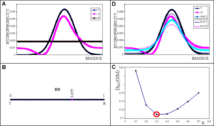
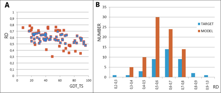
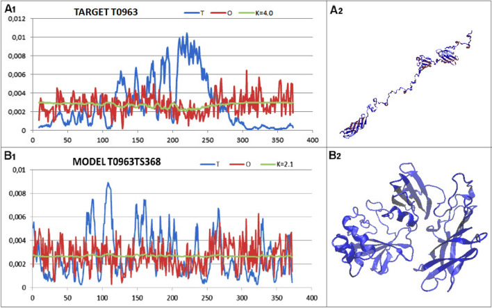
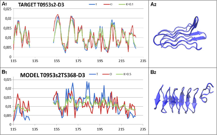
Results: This work was an attempt at explaining the structural variation by considering the contribution of the environment to protein structuring. The application of the fuzzy oil drop (FOD) model to assess the validity of the selected models provided in the CASP13, CASP14 and CASP15 projects reveals the need for an environmental factor to determine the 3D structure of proteins. Consideration of the external force field in the form of polar water (Fuzzy Oil Drop) and a version modified by the presence of the hydrophobic compounds, FOD-M (FOD-Modified) reveals that the protein folding process is environmentally dependent. An analysis of selected models from the CASP competitions indicates the need for structure prediction as dependent on the consideration of the protein folding environment.

Conclusions: The conditions governed by the environment direct the protein folding process occurring in a certain environment. Therefore, the variation of the external force field should be taken into account in the models used in protein structure prediction.

Keywords: CASP; Folding environment; Folding simulation in Silico; Protein folding.


Conflict of interest statement

The authors declare no conflict of interest.

Figures

Fig. 1
Fig. 1
Graphical visualisation of the FOD-M model assumptions. A An example set of T (blue), O (pink) and R (brown) distributions. B The determined RD value for the example in (A) is 0.633, as shown on the axis. This value is calculated to express the difference expressed by DKL(O|T) and DKL(O|R) according to Eq. 4. C Determination of optimum value for K – the minimum DKL value for different K values results in best fit. The K = 0.3 appears to be the optimal one for presented example. D The juxtaposition of the T (blue), O (pink) and M (cyan) distributions for K = 0.3 illustrates the interpretation of the M distribution, which most closely replicates the O distribution in the example in question. Additional (thin lines) represent the M distributions for K = 0.1 (thin line – pink) and M distribution for K = 0.5 (thin line – blue). The comparison of M distributions visualises the best fit for M distribution for K = 0.3
Fig. 2
Fig. 2
Dependence of the GDT_TS scale score on the status of the model protein structure expressed on the RD scale. The vertical lines are the RD values for the targets. The height of the vertical lines is the maximum score level on the GDT_TS scale. The encircled positions are the results obtained with AlphaFold
Fig. 3
Fig. 3
Example of incompatibility for T0953s2-D3. The blue dots on the x-axis identify the area that does not reproduce the arrangement present in the target protein. The 3D presentation with a highlighted section of the chain where a significant discrepancy between the top-ranked model against the target is present
Fig. 4
Fig. 4
Characteristics of the N-terminal helical domain of biba, a group b Streptococcus immunogenic bacterial adhesin (PDB ID—6POO). A 3D structure left – the target T1030; right – the model T1030TS427-1 B set of T, O and M profiles for the corresponding K representing the target T1030 C set of T, O and M profiles for the corresponding K, representing the model T1030TS427-1
Fig. 5
Fig. 5
Set of T, O and M profiles for the corresponding K, representing A Model T1029TS361-D1-1 B Target T1029
Fig. 6
Fig. 6
Analysis of the models obtained with the Baker-Rosettaserver A relationship of the RD value to the GDT_TS score. The blue dots represent the range of RD of highest representation as shown in B. B number of available targets and delivered models for the RD value ranges
Fig. 7
Fig. 7
Summary of the T, O and M profiles for the corresponding K values. A Target T0963 together with the 3D structure of the native form of the protein in question (PDB ID 6CL6) B – Model T0963TS368 together with the 3D structure proposed using the Baker-Rosettaserver (participant No. 368) [43]
Fig. 8
Fig. 8
LOW A set of the T, O and M profiles for the respective K values, together with a 3D presentation for: A target T0953s2-D3 B model T0953s2TS368-D3
Fig. 9
Fig. 9
Summary of the results based on the examples discussed. The vertical axis expresses the number of models provided for a given range of RD values. A all examples discussed in this work. B target T0953s2-D3 representing the lowest RD = 0.286 for the target status – the range distinguished by the red bar

References

    1. Dill KA, MacCallum JL. The protein-folding problem, 50 years on. Science. 2012;338(6110):1042–1046. doi: 10.1126/science.1219021. - DOI - PubMed
    1. Dill KA, Ozkan SB, Weikl TR, Chodera JD, Voelz VA. The protein folding problem: when will it be solved? Curr Opin Struct Biol. 2007;17(3):342–346. doi: 10.1016/j.sbi.2007.06.001. - DOI - PubMed
    1. https://predictioncenter.org/ (accessed Aug 7, 2023)
    1. MacCallum JL, Pérez A, Schnieders MJ, Hua L, Jacobson MP, Dill KA. Assessment of protein structure refinement in CASP9. Proteins. 2011;79(Suppl 10):74–90. doi: 10.1002/prot.23131. - DOI - PMC - PubMed
    1. Runthala A. Protein structure prediction: challenging targets for CASP10. J Biomol Struct Dyn. 2012;30(5):607–615. doi: 10.1080/07391102.2012.687526. - DOI - PubMed

LinkOut - more resources