

Linking brain structure, cognition, and sleep: insights from clinical data
- PMID: 37950486
- PMCID: PMC10851868
- DOI: 10.1093/sleep/zsad294
Linking brain structure, cognition, and sleep: insights from clinical data
Abstract
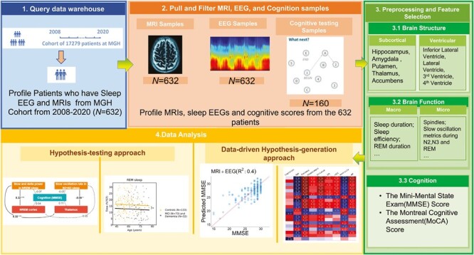
Study objectives: To use relatively noisy routinely collected clinical data (brain magnetic resonance imaging (MRI) data, clinical polysomnography (PSG) recordings, and neuropsychological testing), to investigate hypothesis-driven and data-driven relationships between brain physiology, structure, and cognition.
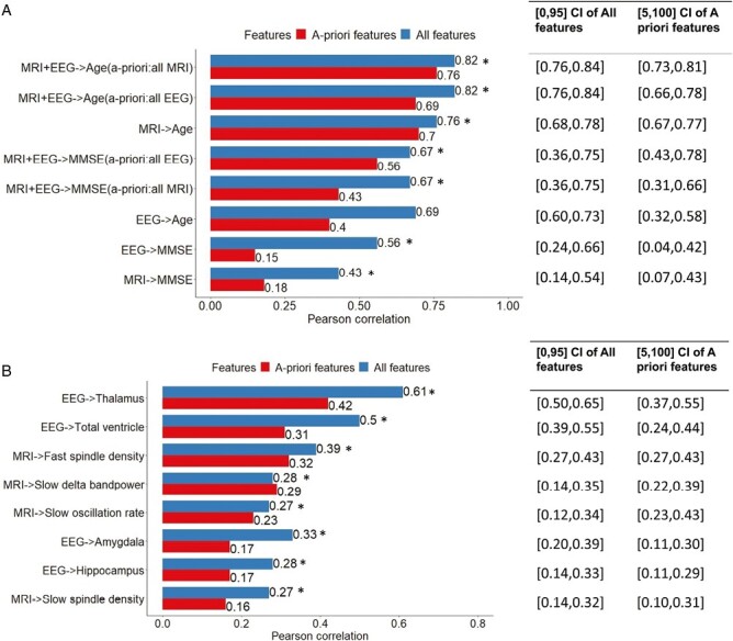
Methods: We analyzed data from patients with clinical PSG, brain MRI, and neuropsychological evaluations. SynthSeg, a neural network-based tool, provided high-quality segmentations despite noise. A priori hypotheses explored associations between brain function (measured by PSG) and brain structure (measured by MRI). Associations with cognitive scores and dementia status were studied. An exploratory data-driven approach investigated age-structure-physiology-cognition links.
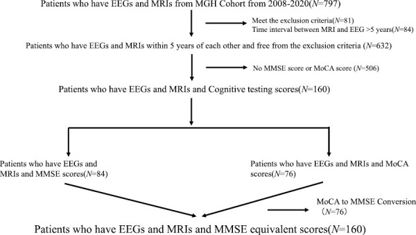
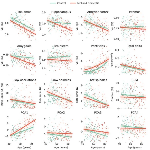
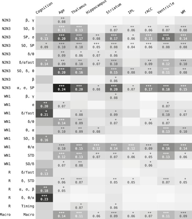
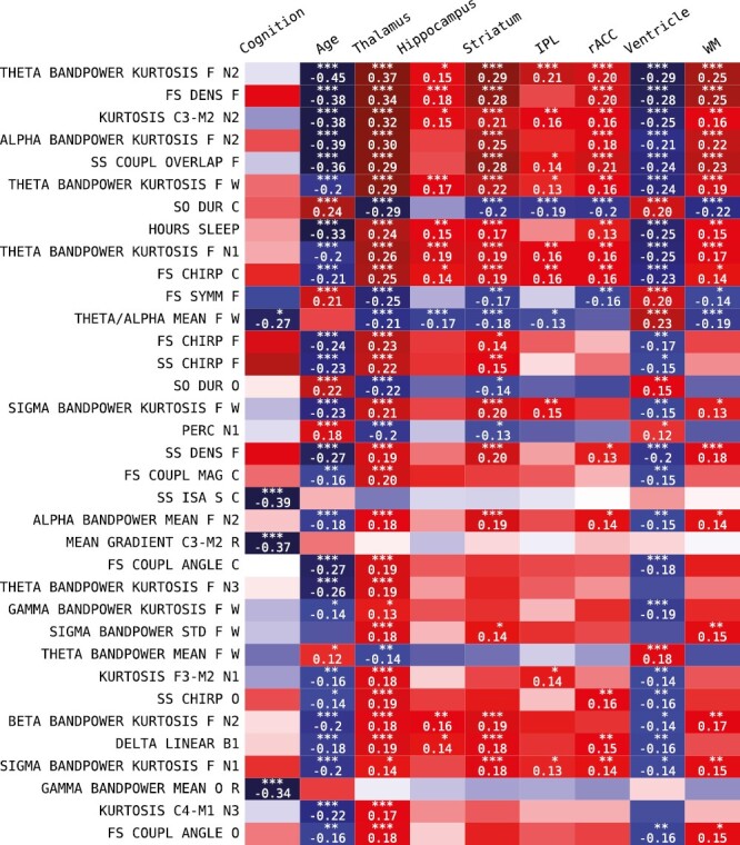
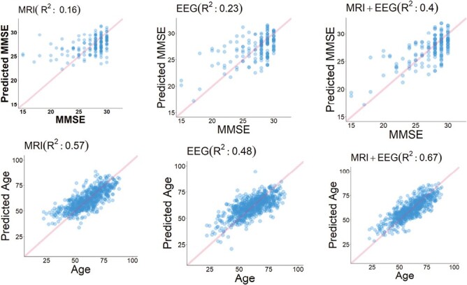
Results: Six hundred and twenty-three patients with sleep PSG and brain MRI data were included in this study; 160 with cognitive evaluations. Three hundred and forty-two participants (55%) were female, and age interquartile range was 52 to 69 years. Thirty-six individuals were diagnosed with dementia, 71 with mild cognitive impairment, and 326 with major depression. One hundred and fifteen individuals were evaluated for insomnia and 138 participants had an apnea-hypopnea index equal to or greater than 15. Total PSG delta power correlated positively with frontal lobe/thalamic volumes, and sleep spindle density with thalamic volume. rapid eye movement (REM) duration and amygdala volume were positively associated with cognition. Patients with dementia showed significant differences in five brain structure volumes. REM duration, spindle, and slow-oscillation features had strong associations with cognition and brain structure volumes. PSG and MRI features in combination predicted chronological age (R2 = 0.67) and cognition (R2 = 0.40).
Conclusions: Routine clinical data holds extended value in understanding and even clinically using brain-sleep-cognition relationships.
Keywords: brain health; cognition; electroencephalography; magnetic resonance imaging; polysomnography; sleep.
© The Author(s) 2023. Published by Oxford University Press on behalf of Sleep Research Society. All rights reserved. For permissions, please e-mail: journals.permissions@oup.com.
Figures
Comment in
-
Multimodal integration of sleep electroencephalogram, brain imaging, and cognitive assessments: approaches using noisy clinical data.Sleep. 2024 Feb 8;47(2):zsad305. doi: 10.1093/sleep/zsad305. Sleep. 2024. PMID: 38019853 Free PMC article. No abstract available.
References
Publication types
MeSH terms
Grants and funding
- R01NS102190/GF/NIH HHS/United States
- R01 NS102574/NS/NINDS NIH HHS/United States
- R01 NS107291/NS/NINDS NIH HHS/United States
- RF1 AG064312/AG/NIA NIH HHS/United States
- RF1 NS120947/NS/NINDS NIH HHS/United States
- R01 AG073410/AG/NIA NIH HHS/United States
- R01 HL161253/HL/NHLBI NIH HHS/United States
- R01 NS126282/NS/NINDS NIH HHS/United States
- R01 AG073598/AG/NIA NIH HHS/United States
- RF1 MH123195/MH/NIMH NIH HHS/United States
- R01 AG070988/AG/NIA NIH HHS/United States
- R01 EB031114/EB/NIBIB NIH HHS/United States
- UM1 MH130981/MH/NIMH NIH HHS/United States
- P41 EB015902/EB/NIBIB NIH HHS/United States
LinkOut - more resources
Full Text Sources
Medical
