. 2023 Dec 11;24(12):5605-5619.
doi: 10.1021/acs.biomac.3c00596. Epub 2023 Nov 11.

Preparation and Characterization of Softwood and Hardwood Nanofibril Hydrogels: Toward Wound Dressing Applications

Affiliations

Preparation and Characterization of Softwood and Hardwood Nanofibril Hydrogels: Toward Wound Dressing Applications

Yağmur Baş et al. Biomacromolecules. .

Abstract

Hydrogels of cellulose nanofibrils (CNFs) are promising wound dressing candidates due to their biocompatibility, high water absorption, and transparency. Herein, two different commercially available wood species, softwood and hardwood, were subjected to TEMPO-mediated oxidation to proceed with delignification and oxidation in a one-pot process, and thereafter, nanofibrils were isolated using a high-pressure microfluidizer. Furthermore, transparent nanofibril hydrogel networks were prepared by vacuum filtration. Nanofibril properties and network performance correlated with oxidation were investigated and compared with commercially available TEMPO-oxidized pulp nanofibrils and their networks. Softwood nanofibril hydrogel networks exhibited the best mechanical properties, and in vitro toxicological risk assessment showed no detrimental effect for any of the studied hydrogels on human fibroblast or keratinocyte cells. This study demonstrates a straightforward processing route for direct oxidation of different wood species to obtain nanofibril hydrogels for potential use as wound dressings, with softwood having the most potential.


Conflict of interest statement

The authors declare no competing financial interest.

Figures
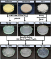
Figure 1
Figure 1
Photographs of the production steps of TO-SWNF, TO-HWNF, and TO-CNF.
Figure 2
Figure 2
(A) Updegraff cellulose and lignin portions of SW, HW, TO-SW, and TO-HW. Lignin was determined using two different analytical methods, namely, Klason lignin assay for SW and TO-SW samples and acetyl bromide assay for HW and TO-HW samples. (B) Main hemicellulosic monosugar composition of the noncrystalline fractions of SW, HW, TO-C, and oxidized wood samples. Values belong to the noncrystalline polysaccharides in the cell wall of SW and HW, and the noncrystalline parts of TO-C, TO-SW, and TO-HW.
Figure 3
Figure 3
SEM images of (A) TO-SW, (B) TO-HW, and (C) TO-C fibers prior to fibrillation.
Figure 4
Figure 4
Polarized optical micrographs of (a) TO-SW, (b) TO-HW, (c) TO-C, and (d) TO-SWNF, (e) TO-HWNF, and (f) TO-CNF. Scale: 500 μm.
Figure 5
Figure 5
AFM height images and corresponding height distribution profiles of (a) TO-SWNF, (b) TO-HWNF, and (c) TO-CNF.
Figure 6
Figure 6
Photographs of (a) TO-SWNF, (b) TO-HWNF, (c) TO-CNF as dry networks, and (d) TO-SWNF, (e) TO-HWNF, and (f) TO-CNF as water-absorbed networks.
Figure 7
Figure 7
SEM surface images of (a) TO-SWNF, (b) TO-HWNF, and (c) TO-CNF networks. (d) FTIR spectra, (e) water absorption profiles, (f) mass loss curves, and (g) derivative weight curves of networks.
Figure 8
Figure 8
(a) Ultimate tensile strength, (b) elongation at break, and (c) elastic modulus of networks in wet conditions. (d) Photograph of wet TO-SWNF network applied on hand.
Figure 9
Figure 9
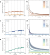
Storage and loss moduli of (a) TO-SWNF, (c) TO-HWNF, and (e) TO-CNF networks as a function of time. Axial force measurement of (b) TO-SWNF, (d) TO-HWNF, and (f) TO-CNF networks at equilibrium water absorption as a function of step time in the compression–stress relaxation test. Dashed lines in (a, c, e) correspond to the start and end of relaxation times for applied compression to axial force levels 0.1, 0.5, 1, 2, 4, and 6 N.
Figure 10
Figure 10
Graphs illustrating proliferation over time following exposure to TO-HWNF, TO-SWNF, and TO-CNF, as well as nontreated control. (A, C) Keratinocytes and (B, D) fibroblasts.
Figure 11
Figure 11
Graphs illustrating average cell speed over time following exposure to TO-HWNF, TO-SWNF, and TO-CNF, as well as nontreated control. (A) Keratinocytes and (B) fibroblasts.

References

    1. Moon R. J.; Martini A.; Nairn J.; Simonsen J.; Youngblood J. Cellulose Nanomaterials Review: Structure, Properties and Nanocomposites. Chem. Soc. Rev. 2011, 40, 3941–3994. 10.1039/c0cs00108b. - DOI - PubMed
    1. Nogi M.; Iwamoto S.; Nakagaito A. N.; Yano H. Optically Transparent Nanofiber Paper. Adv. Mater. 2009, 21, 1595–1598. 10.1002/adma.200803174. - DOI
    1. Abe K.; Yano H. Cellulose Nanofiber-Based Hydrogels with High Mechanical Strength. Cellulose 2012, 19, 1907–1912. 10.1007/s10570-012-9784-3. - DOI
    1. Basti A. T. K.; Jonoobi M.; Sepahvand S.; Ashori A.; Siracusa V.; Rabie D.; Mekonnen T. H.; Naeijian F. Employing Cellulose Nanofiber-Based Hydrogels for Burn Dressing. Polymers 2022, 14, 120710.3390/polym14061207. - DOI - PMC - PubMed
    1. Kolakovic R.; Peltonen L.; Laukkanen A.; Hirvonen J.; Laaksonen T. Nanofibrillar Cellulose Films for Controlled Drug Delivery. Eur. J. Pharm. Biopharm. 2012, 82, 308–315. 10.1016/j.ejpb.2012.06.011. - DOI - PubMed

Publication types