. 2023 Nov 11;80(12):357.
doi: 10.1007/s00018-023-05005-1.

MiR-326-mediated overexpression of NFIB offsets TGF-β induced epithelial to mesenchymal transition and reverses lung fibrosis

Affiliations

MiR-326-mediated overexpression of NFIB offsets TGF-β induced epithelial to mesenchymal transition and reverses lung fibrosis

Bijay Pattnaik et al. Cell Mol Life Sci. .

Abstract

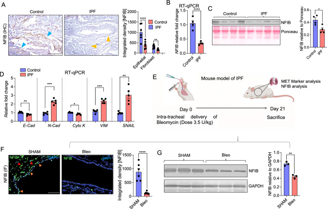
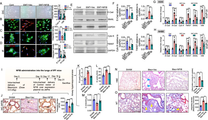
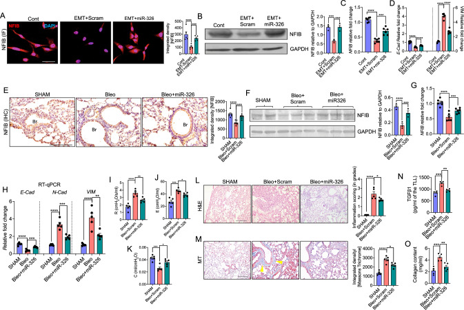
Idiopathic Pulmonary Fibrosis (IPF) is a progressively fatal and incurable disease characterized by the loss of alveolar structures, increased epithelial-mesenchymal transition (EMT), and aberrant tissue repair. In this study, we investigated the role of Nuclear Factor I-B (NFIB), a transcription factor critical for lung development and maturation, in IPF. Using both human lung tissue samples from patients with IPF, and a mouse model of lung fibrosis induced by bleomycin, we showed that there was a significant reduction of NFIB both in the lungs of patients and mice with IPF. Furthermore, our in vitro experiments using cultured human lung cells demonstrated that the loss of NFIB was associated with the induction of EMT by transforming growth factor beta (TGF-β). Knockdown of NFIB promoted EMT, while overexpression of NFIB suppressed EMT and attenuated the severity of bleomycin-induced lung fibrosis in mice. Mechanistically, we identified post-translational regulation of NFIB by miR-326, a miRNA with anti-fibrotic effects that is diminished in IPF. Specifically, we showed that miR-326 stabilized and increased the expression of NFIB through its 3'UTR target sites for Human antigen R (HuR). Moreover, treatment of mice with either NFIB plasmid or miR-326 reversed airway collagen deposition and fibrosis. In conclusion, our study emphasizes the critical role of NFIB in lung development and maturation, and its reduction in IPF leading to EMT and loss of alveolar structures. Our study highlights the potential of miR-326 as a therapeutic intervention for IPF. The schema shows the role of NFIB in maintaining the normal epithelial cell characteristics in the lungs and how its reduction leads to a shift towards mesenchymal cell-like features and pulmonary fibrosis. A In normal lungs, NFIB is expressed abundantly in the epithelial cells, which helps in maintaining their shape, cell polarity and adhesion molecules. However, when the lungs are exposed to factors that induce pulmonary fibrosis, such as bleomycin, or TGF-β, the epithelial cells undergo epithelial to mesenchymal transition (EMT), which leads to a decrease in NFIB. B The mesenchymal cells that arise from EMT appear as spindle-shaped with loss of cell junctions, increased cell migration, loss of polarity and expression of markers associated with mesenchymal cells/fibroblasts. C We designed a therapeutic approach that involves exogenous administration of NFIB in the form of overexpression plasmid or microRNA-326. This therapeutic approach decreases the mesenchymal cell phenotype and restores the epithelial cell phenotype, thus preventing the development or progression of pulmonary fibrosis.

Keywords: Epithelial–mesenchymal transition; Idiopathic pulmonary fibrosis; NFIB; miR-326.


Conflict of interest statement

The authors declare that they have no competing interests pertaining to this work.

Figures

Fig. 1
Fig. 1
Expression of NFIB is decreased in both human lung samples from IPF patients and in two independent mice models of pulmonary fibrosis. Human lung tissue samples were collected from control and IPF in Mayo Clinic, Rochester, USA. Expression of NFIB was determined by A Immunohistochemistry with quantitative analysis (right panel; N = 6), B Relative intensity of NFIB expression in fold change was determined by RT-qPCR (N = 4). C Immunoblot in total lung lysate was performed. Ponceau staining was used as loading control for NFIB (N = 4) (right panel). Densitometry of western blots was performed by ImageJ software. D RT-qPCR analysis showing relative expression of epithelial markers (E-Cad, and Cyto K) and mesenchymal markers (N-Cad, VIM, and SNAIL) were analyzed in IPF along with control lung tissue (N = 5). E Schematic representation of bleomycin induced-PF mice model. Mice were sensitized sensitization/treated with PBS: SHAM (Vehicle), or bleomycin (mice treated with 3.5 units (U)/kg of bleomycin) on Day 0. and sacrificed on Day 21 for EMT marker analysis and NFIB expression F Representative immunofluorescence images of NFIB expression in bleomycin-induced mice lung sections (N = 6). Quantitative analysis of the images is represented as integrated density (right panel). G Representative immunoblot in total lung lysate obtained from SHAM (control) and Bleomycin (Bleo)-induced mice (N = 3). Densitometry of western blots (right panel). Each experiment was repeated 3 times independently. Data represents mean ± SE; from three independent experiments; *p < 0.05; **p < 0.01; ***p < 0.001; ****p < 0.0001. GAPDH was used as the loading control. Non-parametric t test was used for statistical analysis. Scale bar = 200 μm
Fig. 2
Fig. 2
Loss of NFIB promotes TGF-β1 + IL-1β induced EMT. A Human alveolar epithelial cells (A549) were treated with a combination of human recombinant TGF-β1 (10 ng/ml) and IL-1β (5 ng/ml) for 72 h, and NFIB expression was determined by RT-qPCR (N = 8). B Representative immunofluorescence images of cells undergoing EMT and stained with anti-NFIB (green) and DAPI (blue). Quantitative analysis of the images represented as integrated density (right panel) (N = 10). C Representative immunoblot of NFIB in total protein obtained from cells from Control (Cont) and cells which have undergone EMT. Densitometry of western blots (right panel) (N = 3). DF Representative images of stable cell lines derived from transfection of scrambled (Scram) and shRNA targeting NFIB (NFIBshRNA). These stable cells were used as Cont cells or treated to undergo EMT (N = 5). D the morphology of the cells under various conditions. E Invasion assay was performed by staining the cells with crystal violet and assessed in all groups. F immunofluorescence images of cells stained with anti-cytokeratin (Cyto K; green), anti-vimentin (VIM; red) and DAPI (blue). G Quantitative analysis of (E) represented as integrated density (N = 5). H RT-qPCR analysis showing relative fold change expression of Cyto K and VIM in indicated groups (N = 4). I Phase contrast images of wound healing assay and migration of cells was assessed in all groups after 12 and 24 h of scratch. J Quantitative analysis of the cells migrated in wound healing assay at 12 and 24 h (N = 4). Each experiment was repeated 3 times independently with N = 5 and data represents mean ± SE; *p < 0.05; **p < 0.01; ***p < 0.001; ****p < 0.0001; ns (non-significant). Scale bar; D, E, I: 100 μm; F: 50 μm. Non-parametric t test was used for statistical analysis
Fig. 3
Fig. 3
Exogenous administration of NFIB prevents EMT-like condition and development of pulmonary fibrosis. A Representative bright field images of Cont, EMT-induced cells transfected with vector (EMT + Vec), or NFIB plasmid (EMT + NFIB) (N = 6). B Representative immunofluorescence images stained with epithelial marker, anti-cytokeratin (green) and mesenchymal marker, anti-vimentin (red) (N = 6). C Representative immunofluorescence images stained with epithelial marker anti-β-Catenin (green) (N = 6). D Representative immunofluorescence images stained with epithelial marker anti-E-Cadherin (E-Cad; green) and anti-N-Cadherin (N-Cad; red) showing transition from E-Cadherin to N-Cad expression during EMT. The right panels show the quantitative analysis of the images from (B), (C) and (D) and is represented as integrated density (N = 6). E Representative immunoblot of various epithelial and mesenchymal markers in EMT-induced cells transfected with either vector or NFIB plasmid. GAPDH was used as loading control. F The right panel shows the densitometry analysis of the blots (N = 4). G and H RT-qPCR analysis showing relative fold change of NFIB showing in A549 and NHBE (Normal Human Bronchial Epithelial Cells) cells (N = 5). I Schematic representation of the NFIB plasmid administration into the lungs of bleomycin-induced PF mice. J Representative immunohistochemistry images of NFIB in bleomycin mice lungs treated with either vector or NFIB plasmid. The right panels represented as integrated density (N = 5). K Airway resistance was increased in bleomycin treated mice and shows improvement with administration of NFIB plasmid (N = 5). L Airway elastance was increased in bleomycin treated mice and shows improvement NFIB plasmid administered mice (N = 5). M Compliance was reduced in bleomycin treated mice and shows improvement NFIB plasmid administered mice (N = 5). N Representative H&E images of the bleomycin mice treated with vector or with NFIB showing inflammatory cell infiltration. Right panel shows the inflammation scoring (N = 5). O Representative MT stained images of the bleomycin mice treated with vector or with NFIB showing collagen deposition. Right panel shows the integrated density of masons trichrome staining (N = 5). Each experiment was repeated 3 times independently and data represents mean ± SE; *p < 0.05; ***p < 0.01; ***p < 0.001; ****p < 0.0001. Scale bar; A: 100 μm; BD: 50 μm; J, N, O: 200 μm. Non-parametric t test was used for statistical analysis
Fig. 4
Fig. 4
miR-326 upregulates NFIB expression and attenuates EMT-like conditions and lung fibrosis. A Representative immunofluorescence images of cells treated with Scram or miR-326 under EMT condition, followed by staining with anti-NFIB (red) and DAPI (blue). Right panel shows the respective quantitative analysis of the images represented as integrated density of NFIB (N = 5). B Representative immunoblot of cells treated with Scram or miR-326 under EMT condition (N = 5). C Relative NFIB RNA expression in EMT induced cells treated with Scram or miR-326 mimic (N = 6). D Relative expression of epithelial and mesenchymal markers including E-Cad and VIM shows in A549 cells treated with Scram or miR-326 under EMT condition (N = 5). E Representative immunohistochemistry images of mice lung sections of bleomycin induced fibrosis model, and probed for anti-NFIB. Right panel shows the quantitative analysis (N = 5). F Representative immunoblot of NFIB in total protein isolated from mice lungs treated with scram or miR-326 under bleomycin-induced condition. Right panel shows the densitometry of the images (N = 3). G Relative expression of NFIB shown in fold change (N = 6). H RT-qPCR analysis showing relative fold change expression of E-Cad, N-Cad, and VIM shows in total RNA isolated from mice lungs treated with scram or miR-326 under bleomycin-induced condition (N = 5). I Airway resistance was increased in bleomycin treated mice and shows improvement with administration of miR-326 mimic (N = 5). J Airway elastance was increased in bleomycin treated mice and shows improvement miR-326 mimic administered mice (N = 5). K Compliance was reduced in bleomycin-treated mice and shows improvement miR-326 mimic administered mice (N = 5). L Representative H&E images of mice treated with Scram or miR-326 under bleomycin-induced condition. Right panel shows the inflammation scoring (N = 5). M Representative MT stained images of the bleomycin mice treated with vector or with miR-326 mimic showing collagen deposition. Right panel shows the integrated density of masons trichrome staining (N = 5). N TGF-β1 levels in the total lung lysate (TLL) prepared from mice lungs, and plotted as picogram per microliter of the TLL (N = 4). O Collagen content measured in the indicated groups and represented as mg/ml (N = 5). Data are presented as mean ± SE; *p < 0.05; ***p < 0.01; ***p < 0.001; ****p < 0.0001. Scale bar; A: 50 μm; E. L, M: 200 μm. Non-parametric t test was used for statistical analysis
Fig. 5
Fig. 5
miR-326 mediates upregulation of NFIB via RNA-binding protein HuR. A Prediction by miRanda tool shows two potential binding sites of miR-326 at NFIB 3ʹUTR. B Two different plasmids with 3’UTR of NFIB were generated; NFIB 3ʹUTR-A and 3ʹUTR-B. Scram or miR-326 mimics were transfected in A549 cells and binding of miR-326 to NFIB 3ʹUTR-A is shown, as validated by luciferase assay. pEZX was used as vector control (N = 6). C Similarly, cells were transfected with Scram or miR-326 and NFIB transcript level was evaluated by RT-qPCR (N = 4). D Representative immunoblot of NFIB in cells from Control or transfected with Scram or miR-326. Lamin was used as loading control. The right panel shows the densitometry analysis of the blots (N = 4). E Schematic representation of the RNA immunoprecipitation of NFIB with anti-HuR. F Binding of HuR at 3’UTR of NFIB was validated by RNA immunoprecipitation (RIP). Human IgG was used as IP control and SNRNP70 was used as positive control (N = 4). G Plasmid expressing HuR was overexpressed in A549 and expression of NFIB was determined by RT-qPCR (N = 4). H Half-life of NFIB RNA was measured after the transfection of miR-326 mimics and/or HuR plasmid, followed by actinomycin D treatment at different time points (N = 4). I Stability of NFIB was determined upon transfection of miR-326 mimics and HuR overexpression plasmid by luciferase assay. A set of plasmids (vector or HuR) or miR-326 wild-type and deletion mutants were transfected with or without wild-type or deleted mutants of HuR (N = 4). J Similarly, RT-qPCR was used to determine the presence of NFIB based on the expression levels. The specific effect of miR-326 on NFIB expression was determined by anti-miR-326 mimetic (N = 4). Each experiment was repeated 4 times independently and data represents mean ± SE; *p < 0.05; **p < 0.01; ***p < 0.001; ****p < 0.0001. Non-parametric t test (C, F, G) was used for statistical analysis and one-way ANOVA (B, H, I, J)

References

    1. Pardo A, Selman M, Kaminski N. Approaching the degradome in idiopathic pulmonary fibrosis. Int J Biochem Cell Biol. 2008;40(6–7):1141–1155. - PubMed
    1. Todd NW, Luzina IG, Atamas SP. Molecular and cellular mechanisms of pulmonary fibrosis. Fibrogenes Tissue Repair. 2012;5(1):1. - PMC - PubMed
    1. Shi W, Xu J, Warburton D. Development, repair and fibrosis: What is common and why it matters series. Respirology. 2009;14(5):656–665. - PMC - PubMed
    1. Selman M, Pardo A. Idiopathic pulmonary fibrosis: an epithelial/fibroblastic cross-talk disorder. Respir Res. 2001;3:1–8. - PMC - PubMed
    1. Willis BC, DuBois RM, Borok Z. Epithelial origin of myofibroblasts during fibrosis in the lung. Proc Am Thorac Soc. 2006;3(4):377–382. - PMC - PubMed

MeSH terms

LinkOut - more resources