. 2023 Nov 11;14(1):7315.
doi: 10.1038/s41467-023-42791-5.

An on-demand bioresorbable neurostimulator

Affiliations

An on-demand bioresorbable neurostimulator

Dong-Min Lee et al. Nat Commun. .

Abstract

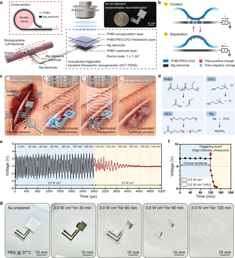
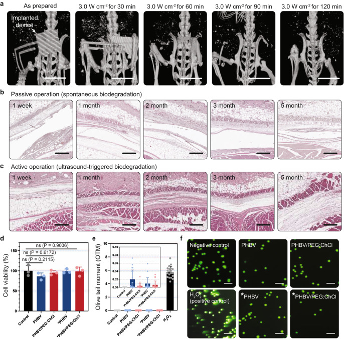
Bioresorbable bioelectronics, with their natural degradation properties, hold significant potential to eliminate the need for surgical removal. Despite notable achievements, two major challenges hinder their practical application in medical settings. First, they necessitate sustainable energy solutions with biodegradable components via biosafe powering mechanisms. More importantly, reliability in their function is undermined by unpredictable device lifetimes due to the complex polymer degradation kinetics. Here, we propose an on-demand bioresorbable neurostimulator to address these issues, thus allowing for clinical operations to be manipulated using biosafe ultrasound sources. Our ultrasound-mediated transient mechanism enables (1) electrical stimulation through transcutaneous ultrasound-driven triboelectricity and (2) rapid device elimination using high-intensity ultrasound without adverse health effects. Furthermore, we perform neurophysiological analyses to show that our neurostimulator provides therapeutic benefits for both compression peripheral nerve injury and hereditary peripheral neuropathy. We anticipate that the on-demand bioresorbable neurostimulator will prove useful in the development of medical implants to treat peripheral neuropathy.


Conflict of interest statement

D.-M.L., M.K., and S.-W.K. are inventors on the patent (KR/ 10-2348997) and patent application (US/ 17/515,675) filed through the Sungkyunkwan University Research and Business Foundation which cover the on-demand bioresorbable neurostimulator for peripheral nerve electrotherapy used in this work. Energymining Co., Ltd. acquired the patent and patent application through technology transfer. The remaining authors declare no competing interests.

Figures

Fig. 1
Fig. 1. Concept and design of the on-demand bioresorbable neurostimulator.
a The overall structure of the neurostimulator with an expanded view of ACT-TENG and a bioresorbable cuff electrode. The inset displays a photograph of the device. b Schematic of the ACT–TENG operating mechanism. c Proposed clinical protocol of peripheral nerve electrotherapy using the on-demand bioresorbable neurostimulator. d Hydrolytic degradation processes of the constituent materials. e On-demand transience demonstration with long-term electrical characterization for the output of ACT–TENG. f The summation plots of (e). HIU represents high-intensity ultrasound. g HIU-triggered transient performance of the device immersed in diluted PBS solution (pH 7.4).
Fig. 2
Fig. 2. In vivo HIU-triggered transience of the neurostimulator and its biosafety.
a Micro-CT images showing in vivo transient performances upon the HIU-driven triggering event (scale bar: 10 mm). b, c H&E-stained tissue images exhibiting inflammation responses with spontaneous biodegradation (b) and HIU-triggered transience (c). d Relative cell viability of human fibroblasts (CRL-1502) on the as-prepared and HIU-triggered materials (n = 24 for each group). The asterisk mark (*) indicates the HIU-induced degraded materials. Data are presented as mean values ± SD (standard deviation). The scale bar is equivalent to 100 μm. e, f The olive tail moment (OTM) of the as-prepared and HIU-triggered materials (n = 20 for each group) (e) and their fluorescent images (f), measured by comet assay. The asterisk mark (*) indicates the HIU-induced degraded materials. Data are presented as mean values ± SD. The scale bar is equivalent to 100 μm. P-values are evaluated through a two-sided t-test; ns = non significant.
Fig. 3
Fig. 3. Experimental settings for in vivo electrotherapy and the recorded NCS results for compression peripheral nerve injury model and C22 mouse (hereditary peripheral neuropathy, CMT1A) model.
a Schematic of the experimental setup for in vivo electrotherapy. b, c In vitro cell experiments were performed to validate the electrical stimulation conditions (e.g., amplitude, treatment duration) required for neuroregeneration. b Optical microscopic images of iPSC-driven motor neurons. c Axon length plots along the culturing time (n = 3 for each group). Data are presented as mean values ± SD. Based on the results of in vitro cell experiments, electrical impulses of 1.3 V mm−1 (20 kHz sinusoidal waveform, 5 min daily) are effective for accelerating the growth of iPSC-driven motor neurons. Thus, we adopted those electrical stimulation conditions in our in vivo demonstrations described in (a). d Recorded nerve conduction velocities and action potentials after the in vivo electrotherapy for the compression peripheral nerve injury mouse model (Wild type: n = 5; Injury model: n = 5; ESE-untreated: n = 5; ESE-treated: n = 4). Data are presented as mean values ± SD. ESE represents electrical stimulation events. e Recorded nerve conduction velocities and action potentials after the in vivo electrotherapy for the C22 mouse model (Wild type: n = 5; C22 model: n = 5; ESE-untreated: n = 5; ESE-treated: n = 5). Data are presented as mean values ± SD. P-values are evaluated through two-sided t-test; *P < 0.05; **P < 0.01; ***P < 0.001; ****P < 0.0001.
Fig. 4
Fig. 4. Histopathological demonstration and statistical analyses for the therapeutic effects on both compression peripheral nerve injury and CMT1A (hereditary peripheral neuropathy).
a Semi-thin sections (scale bar: 30 μm) and electron microscopy images (scale bar: 2 μm) to observe the toluidine blue-stained sciatic nerve cross-section. b Histogram representing the inner diameter distribution of the myelinated axons (Wild type: n = 3; Injury model: n = 3; ESE-treated: n = 3). Data are presented as mean values ± SD. c Histopathological demonstration using semi-thin (scale bar: 30 μm) and electron microscopy images (scale bar: 2 μm) of the sciatic nerve cross-section. d, e Percentage of the myelinated axons (d) and the unmyelinated axons (e) (the myelinated axons refer to the axons with a diameter greater than 5 μm) (Wild type: n = 3; C22 model: n = 3; ESE-untreated: n = 3; ESE-treated: n = 3). Data are presented as mean values ± SD. f Histogram displaying the diameter distribution (Wild type: n = 3; C22 model: n = 3; ESE-untreated: n = 3; ESE-treated: n = 3). Data are presented as mean values ± SD. g Scatterplots representing the correlation of g-ratio and axon diameter (Wild type: n = 900; C22 model: n = 800; ESE-untreated: n = 200; ESE-treated: n = 416). P-values are evaluated through a two-sided t-test; ns = non significant; *P < 0.05; ****P < 0.0001.

References

    1. Miller G. On the origin of the nervous system. Science. 2009;325:24–26. doi: 10.1126/science.325_24. - DOI - PubMed
    1. Robinson, A. J. & Snyder-Mackler, L. Clinical Electrophysiology: Electrotherapy and Electrophysiologic Testing (Lippincott Williams & Wilkins, 2008).
    1. Rogers JA. Electronics for the human body. JAMA. 2015;313:561–562. doi: 10.1001/jama.2014.17915. - DOI - PubMed
    1. Nadeau P, et al. Prolonged energy harvesting for ingestible devices. Nat. Biomed. Eng. 2017;1:0022. doi: 10.1038/s41551-016-0022. - DOI - PMC - PubMed
    1. Koo J, et al. Wirelessly controlled, bioresorbable drug delivery device with active valves that exploit electrochemically triggered crevice corrosion. Sci. Adv. 2020;6:eabb1093. doi: 10.1126/sciadv.abb1093. - DOI - PMC - PubMed

Publication types