. 2023 Nov 11;13(1):19692.
doi: 10.1038/s41598-023-46461-w.

COVID-19 screening in low resource settings using artificial intelligence for chest radiographs and point-of-care blood tests

Affiliations

COVID-19 screening in low resource settings using artificial intelligence for chest radiographs and point-of-care blood tests

Keelin Murphy et al. Sci Rep. .

Abstract

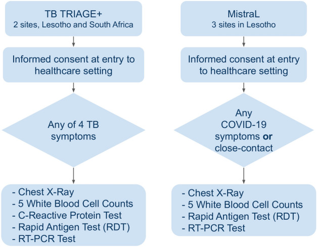
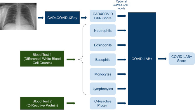
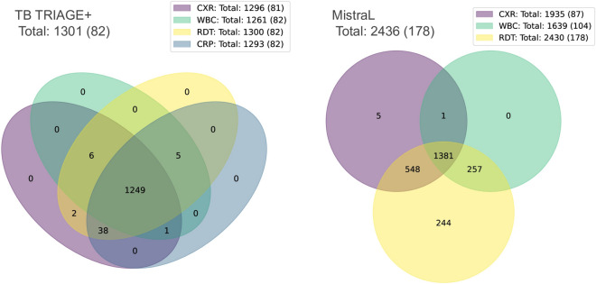
Artificial intelligence (AI) systems for detection of COVID-19 using chest X-Ray (CXR) imaging and point-of-care blood tests were applied to data from four low resource African settings. The performance of these systems to detect COVID-19 using various input data was analysed and compared with antigen-based rapid diagnostic tests. Participants were tested using the gold standard of RT-PCR test (nasopharyngeal swab) to determine whether they were infected with SARS-CoV-2. A total of 3737 (260 RT-PCR positive) participants were included. In our cohort, AI for CXR images was a poor predictor of COVID-19 (AUC = 0.60), since the majority of positive cases had mild symptoms and no visible pneumonia in the lungs. AI systems using differential white blood cell counts (WBC), or a combination of WBC and C-Reactive Protein (CRP) both achieved an AUC of 0.74 with a suggested optimal cut-off point at 83% sensitivity and 63% specificity. The antigen-RDT tests in this trial obtained 65% sensitivity at 98% specificity. This study is the first to validate AI tools for COVID-19 detection in an African setting. It demonstrates that screening for COVID-19 using AI with point-of-care blood tests is feasible and can operate at a higher sensitivity level than antigen testing.


Conflict of interest statement

The authors declare no competing interests.

Figures

Figure 1
Figure 1
The recruitment process for participants included in this study.
Figure 2
Figure 2
Schematic illustration of the AI systems (dark blue boxes) used in this study. The green boxes indicate a point-of-care blood-test procedure. The pale blue boxes indicate numeric values which are inputs or outputs of the systems as shown. The COVID-LAB+ system does not require all the indicated inputs to be present.
Figure 3
Figure 3
Numbers of participants enrolled, excluded and finally included in our analysis. Figures in brackets indicate the subset which had a positive RT-PCR result.
Figure 4
Figure 4
Information on the numbers of participants recruited in each trial and how many results were recorded for the various tests available. Figures in brackets indicate numbers with a positive RT-PCR test. CXR, chest X-Ray; WBC, white blood cell differential count; RDT, rapid diagnostic test (antigen based); CRP, C-reactive protein test.
Figure 5
Figure 5
COVID-19 prediction using X-Ray analysis only. The ROC curves show the results for CAD4COVID-XRay using all available CXR images (orange) and a subset selected for radiological reading (blue). The blue points indicate the performance using the scores from the radiologist on this same subset at two cutoff points (i.e. scores>2 considered positive and scores>1 considered positive).
Figure 6
Figure 6
ROC results for the COVID-Lab+ system, run using different combinations of input parameters. Note that different curves are created from different populations, depending on data available for specific input parameters. The sensitivity and specificity obtained by antigen-based testing is additionally plotted. WBC, White blood cell counts; CRP, C-Reactive Protein; RDT, rapid diagnostic test (antigen based).

References

    1. Aljondi R, Alghamdi S. Diagnostic value of imaging modalities for COVID-19: Scoping review. J. Med. Internet Res. 2020;22:e19673. doi: 10.2196/19673. - DOI - PMC - PubMed
    1. Kovács A, et al. The sensitivity and specificity of chest CT in the diagnosis of COVID-19. Eur. Radiol. 2021;31:2819–2824. doi: 10.1007/s00330-020-07347-x. - DOI - PMC - PubMed
    1. Long C, et al. Diagnosis of the Coronavirus disease (COVID-19): rRT-PCR or CT? Eur. J. Radiol. 2020;126:108961. doi: 10.1016/j.ejrad.2020.108961. - DOI - PMC - PubMed
    1. Prokop M, et al. CO-RADS: A categorical CT assessment scheme for patients suspected of having COVID-19-definition and evaluation. Radiology. 2020;296:E97–E104. doi: 10.1148/radiol.2020201473. - DOI - PMC - PubMed
    1. Salameh J-P, et al. Thoracic imaging tests for the diagnosis of COVID-19. Cochrane Database Syst. Rev. 2020;2020:896. doi: 10.1002/14651858.CD013639.pub2. - DOI - PubMed

Publication types