. 2023 Nov 28;42(11):113421.
doi: 10.1016/j.celrep.2023.113421. Epub 2023 Nov 11.

Cochlear organoids reveal transcriptional programs of postnatal hair cell differentiation from supporting cells

Affiliations

Cochlear organoids reveal transcriptional programs of postnatal hair cell differentiation from supporting cells

Gurmannat Kalra et al. Cell Rep. .

Abstract

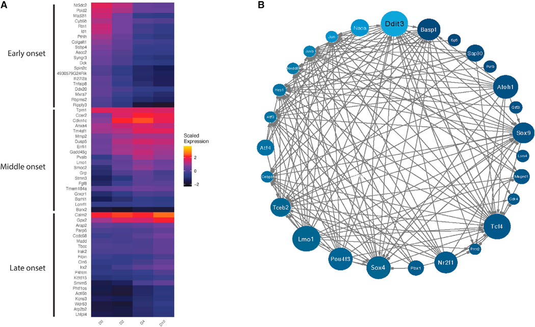
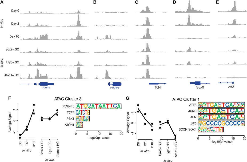
We explore the changes in chromatin accessibility and transcriptional programs for cochlear hair cell differentiation from postmitotic supporting cells using organoids from postnatal cochlea. The organoids contain cells with transcriptional signatures of differentiating vestibular and cochlear hair cells. Construction of trajectories identifies Lgr5+ cells as progenitors for hair cells, and the genomic data reveal gene regulatory networks leading to hair cells. We validate these networks, demonstrating dynamic changes both in expression and predicted binding sites of transcription factors (TFs) during organoid differentiation. We identify known regulators of hair cell development, Atoh1, Pou4f3, and Gfi1, and the analysis predicts the regulatory factors Tcf4, an E-protein and heterodimerization partner of Atoh1, and Ddit3, a CCAAT/enhancer-binding protein (C/EBP) that represses Hes1 and activates transcription of Wnt-signaling-related genes. Deciphering the signals for hair cell regeneration from mammalian cochlear supporting cells reveals candidates for hair cell (HC) regeneration, which is limited in the adult.

Keywords: ATAC sequencing; CP: Stem cell research; cochlea; gene expression analysis; sensory hair cells; single-cell RNA sequencing.


Conflict of interest statement

Declaration of interests The authors declare no competing interests.

Figures

Figure 1.
Figure 1.. Marker genes for cochlear and utricular cell types derived by scRNA-seq integration
(A) Anatomical organization of transcriptionally defined cell types in the mammalian cochlea and utricle. (B) Cell types from P2 and P7 mouse utricle were identified by UMAP clustering of scRNA-seq expression data. (C) Marker genes for the principal cell types obtained by UMAP clustering of the mouse utricle were identified. (D) Robust marker genes for each cochlear and utricular cell type were defined by integration of multiple scRNA-seq datasets at specific developmental timepoints. HC, hair cell; SC, supporting cell; IPhC, inner phalangeal cell; IPC, inner pillar cell; OPC, outer pillar cell; GER, greater epithelial ridge; LER, lesser epithelial ridge; OS, outer sulcus; SGN, spiral ganglion neuron. Ube2c (ubiquitin-conjugating enzyme E2 C) and Oc90 (otoconin-90) are genes previously identified as markers for non-sensory epithelial cells.
Figure 2.
Figure 2.. scRNA-seq characterization of in vitro cell types in cochlear organoids
(A) Integrated scRNA-seq data from days 0 and 10 of organoid differentiation, labeled by sample and time point, were used to obtain UMAP clusters. (B) Cells obtained at days 0 and 10 of differentiation are plotted onto the UMAP. (C) Marker expression is plotted for the epithelial marker Epcam; supporting cell marker Hes1; cochlear interdental cell marker and vestibular supporting cell marker Otoa; and canonical hair cell marker Pou4f3. (D) Distribution of epithelial markers across UMAP clusters with expression patterns of hair cells, interdental/supporting cells, and GER. (E) Pairwise correlations of marker genes of in vitro clusters vs. in vivo cochlear and utricular cell types. Color intensity indicates Pearson correlations of cell-type specificity scores for up to 300 genes per cell type. For an interactive version of the trajectory analysis, see https://umgear.org/lgr5org.
Figure 3.
Figure 3.. Molecular characterization and pseudotemporal trajectories of hair cells in cochlear organoids
(A) Volcano plot indicating genes differentially expressed between two clusters of hair cells in organoids: clusters 7 (primarily in day 0 organoids) and 8 (primarily in day 10 organoids). (B) Aggregate expression of markers specifically expressed in E14, E16, P1, or P7 cochlear hair cells in clusters 7 and 8. Orange indicates high expression, whereas gray indicates low expression. (C) Aggregate expression of genes expressed in cochlear and non-cochlear Atoh1-dependent cell lineages in clusters 7 and 8. (D) Monocle pseudotime trajectory delineates transdifferentiation of Lgr5+ supporting cells (cluster 4) to hair cells (clusters 7 and 8). Trajectory labeled by cluster number (left) or pseudotime (right). For an interactive version of the trajectory analysis, see https://umgear.org/lgr5org. (E) Expression of known marker genes with dynamic expression across pseudotime.
Figure 4.
Figure 4.. Clustering analysis of bulk RNA-seq data from days 0, 2, 4, and 10
(A) Principal-component analysis (PCA) of the day 0, 2, 4, and 10 bulk RNA-seq samples and their replicates. (B) PCA of Atoh1+, Lgr5+, and Sox2+ cells and their replicates from P2 mouse cochlea. (C) Expression of select genes in day 0, 2, 4, and 10 samples. (D) K-means clustering of day 0, 2, 4, and 10 bulk RNA-seq samples, showing eight gene expression patterns. (E) Expression of select genes in groups 1, 3, and 4 from (D).
Figure 5.
Figure 5.. Gene regulatory network of transcription factors that drive the transdifferentiation of Lgr5+ cochlear progenitor cells to hair cells
Each TF is colored by its activity (A) in the pseudotime course from Figure 3E, and its size in the diagram (B) reflects the magnitude of its outdegree.
Figure 6.
Figure 6.. Chromatin accessibility dynamics during transdifferentiation in cochlear organoids
(A–E) Chromatin accessibility at the promoters of the known and predicted key regulator TFs Atoh1 (A), Pou4f3 (B), Tcf4 (C), Sox9 (D), and Atf3 (E). (F and G) Average accessibility pattern for peaks within clusters 3 (F) and 1 (G) and enrichment of peaks for sequence motifs recognized by key regulator TFs with dynamic expression in the RNA-based gene regulatory network model.

References

    1. McLean WJ, Yin X, Lu L, Lenz DR, McLean D, Langer R, Karp JM, and Edge ASB (2017). Clonal Expansion of Lgr5-Positive Cells from Mammalian Cochlea and High-Purity Generation of Sensory Hair Cells. Cell Rep. 18, 1917–1929. 10.1016/j.celrep.2017.01.066. - DOI - PMC - PubMed
    1. Clevers H. (2016). Modeling Development and Disease with Organoids. Cell 165, 1586–1597. 10.1016/j.cell.2016.05.082. - DOI - PubMed
    1. Roccio M, and Edge ASB (2019). Inner ear organoids: new tools to understand neurosensory cell development, degeneration and regeneration. Development 146, dev177188. 10.1242/dev.177188. - DOI - PMC - PubMed
    1. Lenz DR, Gunewardene N, Abdul-Aziz DE, Wang Q, Gibson TM, and Edge ASB (2019). Applications of Lgr5-Positive Cochlear Progenitors (LCPs) to the Study of Hair Cell Differentiation. Front. Cell Dev. Biol 7, 14. 10.3389/fcell.2019.00014. - DOI - PMC - PubMed
    1. Kolla L, Kelly MC, Mann ZF, Anaya-Rocha A, Ellis K, Lemons A, Palermo AT, So KS, Mays JC, Orvis J, et al. (2020). Characterization of the development of the mouse cochlear epithelium at the single cell level. Nat. Commun 11, 2389. 10.1038/s41467-020-16113-y. - DOI - PMC - PubMed

Publication types

MeSH terms

Substances