. 2023 Dec 11;51(22):12224-12241.
doi: 10.1093/nar/gkad952.

TATDN2 resolution of R-loops is required for survival of BRCA1-mutant cancer cells

Affiliations

TATDN2 resolution of R-loops is required for survival of BRCA1-mutant cancer cells

Aruna S Jaiswal et al. Nucleic Acids Res. .

Abstract

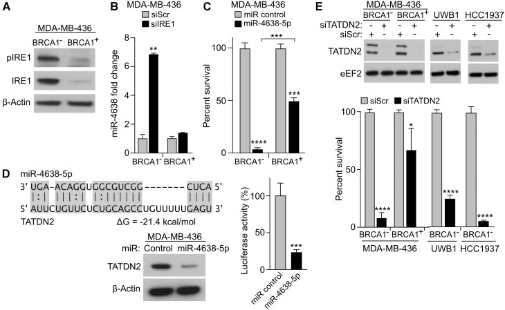
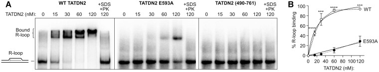
BRCA1-deficient cells have increased IRE1 RNase, which degrades multiple microRNAs. Reconstituting expression of one of these, miR-4638-5p, resulted in synthetic lethality in BRCA1-deficient cancer cells. We found that miR-4638-5p represses expression of TATDN2, a poorly characterized member of the TATD nuclease family. We discovered that human TATDN2 has RNA 3' exonuclease and endonuclease activity on double-stranded hairpin RNA structures. Given the cleavage of hairpin RNA by TATDN2, and that BRCA1-deficient cells have difficulty resolving R-loops, we tested whether TATDN2 could resolve R-loops. Using in vitro biochemical reconstitution assays, we found TATDN2 bound to R-loops and degraded the RNA strand but not DNA of multiple forms of R-loops in vitro in a Mg2+-dependent manner. Mutations in amino acids E593 and E705 predicted by Alphafold-2 to chelate an essential Mg2+ cation completely abrogated this R-loop resolution activity. Depleting TATDN2 increased cellular R-loops, DNA damage and chromosomal instability. Loss of TATDN2 resulted in poor replication fork progression in the presence of increased R-loops. Significantly, we found that TATDN2 is essential for survival of BRCA1-deficient cancer cells, but much less so for cognate BRCA1-repleted cancer cells. Thus, we propose that TATDN2 is a novel target for therapy of BRCA1-deficient cancers.


Figures

Graphical Abstract
Graphical Abstract
Figure 1.
Figure 1.
MiR-4638-5p reconstitution in BRCA1-deficient cells results in synthetic lethality. (A) The RNase IRE1 (both total and phosphorylated) is constitutively over-expressed in BRCA1-deficient MDA-MB-436 cells. (B) Depleting IRE1 results in increased levels of miR-4638-5p in BRCA1-deficient but not WT BRCA1-repleted MDA-MB-436 cells. (C) Reconstituting miR-4638–5p expression is synthetic lethal in BRCA1-deficient but not WT BRCA1-repleted cells. (D) miR-4638-5p has high affinity binding sites in the 3′ UTR of TATDN2 (position 3231 to 3259 in NM_014760.4). Western blot demonstrates miR-4638-5p regulates the expression of TATDN2. Transfection of miR-4638-5p in MDA-MB436 cells represses luciferase activity when the reporter contains the 3′ UTR of TATDN2. (E) Depletion of TATDN2 in the BRCA1-deficient MDA-MB-436, HCC1937 (breast cancer) and UWB1.289 (ovarian cancer) cell lines generate synthetic lethality, but 8-fold less of an effect is seen in BRCA1-repleted MDA-MB-436 cells. Western analysis demonstrates depletion of TATDN2, which has two isoforms in some cell lines, both specifically depleted. For all figures, *P< 0.05, **P< 0.01, ***P< 0.001, ****P< 0.0001.
Figure 2.
Figure 2.
TATDN2 is a structure-specific RNase. (A) Schematic of 68 nt RNA hairpin (above) and denaturing gel ribonuclease assay (below) showing TATDN2 RNA hairpin 3′ exonuclease and endonuclease activity. Arrows indicate approximate cleavage positions in RNA hairpin. (B) The three-dimensional structure of TATDN2 catalytic site predicted by AlphaFold and by comparison to bacterial TatD and human TATDN1 and 3 (2,4), showing the acidic amino acids predicted to chelate the required divalent cation essential for nuclease activity of all DNaseI family members. (C, D) Activity of recombinant TATDN2-WT, E593A, D707A, E705A mutant proteins on R-loops without an RNA overhang (lanes 1–9), with a 5′ RNA overhang (lanes 10–18), or with 3′-RNA overhang (lanes 19–27); products separated in a native polyacrylamide gel (C) or denaturing polyacrylamide gel (D) to assess the fraction and size of cleaved RNA products from R-loops. E593A and E705A mutations in TATDN2 protein lack R-loop RNA exonuclease activity, while the D707A mutant has decreased activity. (E) Quantitation of R-loop activity expressed as a percentage of cleaved RNA in R-loops (n = 3).
Figure 3.
Figure 3.
TATDN2 R-loop binding. (A) WT, E593A and N-terminal truncated (490–761) TATDN2 were incubated with an R-loop substrate and binding was determined by EMSA. (B) Plot of WT and E593A mutant TATDN2 R-loop binding measured by scanning densitometry. The Kd for WT TATDN2 is 19.3 nM; Kd was not calculated for the weakly binding E593A mutant, nor the non-binding 490–761 truncation mutant.
Figure 4.
Figure 4.
TATDN2 is specific for RNA in R-loops. (A) TATDN2 activity on R-loops (lanes # 1–4), D-loops (lanes # 5–8) and RNA–DNA duplexes (lanes # 9–12) was analyzed in a native gel. TATDN2 nucleolytic activity is specific for RNA in a R-loop substrate. (B) Quantitation of cleaved products from scans of gels as in panel A (n = 3). Statistics are shown for R-loop versus RNA–DNA duplex. (C) TATDN2 R-loop cleavage products resolved by size in a denaturing gel. (D) Quantitation of the cleaved RNA in R-loop (lane #4 of panel C) and RNA–DNA duplex (lane #12 of panel C); gel scan images were generated with ImageJ. TATDN2 only has RNase activity on R-loops, not D-loops or RNA–DNA hybrids.
Figure 5.
Figure 5.
TATDN2 depletion increases R-loop formation in vivo. (A) Representative confocal immunofluorescent microscopic images of R-loops detected by S9.6 antibodies. (B) Quantitation of R-loop formation in MDA-MB-436 cells. Data expressed as a graphical representation of S9.6 confocal immunofluorescence intensity in BRCA1-deficient and WT repleted MDA-MB-436 cells with or without TATDN2 depletion; data are from >100 cells scanned per condition. The BRCA1-deficient cells showed increased R-loops without further stress when compared with the WT BRCA1 repleted cells. Statistically significant increases in R-loops are seen in both BRCA1-deficient and WT-repleted cells after TATDN2 depletion. (C) DRIP-qPCR analysis showing that depletion of TATDN2 increases R-loops at the APOE gene locus, and further increased by BRCA1-deficiency (n = 3). The EGR-1 locus does not form R-loops and serves as a negative control. (D) Loss of APOE signal after treatment with RNaseH1 demonstrates that these are indeed R-loops. There are no statistical differences between any of the data after RNaseH1 treatment.
Figure 6.
Figure 6.
TATDN2 depletion suppresses replication fork progression when R-loops are increased in BRCA1-deficient MDA-MB-436 cells. (A) Schematic of the DNA fiber experiment and representative fiber images. R-loop formation was stimulated by ARV-825 over 6 h then removed. (B) CIdU track lengths represent replication fork progression when ARV-825 is generating R-loops. (C) IdU track lengths represent replication fork progression after release from R-loop stress. TATDN2 depletion decreased replication fork progression both when ARV-825 is present and also after release from that R-loop stress. (D) Average replication track lengths in during (CldU) and after ARV-825 treatment (IdU). A total of 74–171 (average 118) fibers scored per condition.
Figure 7.
Figure 7.
TATDN2 is required for genomic stability. (A) Alkaline Comet assays show increased genomic SS breaks in BRCA1-deficient MDA-MB-436 cells after depletion of TATDN2. (B) Depletion of TATDN2 alone or with chromosomal segregation stress induced by the decatenation inhibitor ICRF-193 results in increased γ-H2AX and BLM foci in BRCA1 WT MCF7 cells. (C) Depletion of TATDN2 alone or with ICRF-193 exposure increases anaphase arrest in BRCA1-deficient MDA-MB-436 breast cancer cells. (D) Depletion of TATDN2 increases chromosome segregation defects in BRCA1-deficient MDA-MB-436 cells, which results in retained chromosomes seen as micronuclei and shared chromosomes seen as nuclear bridges.

References

    1. Lee K.Y., Cheon S.H., Kim D.G., Lee S.J., Lee B.J.. A structural study of TatD from Staphylococcus aureus elucidates a putative DNA-binding mode of a Mg2+-dependent nuclease. IUCrJ. 2020; 7:509–521. - PMC - PubMed
    1. Chen Y.C., Li C.L., Hsiao Y.Y., Duh Y., Yuan H.S.. Structure and function of TatD exonuclease in DNA repair. Nucleic Acids Res. 2014; 42:10776–10785. - PMC - PubMed
    1. Singh D., Rahi A., Kumari R., Gupta V., Gautam G., Aggarwal S., Rehan M., Bhatnagar R.. Computational and mutational analysis of TatD DNase of Bacillus anthracis. J. Cell. Biochem. 2019; 120:11318–11330. - PubMed
    1. Dorival J., Eichman B.F.. Human and bacterial TatD enzymes exhibit apurinic/apyrimidinic (AP) endonuclease activity. Nucleic Acids Res. 2023; 51:2838–2849. - PMC - PubMed
    1. Piovesan D., Monzon A.M., Tosatto S.C.E.. Intrinsic protein disorder and conditional folding in AlphaFoldDB. Protein Sci. 2022; 31:e4466. - PMC - PubMed

Publication types