. 2024 Mar;244(3):424-437.
doi: 10.1111/joa.13969. Epub 2023 Nov 12.

Intracortical remodelling increases in highly loaded bone after exercise cessation

Affiliations

Intracortical remodelling increases in highly loaded bone after exercise cessation

Raniere Gaia Costa da Silva et al. J Anat. 2024 Mar.

Abstract

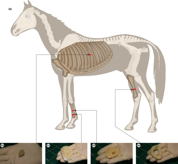
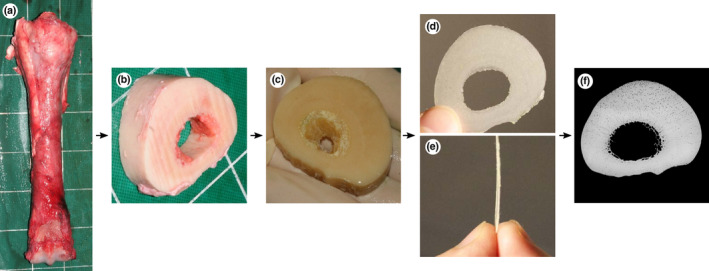
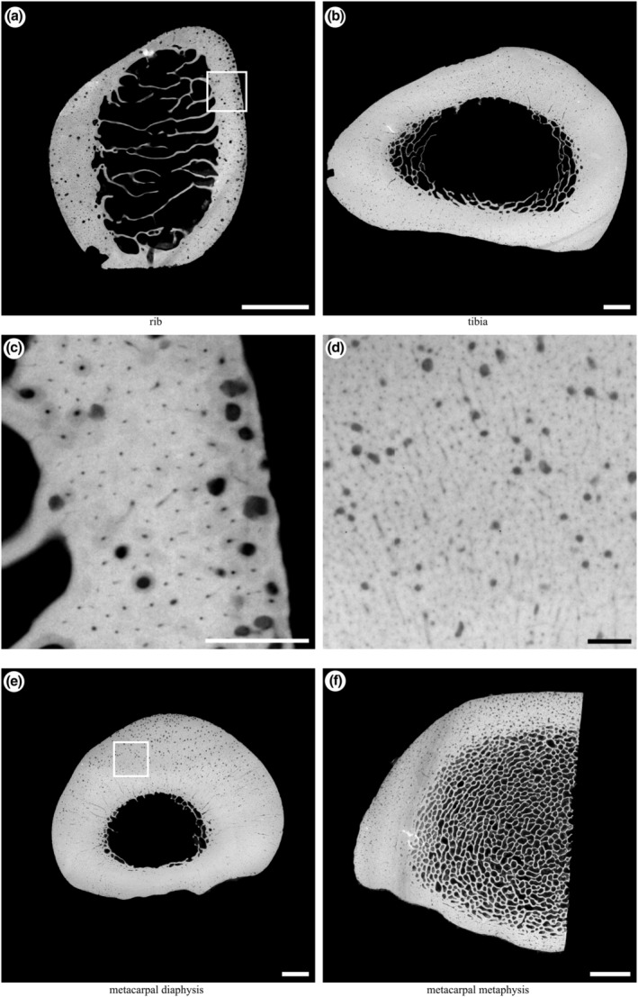
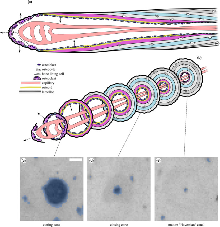
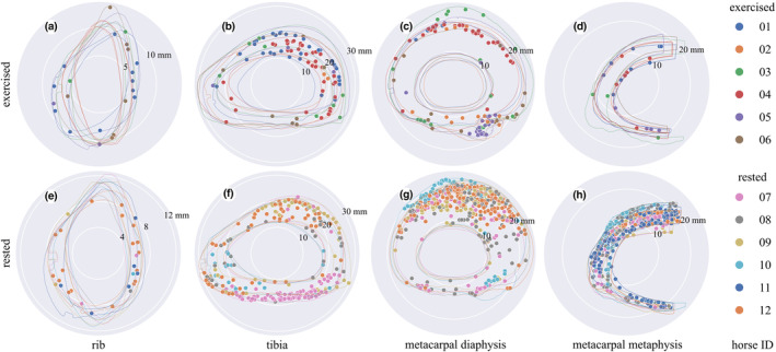
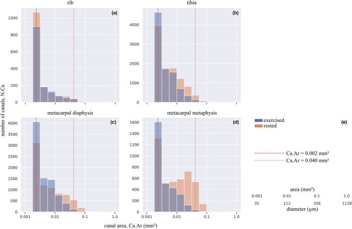
Resorption within cortices of long bones removes excess mass and damaged tissue and increases during periods of reduced mechanical loading. Returning to high-intensity exercise may place bones at risk of failure due to increased porosity caused by bone resorption. We used point-projection X-ray microscopy images of bone slices from highly loaded (metacarpal, tibia) and minimally loaded (rib) bones from 12 racehorses, 6 that died during a period of high-intensity exercise and 6 that had a period of intense exercise followed by at least 35 days of rest prior to death, and measured intracortical canal cross-sectional area (Ca.Ar) and number (N.Ca) to infer remodelling activity across sites and exercise groups. Large canals that are the consequence of bone resorption (Ca.Ar >0.04 mm2 ) were 1.4× to 18.7× greater in number and area in the third metacarpal bone from rested than exercised animals (p = 0.005-0.008), but were similar in number and area in ribs from rested and exercised animals (p = 0.575-0.688). An intermediate relationship was present in the tibia, and when large canals and smaller canals that result from partial bony infilling (Ca.Ar >0.002 mm2 ) were considered together. The mechanostat may override targeted remodelling during periods of high mechanical load by enhancing bone formation, reducing resorption and suppressing turnover. Both systems may work synergistically in rest periods to remove excess and damaged tissue.

Keywords: bone; exercise; resorption; rest.


Conflict of interest statement

The authors have no conflict of interest.

Figures

FIGURE 1
FIGURE 1
Sample collection sites mapped to (a) the whole horse: (b) mid‐diaphysis of the left tenth rib (c) lateral distal metaphysis of the right third metacarpal bone; (d) mid‐diaphysis of the right third metacarpal bone; (e) distal third region of the right tibia. This work is a derivative of “Horse anatomy.svg” by Wikipedian Prolific and Wilfredor used under Creative Commons Attribution‐Share Alike 3.0 Unported licence.
FIGURE 2
FIGURE 2
Bone processing from post‐mortem collection to microradiograph: (a) dorsal view of the right third metacarpal bone immediately post‐mortem, (b) transverse 20 mm thick slice cut with a band‐saw, (c) slice after 5 days of immersion in bacterial pronase detergent and subsequent fixation for 7 days in 70% ethanol, (d) proximodistal and (e) lateral view of 250 μm thick sections cut with an annular saw, (f) microradiograph of 250 μm thick section. Lateral to the left, dorsal to the top (c, d, f).
FIGURE 3
FIGURE 3
Example microradiographs obtained from each section: (a) transverse section of the mid‐diaphysis of the left tenth rib (5× magnification); (b) transverse section of the distal third region of the right tibia (2×magnification); (e) transverse section of the mid‐diaphysis of the right third metacarpal (2× magnification); (f) transverse section of the lateral half of the distal metaphysis of the right third metacarpal (3× magnification). Box in (a) magnified in (c); box in (e) magnified in (d). Scale bars: a, b, e, f, 5 mm; c, d, 1 mm. Lateral to left, cranial/dorsal to top.
FIGURE 4
FIGURE 4
Illustration of secondary osteonal remodelling “basic multicellular unit” in cortical bone in (a) longitudinal section and (b) a series of transverse sections along its length. Representative microradiographs of the (c) cutting cone, (d) closing cone and (e) mature Haversian canal, shown with binary masks derived from processing with Fiji's Analyze Particles overlaid in blue. In this study, we classify canals into large (Ca.Ar >0.04 mm2; (c)) and combined large and small (Ca.Ar >0.002 mm2; (c + d)) and ignore the smallest canals (Ca.Ar ≤0.002 mm2; (e)). Scale bar 100 μm. Modified from (Doube, 2022) under CC‐BY licence terms.
FIGURE 5
FIGURE 5
Polar plots of the largest canals representing resorption spaces or cutting cones (Ca.Ar >0.04 mm2) in bones from the exercised (a, b, c, d) and rested (e, f, g, h) groups, centred on each sample's centre of mass. Canals are indicated by solid dots, and periosteal and endosteal contours are indicated by outlines, all colour‐coded by horse ID. Note the larger number of large canals in the rested metacarpal and tibial sites and their anatomical distribution in the caudal tibia, dorsal metacarpal diaphysis and throughout the metacarpal metaphysis. Cranial / dorsal to the top, medial to the right. Scale in mm given by numbers indicating the radius of the white circles on each plot. See Tables 5 and 6 for statistical summaries and comparisons.
FIGURE 6
FIGURE 6
Canal number (N.Ca) summed over all samples within exercise and rested groups versus canal area (Ca.Ar) for canals with Ca.Ar >0.002 mm2, for each sample site. Note the greater number of large canals in the rested metacarpal metaphysis (d) and the similar distributions of canal sizes in rested and exercised ribs (a). See Tables 3, 4, 5, 6 for statistical summaries and comparisons. Vertical dashed line at 0.04 mm2 indicates the cut‐off value between large (resorption spaces; illustrated in Figure 5) and small (partially infilled) canals. Vertical dashed line at 0.002 mm2 indicates the cut‐off value between completely infilled mature Haversian canals and closing cones. Conversion between area and diameter assuming a circle provided in (e).

References

    1. Allen, M.R. , Iwata, K. , Phipps, R. & Burr, D.B. (2006) Alterations in canine vertebral bone turnover, microdamage accumulation, and biomechanical properties following 1‐year treatment with clinical treatment doses of risedronate or alendronate. Bone, 39, 872–879. Available from: 10.1016/j.bone.2006.04.028 - DOI - PubMed
    1. Andreasen, C.M. , Bakalova, L.P. , Brüel, A. , Hauge, E.M. , Kiil, B.J. , Delaisse, J.‐M. et al. (2020) The generation of enlarged eroded pores upon existing intracortical canals is a major contributor to endocortical trabecularization. Bone, 130, 115127. Available from: 10.1016/j.bone.2019.115127 - DOI - PubMed
    1. Andreasen, C.M. , Delaisse, J.‐M. , van der Eerden, B.C. , van Leeuwen, J.P. , Ding, M. & Andersen, T.L. (2018) Understanding age‐induced cortical porosity in women: the accumulation and coalescence of eroded cavities upon existing intracortical canals is the Main contributor. Journal of Bone and Mineral Research, 33, 606–620. Available from: 10.1002/jbmr.3354 - DOI - PubMed
    1. Armstrong, D.W. , Rue, J.‐P.H. , Wilckens, J.H. & Frassica, F.J. (2004) Stress fracture injury in young military men and women. Bone, 35, 806–816. Available from: 10.1016/j.bone.2004.05.014 - DOI - PubMed
    1. Bakalova, L.P. , Andreasen, C.M. , Thomsen, J.S. , Brüel, A. , Hauge, E.‐M. , Kiil, B.J. et al. (2018) Intracortical bone mechanics are related to pore morphology and remodeling in human bone. Journal of Bone and Mineral Research, 33, 2177–2185. Available from: 10.1002/jbmr.3561 - DOI - PubMed

Publication types