Review
. 2023 Nov 30;27(4):329-341.
doi: 10.14701/ahbps.23-046. Epub 2023 Nov 13.

Vascular tumors of the liver: A brief review

Affiliations
Review

Vascular tumors of the liver: A brief review

Sujata Sarangi et al. Ann Hepatobiliary Pancreat Surg. .

Abstract

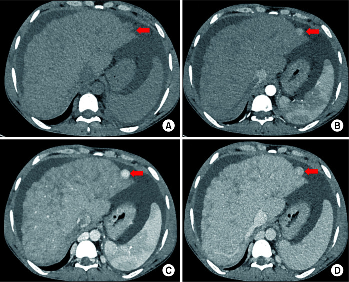
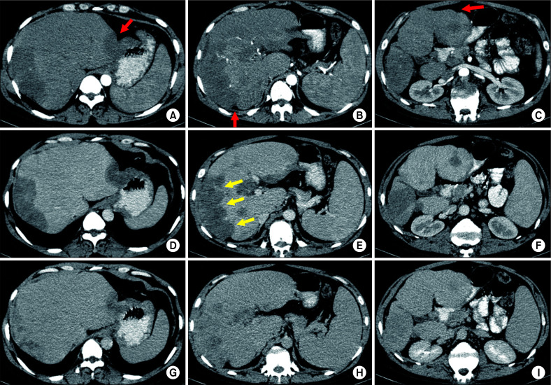
Vascular tumors of the liver are mesenchymal lesions from endothelial cells. They range from common benign lesions such as haemangioma, intermediate tumors like Kaposi sarcoma, and perivascular epithelioid cell tumor to malignant tumors such as hepatic epithelioid hemangioendothelioma and hepatic angiosarcoma in adults. Pediatric vascular tumors of the liver also include benign, locally aggressive, borderline, and malignant masses with haemangiomas being the most common benign tumors and epithelioid hemangioendothelioma being an uncommon pediatric malignancy. The list of these lesions is completed by nodular regenerative hyperplasia, solitary fibrous tumour, and hepatic small vessel neoplasms (HSVN). Some of these tumors are uncommon and rare. This review article aimed to enumerate hepatic vascular tumors along with their imaging, histopathology, molecular findings for accurate diagnosis that can result in better management.

Keywords: Diagnostic; Liver; Vascular tumour.


Conflict of interest statement

CONFLICT OF INTEREST

No potential conflict of interest relevant to this article was reported.

Figures

Fig. 1
Fig. 1
Multiphasic contrast-enhanced computed tomography (CT) in a 52-year-old male patient showing a giant hepatic haemangioma (arrow in A). Axial images in (A) arterial, (B) portal venous, (C) hepatic venous, and (D) 3-minute delayed phases show typical peripheral discontinuous nodular enhancement with progressive centripetal filling of contrast. Note that the density of nodular contrast in the lesion matches with the aorta (blood pool).
Fig. 2
Fig. 2
Multiple hepatic haemangiomas on contrast-enhanced computed tomography (CT) in portal venous phase in a 42-year-old female. The coronal image shows two small haemangiomas (red arrows) and one giant haemangioma (yellow arrow).
Fig. 3
Fig. 3
Ultrasound images in an infant with congenital hepatic haemangioma. (A) Colour doppler image in a 19-day-old neonate shows a large hyperechoic mass with internal cystic areas and vascularity. Lesion is delineated by red arrows. Mass is causing splaying of hepatic veins. (B) Follow-up ultrasound at the age of 15 months shows a significant decrease in the size of the mass lesion with increased hyperechoic fibrous component. Mass is delineated by yellow arrows.
Fig. 4
Fig. 4
Contrast-enhanced computed tomography (CT) images in a 28-day-old female neonate showing diffuse infantile hepatic haemangiomas who presented with abdominal distension. Axial images in (A) portal venous, (B) 3-minute delayed phases show typical peripheral nodular enhancement with progressive centripetal filling of contrast in delayed phase. Intervening normal liver parenchyma is seen between lesions.
Fig. 5
Fig. 5
Multiphasic contrast-enhanced computed tomography (CT) in a 45-year-old male patient showing a large regenerative nodule in the background of Budd-Chiari syndrome. Axial images in (A) non-contrast, (B) arterial, (C) portal venous, and (D) hepatic venous phases show a well-defined nodule in segment II (red arrow). Nodule is iso-hypoattenuating in non-contrast CT (A), showing arterial enhancement (B) which homogeneously progresses in portal venous phase and becomes isoattenuating in hepatic venous phase (D) with the background parenchyma. Hepatomegaly, heterogeneous reticular enhancement of liver, splenomegaly, prominent azygous system, and ascites are features of Budd-Chiari syndrome seen in this case due to supra hepatic inferior vena cava (IVC) stenosis.
Fig. 6
Fig. 6
Multiple hepatic epitheloid hemangioendotheliomas on multiphasic contrast-enhanced computed tomography (CT) (all axial images at three different levels) in a 35-year-old male patient. (A, B, C) Arterial phase, (D, E, F) venous phase, (G, H, I) 3-minute delayed phase images showing multiple peripheral subcapsular lesions with progressive enhancement. Lesions are causing subcapsular retraction (red arrows) leading to a nodular outline of the liver. Some lesions are coalescing to form confluent masses along the right lobe (yellow arrows in E).

Similar articles

Cited by

References

    1. Lerut J, Iesari S. Vascular tumours of the liver: a particular story. Transl Gastroenterol Hepatol. 2018;3:62. doi: 10.21037/tgh.2018.09.02. - DOI - PMC - PubMed
    1. WHO Classification of Tumours Editorial Board, author. Soft tissue and bone tumours. 5th ed. Vol 3. International Agency for Research on Cancer; 2020.
    1. Bannoura S, Putra J. Primary malignant vascular tumors of the liver in children: angiosarcoma and epithelioid hemangioendothelioma. World J Gastrointest Oncol. 2021;13:223–230. doi: 10.4251/wjgo.v13.i4.223. - DOI - PMC - PubMed
    1. Bridgewater J, Galle PR, Khan SA, Llovet JM, Park JW, Patel T, et al. Guidelines for the diagnosis and management of intrahepatic cholangiocarcinoma. J Hepatol. 2014;60:1268–1289. doi: 10.1016/j.jhep.2014.01.021. - DOI - PubMed
    1. European Association for the Study of the Liver, author. EASL Clinical Practice Guidelines: management of hepatocellular carcinoma. J Hepatol. 2018;69:182–236. doi: 10.1016/j.jhep.2019.01.020. - DOI - PubMed

LinkOut - more resources