Review
. 2023 Sep 29;5(1):20230019.
doi: 10.1259/bjro.20230019. eCollection 2023.

How to 19F MRI: applications, technique, and getting started

Affiliations
Review

How to 19F MRI: applications, technique, and getting started

Olga Maxouri et al. BJR Open. .

Abstract

Magnetic resonance imaging (MRI) plays a significant role in the routine imaging workflow, providing both anatomical and functional information. 19F MRI is an evolving imaging modality where instead of 1H, 19F nuclei are excited. As the signal from endogenous 19F in the body is negligible, exogenous 19F signals obtained by 19F radiofrequency coils are exceptionally specific. Highly fluorinated agents targeting particular biological processes (i.e., the presence of immune cells) have been visualised using 19F MRI, highlighting its potential for non-invasive and longitudinal molecular imaging. This article aims to provide both a broad overview of the various applications of 19F MRI, with cancer imaging as a focus, as well as a practical guide to 19F imaging. We will discuss the essential elements of a 19F system and address common pitfalls during acquisition. Last but not least, we will highlight future perspectives that will enhance the role of this modality. While not an exhaustive exploration of all 19F literature, we endeavour to encapsulate the broad themes of the field and introduce the world of 19F molecular imaging to newcomers. 19F MRI bridges several domains, imaging, physics, chemistry, and biology, necessitating multidisciplinary teams to be able to harness this technology effectively. As further technical developments allow for greater sensitivity, we envision that 19F MRI can help unlock insight into biological processes non-invasively and longitudinally.


Conflict of interest statement

Competing interests: MD and MS work for Cenya Imaging BV.

Figures

Figure 1.
Figure 1.
Highlights of 19F MRI for biomedical applications. Cell tracking and quantification play a prominent role in 19F MRI research due to the quantitative nature and specificity of 19F MRI. PFC formulations have been used to label cells ex vivo and in situ in order to probe the fate of transplanted cells or to image inflammation. Molecular 19F MRI non-invasively probes physiological parameters such as hypoxia, pH, and redox state. Stimuli-responsive OFF/ON nanoprobes attenuate the MR signal of neighbouring 19F atoms through their influence on the local environment. Silencing of 19F reporters occurs via T2 shortening, which can be achieved by immobilising 19F spins or via PRE. Conformational changes or probe disassembly is induced in the presence of the desired stimuli, thereby restoring T2 along with the 19F MR signal. The fate of 19F agents can be further influenced by functional groups and the structure of a nanocarrier system (e.g., multicore PLGA NP). In addition, multimodality and multispectral MRI add another dimension of information to 19F MRI studies. Fluorescent moieties incorporated in 19F formulations allow subsequent model characterisation via ex vivo cytometric analyses such as flow cytometry and fluorescence microscopy. Radioisotopes combined with fluorinated polymers enable sensitive PET imaging alongside 19F MRI. Different cell populations or targets can be imaged within a single subject with multispectral MRI using 19F agents with discriminating NMR signals. c.s: chemical shift, CT: computed tomography; DC: Dendritic cell; LC: lymphocyte; NK cell: natural killer cell; OFI: optical fluorescence imaging; PET: positron emission tomography; PFC: perfluorocarbon; PLGA NP: poly(lactic-co-glycolic acid) nanoparticles; pO2: partial oxygen tension; PRE: paramagnetic relaxation enhancement; SC: stem cell; TC: tumour cell; USG: ultrasound sonography
Figure 2.
Figure 2.
Pitfalls in 19F MRI for beginners. 1H and 19F MR images were acquired in PFPE phantoms at 7T. 1H/19F images are depicted in greyscale (1H) and pseudocolour (19F). Image (iii) is the correct example. (a) Influence of transmitter gain calibrations on image quality, comparing 19F FSE acquisitions of (i) calibrated and (ii) uncalibrated RF pulse power. (b) and (c) Examples of chemical shift artefacts along the directions of frequency-encoding (iv) (v) and (ix), and slice selection (v) indicated by the red and green arrows, respectively. 19F images are superimposed in pseudocolour on top of the 1H images to illustrate the mismapping of the MR signal when selecting an off-resonance working frequency. PFPE has a major and a minor resonance peak in close proximity to each other. NMR signals from the minor peak are not detected at in vivo tracer concentrations. The chemical shift artefact recorded at a different slice (iv, green arrow) occurred due to imaging with an undiluted PFPE phantom. (c) Main magnetic field (B0) inhomogeneities influence the Larmor frequency of 19F spins, causing distortions (vi), loss of signal due to line broadening (vii), and banding artefacts (ix). The 19F MR image in pseudocolour (v) was acquired with a fast spin echo sequence using erroneous shim values, while the 1H acquisition (greyscale) was acquired with proper shims. The 19F NMR spectrum of PFPE depicted in the upper graph was acquired in an inhomogeneous B0 field and displays line broadening and increased noise compared to the bottom graph, which was acquired in a shimmed system. 1H FSE (viii) and bSSFP 19F (ix) MRI of four tubes filled with serially diluted PFPE in 1% low-melting point agarose. The bSSFP sequence is sensitive to B0 inhomogeneities leading to the appearance of banding artefacts (blue arrow). Tissue-border interactions (in this case, the four tubes and air) perturb B0 homogeneity, which may be challenging to fully correct via shimming. The four tubes with PFPE could instead be embedded within another tube with agarose to minimise tissue-border interactions. The bottom tubes were below the detection limits and, therefore, not visible in the 19F image. bSSFP: balanced steady-state free precession; FSE: fast spin echo; PFPE: perfluoropolyether; RF: radiofrequency
Figure 3.
Figure 3.
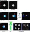
Basic principle of multispectral 19F MRI (a) Schematic representation of NMR spectra of three different fluorinated nanoprobes: I, II and III, each with distinct chemical shifts. The dashed boxes enclose the spectral peaks selected for 19F MRI. (b) Schematic representation of multispectral 19F MRI with phantoms. From top to bottom: 1H MRI for anatomical context, 19F MRI with the working frequency set to the respective nanoprobe (I, II or III) and a merged image with the 19F signals in pseudo-colour.

References

    1. Bassir A, Raynor WY, Park PSU, Werner TJ, Alavi A, Revheim ME. Molecular imaging in atherosclerosis. Clin Transl Imaging 2022; 10: 259–72. doi: 10.1007/s40336-022-00483-y - DOI
    1. Jack CR Jr, Bennett DA, Blennow K, Carrillo MC, Feldman HH, Frisoni GB, et al. A/T/N: an unbiased descriptive classification scheme for Alzheimer disease biomarkers. Neurology 2016; 87: 539–47. doi: 10.1212/WNL.0000000000002923 - DOI - PMC - PubMed
    1. Marciscano AE, Thorek DLJ. Role of noninvasive molecular imaging in determining response. Adv Radiat Oncol 2018; 3: 534–47. doi: 10.1016/j.adro.2018.07.006 - DOI - PMC - PubMed
    1. Bergamasco A, Luyet-Déruaz A, Gollop ND, Moride Y, Qiao Q. Epidemiology of asymptomatic pre-heart failure: a systematic review. Curr Heart Fail Rep 2022; 19: 146–56. doi: 10.1007/s11897-022-00542-5 - DOI - PMC - PubMed
    1. Di Meco A, Vassar R. Early detection and personalized medicine: future strategies against Alzheimer's disease. Prog Mol Biol Transl Sci 2021; 177: 157–73. doi: 10.1016/bs.pmbts.2020.10.002 - DOI - PMC - PubMed