

Pediatric gliosarcoma, a rare central nervous system tumor in children: Case report and literature review
- PMID: 37954329
- PMCID: PMC10637930
- DOI: 10.1016/j.heliyon.2023.e21204
Pediatric gliosarcoma, a rare central nervous system tumor in children: Case report and literature review
Abstract
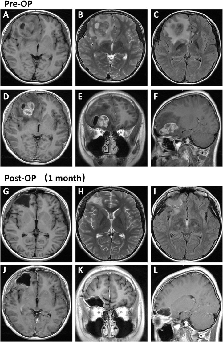
Gliosarcoma is a rare and highly malignant central nervous system tumor that accounts for 1%-8% of glioblastomas; it usually occurs in middle-aged and older adults between 40 and 60 years of age and is rare in children. We report an 11-year-old boy with right frontal lobe gliosarcoma who underwent aggressive gross total resection and postoperative radiotherapy, experienced recurrence and subsequently underwent a second operation. To better understand the disease and explore treatment options, we briefly report this case and review the relevant literature.
Keywords: Cerebral hemispheres; Glioblastoma; Gliosarcoma; Pediatric; Surgery.
© 2023 The Authors.
Conflict of interest statement
The remaining authors declare that the research was conducted in the absence of any commercial or financial relationships that could be construed as a potential conflict of interest.
Figures


Similar articles
-
Gliosarcoma in patients under 20 years of age. A clinicopathologic study of 11 cases and detailed review of the literature.BMC Pediatr. 2021 Feb 26;21(1):101. doi: 10.1186/s12887-021-02556-9. BMC Pediatr. 2021. PMID: 33637068 Free PMC article. Review.
-
Gliosarcoma with multiple extracranial metastases: case report and review of the literature.J Neurooncol. 2007 May;83(1):39-46. doi: 10.1007/s11060-006-9295-x. Epub 2006 Dec 14. J Neurooncol. 2007. PMID: 17171442 Review.
-
Giant parieto-occipital lobe pediatric gliosarcoma: Report of a rare entity and review of literature.Surg Neurol Int. 2018 May 29;9:111. doi: 10.4103/sni.sni_31_18. eCollection 2018. Surg Neurol Int. 2018. PMID: 29930877 Free PMC article.
-
[A Patient with Primary Intraventricular Gliosarcoma and Long-term Survival - a Case Report].Klin Onkol. 2016 Winter;29(6):454-459. doi: 10.14735/amko2016454. Klin Onkol. 2016. PMID: 27951723 Czech.
-
[Pediatric Gliosarcoma: Case Report and Literature Review].Bol Asoc Med P R. 2014;106(3):43-7. Bol Asoc Med P R. 2014. PMID: 25470910 Review. Spanish.
References
-
- D'Arco F., Khan F., Mankad K., Ganau M., Caro-Dominguez P., Bisdas S. Differential diagnosis of posterior fossa tumours in children: new insights. Pediatr. Radiol. 2018;48(13):1955–1963. - PubMed
-
- Ng H.K., Poon W.S. Gliosarcoma of the posterior fossa with features of a malignant fibrous histiocytoma. Cancer. 1990;65(5):1161–1166. - PubMed
Publication types
LinkOut - more resources
Full Text Sources
