. 2023 Nov 6:10:1973-1990.
doi: 10.2147/JHC.S426442. eCollection 2023.

Immune Effects of Cryoablation in Woodchuck Hepatocellular Carcinoma

Affiliations

Immune Effects of Cryoablation in Woodchuck Hepatocellular Carcinoma

Michal Mauda-Havakuk et al. J Hepatocell Carcinoma. .

Abstract

Objectives: Local and systemic immune responses evoked by locoregional therapies such as cryoablation are incompletely understood. The aim of this study was to characterize cryoablation-related immune response and the capacity of immune drugs to augment immunity upon cryoablation for the treatment of hepatocellular carcinoma (HCC) using a woodchuck hepatocellular carcinoma model.

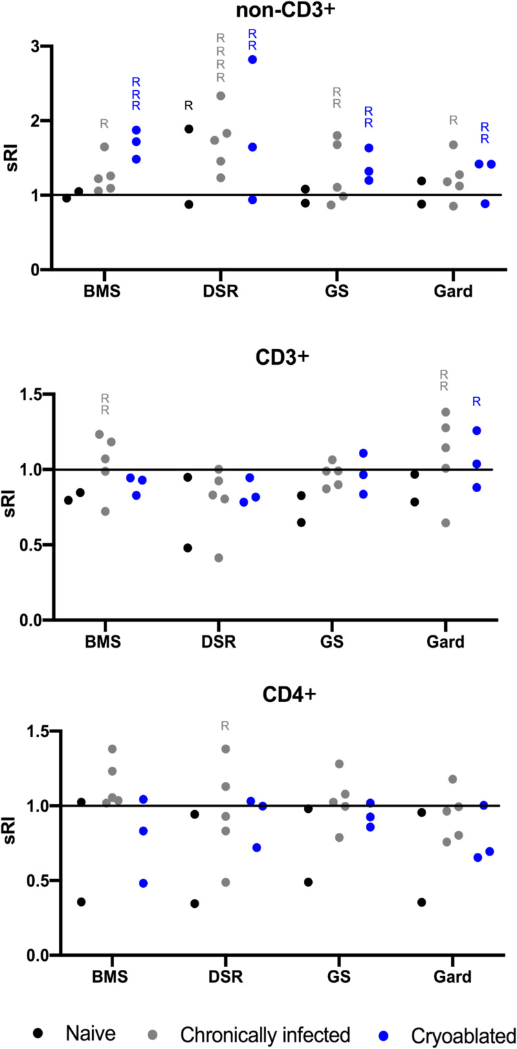
Materials and methods: Twelve woodchucks chronically infected with woodchuck hepatitis virus and with hepatocellular carcinoma underwent imaging with contrast-enhanced CT. Partial cryoablation of tumors in three woodchucks was performed. Fourteen days after cryoablation, liver tissues were harvested and stained with H&E and TUNEL, and immune infiltrates were quantified. Peripheral blood mononuclear cells (PBMC) were collected from ablated and nonablated woodchucks, labeled with carboxyfluorescein succinimidyl ester (CFSE) and cultured with immune-modulating drugs, including a small PD-L1 antagonist molecule (BMS-202) and three TLR7/8 agonists (DSR 6434, GS-9620, gardiquimod). After incubation, cell replication and immune cell populations were analyzed by flow cytometry.

Results: Local immune response in tumors was characterized by an increased number of CD3+ T lymphocytes and natural killer cells in the cryolesion margin compared to other tumor regions. T regulatory cells were found in higher numbers in distant tumors within the liver compared to untreated or control tumors. Cryoablation also augmented the systemic immune response as demonstrated by higher numbers of PBMC responses upon immune drug stimulation in the cryoablation group.

Conclusions: Partial cryoablation augmented immune effects in both treated and remote untreated tumor microenvironments, as well as systemically, in woodchucks with HCC. Characterization of these mechanisms may enhance development of novel drug-device combinations for treatment of HCC.

Keywords: cryoablation; hepatitis B virus; hepatocellular carcinoma; immune effect; tumor microenvironment; woodchuck.


Conflict of interest statement

Dr Andrew S Mikhail reports grants from BTG (Now Boston Scientific), during the conduct of the study. Dr William F Pritchard reports the NIH and Boston Scientific Corporation (previously Biocompatibles UK Ltd) have a Cooperative Research and Development Agreement providing support for this research, non-financial support from Northeastern Wildlife, during the conduct of the study. Dr Bradford J Wood reports non-financial support from Philips, non-financial support from Boston Scientific / BTG Biocompatibles, during the conduct of the study; non-financial support, Cooperative Research and Development Agreement [CRADA] from Siemens, grants, non-financial support from NVIDIA, grants, Cooperative Research and Development Agreement [CRADA] from Celsion/Immunon, personal fees from Philips, non-financial support, negotiating Cooperative Research and Development Agreement [CRADA]. Negotiating licensing agreement from Canon Medical, non-financial support, Equipment MTA from Medtronic, grants, Cooperative Research and Development Agreement [CRADA] from XAct Robotics, non-financial support, Equipment MTA from Angiodynamics, non-financial support, Equipment MTA from Imactis, non-financial support, Equipment MTA from Profound MEdical, grants, non-financial support, Cooperative Research and Development Agreement [CRADA] from ProMaxo, non-financial support, Equipment MTA from QT Imaging, non-financial support, Supplies MTA from Theromics, non-financial support, Equipment MTA from MediView, non-financial support, Equipment MTA from Johnson and Johnson, non-financial support, Equipment MTA from Clinical Laserthermia Systems, non-financial support, Supplies MTA from Varian, non-financial support, Equipment MTA from Civco Medical, non-financial support, Equipment MTA from Combat Medical, non-financial support, Equipment MTA from Galvanize, outside the submitted work; In addition, Dr Bradford J Wood has a patent portfolio available upon request with royalties paid to see above. The authors report no other conflicts of interest in this work.

Figures

Figure 1
Figure 1
Study design and methods. (A) Woodchucks were infected with woodchuck hepatitis virus (WHV) as newborns. At the age of 18–24 months, woodchucks were imaged with computed tomography. Ultrasound guided cryoablation was performed on three animals. Cryoablated woodchucks were imaged and euthanized 14 days after treatment. Whole blood was collected, liver and tumors were sectioned and formalin preserved, and peripheral blood mononuclear cells (PBMC) were purified. (B) Tumors were stained for CD3, CD4, NCAM and FOXP3 markers. Immunohistochemical quantification was performed on 5 distinct regions. Three regions were in the ablated tumors: 1. Ablation margin 2. Ablation zone, and 3. Unablated region. The fourth region was located within a separate, untreated tumor (UT) in ablated animals. The fifth region was located in tumors within control, untreated animals (CT) n=9. (C) PBMC were purified from whole blood, labeled, cultured with immune-modulating drugs for 4 days and stained with CD3 and CD4 antibodies. FACS analysis was performed and data analyzed for cell replication (n=3 cryoablated animals, n=5 control infected animals, n=2 control uninfected animals).
Figure 2
Figure 2
Cryolesion architecture on H&E and TUNEL stains. (A) Representative H&E-stained sections of the 3 cryolesions. In the left panel a defined cryolesion with central coagulative necrosis surrounded with a homogenous rim is demonstrated. The middle panel demonstrates a cryolesion with apparent hemorrhage along the needle track and less homogeneous cryolesion rims (reprinted from. with permission). The right panel shows the ablation zone with liquefactive necrosis surrounded by a rim of cell debris that can be found in the central ablation zone as well as in the periphery. Magnified (B) H&E-stained and (C) TUNEL stained sections of the regions marked in (A) delineating the 3 compartments within the cryolesion: central necrosis (1), cell debris rim (2) and coagulative necrosis outer rim (3). Tumor tissue that was not cryoablated is marked with UT (untreated tumor). TUNEL stain annotation was performed on 4 regions. Two regions in the ablated tumor, the ablation zone and untreated tumor (UT), are shown on panel (D). Spontaneous necrosis and control tumor (CT) in untreated animals were evaluated as well both regions represented in panel (E). (F) TUNEL stain quantification, control animals N=7, cryoablated animals N=3, 1–2 sections per animal, p-value for the entire test is shown on the graph. P-values between pairs of regions are summarized in Table 1.
Figure 3
Figure 3
Local immune effects evaluated with immune cell distribution and quantification within the cryolesions. Rows demonstrate immunohistochemical expression and quantification of immune infiltrates, from top to bottom: CD3+, CD4+, NCAM (natural killers’ cells) and FOXP3 (T-regulatory cells). Whole mount sections for each stain are shown in the left column with magnification of the cryolesion margin zone in the center column. Quantification for each immune stain by regions as defined in methods and Figure 1B is represented on the right column. Control animals N=5, cryoablated animals N=3, 1–2 sections per animal, p-values for the entire test are shown on the graphs. P-values between pairs of regions are summarized in Table 1.
Figure 4
Figure 4
Replication magnitude of PBMC upon nonspecific stimulation in naïve, chronically infected, and cryoablated woodchucks. Evaluation of lymphocyte replication using carboxyfluorescein succinimidyl ester (CFSE) assay in naïve, chronically infected and cryoablated woodchucks for non-CD3+, CD3+, and CD4+ cells. Data represent mean ± SEM. *p<0.05 one-way ANOVA.
Figure 5
Figure 5
Immune-modulating drug effects on PBMC replication. Evaluation of drug-induced lymphocyte replication using carboxyfluorescein succinimidyl ester (CFSE) assay in naïve, chronically infected and cryoablated woodchucks. The standardized replication index (sRI) is presented for all drugs for non-CD3+ (A), CD3+ (B), and CD4+ (C) sub-populations. The test of whether the sRIs were greater than 1 was based on a t-test applied to the sRI’s least-squares means and their corresponding standard error, which were obtained from the statistical models. The significant differences between groups were calculated by one-way ANOVA (Tukey’s multiple comparison test). p<0.05 was considered as statistically significant and marked by *P-values are summarized in Table 2. BMS= BMS-202; DSR= DSR-6434; GS= GS-9620; Gard= gardiquimod.
Figure 6
Figure 6
Systemic immune responses by individuals. PBMC from individual woodchucks were evaluated for responses to four experimental immune-modulating drugs. Each combination of woodchuck PBMC sample and a drug comprised a test. Responses were defined as any test with standardized replication index (sRI) least-squares mean greater than 1 and unadjusted p-value < 0.0025 (= 0.05/20 tests). Drug concentrations for each animal were combined and every dot represents the mean sRI. Samples from the same animal cohort are color coded. Any test that fulfilled the response criteria was marked with “R”. For example, in the non-CD3+ cell population (top graph) upon DSR 6434 stimulation, 1, 4 and 2 responses were found in the naïve, chronically infected untreated, and the cryoablated woodchucks, respectively. BMS= BMS-202; DSR= DSR-6434; GS= GS-9620; Gard= gardiquimod.

References

    1. Cazzato RL, Garnon J, Ramamurthy N, et al. Percutaneous image-guided cryoablation: current applications and results in the oncologic field. Med Oncol. 2016;33:140. doi:10.1007/s12032-016-0848-3 - DOI - PubMed
    1. Greten TF, Mauda-Havakuk M, Heinrich B, et al. Combined locoregional-immunotherapy for liver cancer. J Hepatol. 2019;70:999–1007. doi:10.1016/j.jhep.2019.01.027 - DOI - PMC - PubMed
    1. Erinjeri JP, Fine GC, Adema GJ, et al. Immunotherapy and the interventional oncologist: challenges and opportunities-a society of interventional oncology white paper. Radiology. 2019;292:25–34. doi:10.1148/radiol.2019182326 - DOI - PMC - PubMed
    1. Llovet JM, De Baere T, Kulik L, et al. Locoregional therapies in the era of molecular and immune treatments for hepatocellular carcinoma. Nat Rev Gastroenterol Hepatol. 2021;18:293–313. doi:10.1038/s41575-020-00395-0 - DOI - PubMed
    1. Jansen MC, van Hillegersberg R, Schoots IG, et al. Cryoablation induces greater inflammatory and coagulative responses than radiofrequency ablation or laser induced thermotherapy in a rat liver model. Surgery. 2010;147:686–695. doi:10.1016/j.surg.2009.10.053 - DOI - PubMed