Review
. 2023 Oct 26:14:1234535.
doi: 10.3389/fimmu.2023.1234535. eCollection 2023.

Cutaneous manifestations of inflammatory bowel disease: basic characteristics, therapy, and potential pathophysiological associations

Affiliations
Review

Cutaneous manifestations of inflammatory bowel disease: basic characteristics, therapy, and potential pathophysiological associations

Ronghua He et al. Front Immunol. .

Abstract

Inflammatory bowel disease (IBD) is a chronic inflammatory disease typically involving the gastrointestinal tract but not limited to it. IBD can be subdivided into Crohn's disease (CD) and ulcerative colitis (UC). Extraintestinal manifestations (EIMs) are observed in up to 47% of patients with IBD, with the most frequent reports of cutaneous manifestations. Among these, pyoderma gangrenosum (PG) and erythema nodosum (EN) are the two most common skin manifestations in IBD, and both are immune-related inflammatory skin diseases. The presence of cutaneous EIMs may either be concordant with intestinal disease activity or have an independent course. Despite some progress in research on EIMs, for instance, ectopic expression of gut-specific mucosal address cell adhesion molecule-1 (MAdCAM-1) and chemokine CCL25 on the vascular endothelium of the portal tract have been demonstrated in IBD-related primary sclerosing cholangitis (PSC), little is understood about the potential pathophysiological associations between IBD and cutaneous EIMs. Whether cutaneous EIMs are inflammatory events with a commonly shared genetic background or environmental risk factors with IBD but independent of IBD or are the result of an extraintestinal extension of intestinal inflammation, remains unclear. The review aims to provide an overview of the two most representative cutaneous manifestations of IBD, describe IBD's epidemiology, clinical characteristics, and histology, and discuss the immunopathophysiology and existing treatment strategies with biologic agents, with a focus on the potential pathophysiological associations between IBD and cutaneous EIMs.

Keywords: Crohn’s disease; cutaneous manifestations; extraintestinal manifestations (EIMs); inflammatory bowel disease; pathophysiological associations; ulcerative colitis.


Conflict of interest statement

The authors declare that the research was conducted in the absence of any commercial or financial relationships that could be construed as a potential conflict of interest.

Figures

Figure 1
Figure 1
Partial pathophysiological associations between IBD and cutaneous EIMs. TNF-α, tumor necrosis factor-α; IFN-γ, interferon-γ; IL, interleukin; CXCL, C-X-C motif chemokine ligand; ILCs, innate lymphoid cells; CLA, cutaneous lymphocyte-associated antigen; LFA-1, lymphocyte function-associated antigen-1; ICAM-1, intercellular adhesion molecule-1; Th1, type 1 helper T; Th17, type 17 helper T.
Figure 2
Figure 2
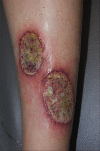
Ulcerative PG, the most common type, presents as a rapidly enlarged deep ulceration with erythematous borders. Reproduced with permission from (62). © 2023 Indian Journal of Dermatology, Venereology and Leprology - Published by Scientific Scholar.
Figure 3
Figure 3
Histopathology of ulcerative PG. (A) Low-power magnification (H&E staining, ×125) reveals diffuse epidermal necrotic ulcer with massive neutrophilic infiltrations and dermal granulation tissue. (B) High-power magnification (H&E staining, ×2000) demonstrates that the majority of lesional cells consist of neutrophils. Reproduced with permission from (62). © 2023 Indian Journal of Dermatology, Venereology and Leprology - Published by Scientific Scholar.
Figure 4
Figure 4
A typical EN shows symmetrical, raised, erythematous, tender non-ulcerative subcutaneous nodules on the tibial anterior region. Reproduced with permission from (57). © 2015 Crohn’s & Colitis Foundation of America, Inc.
Figure 5
Figure 5
Histopathology of EN shows septal panniculitis and a mixed inflammatory infiltrate predominantly composed of lymphocytes, histiocytes, and neutrophils (H&E staining, ×40). Inset: Miescher’s radial granuloma (H&E staining, ×200). Reproduced with permission from (84). © 2016 The International Society of Dermatology.

References

    1. Chang JT. Pathophysiology of inflammatory bowel diseases. N Engl J Med (2020) 383:2652–64. doi: 10.1056/NEJMra2002697 - DOI - PubMed
    1. Ng SC, Shi HY, Hamidi N, Underwood FE, Tang W, Benchimol EI, et al. . Worldwide incidence and prevalence of inflammatory bowel disease in the 21st century: a systematic review of population-based studies. Lancet (2017) 390:2769–78. doi: 10.1016/S0140-6736(17)32448-0 - DOI - PubMed
    1. Windsor JW, Kaplan GG. Evolving epidemiology of IBD. Curr Gastroenterol Rep (2019) 21:40. doi: 10.1007/s11894-019-0705-6 - DOI - PubMed
    1. Ananthakrishnan AN. Epidemiology and risk factors for IBD. Nat Rev Gastroenterol Hepatol (2015) 12:205–17. doi: 10.1038/nrgastro.2015.34 - DOI - PubMed
    1. Mak WY, Zhao M, Ng SC, Burisch J. The epidemiology of inflammatory bowel disease: East meets west. J Gastroenterol Hepatol (2020) 35:380–89. doi: 10.1111/jgh.14872 - DOI - PubMed

Publication types

MeSH terms