. 2023 Oct 6;4(6):zqad058.
doi: 10.1093/function/zqad058. eCollection 2023.

Breathing and Oxygen Carrying Capacity in Ts65Dn and Down Syndrome

Affiliations

Breathing and Oxygen Carrying Capacity in Ts65Dn and Down Syndrome

Lara R DeRuisseau et al. Function (Oxf). .

Abstract

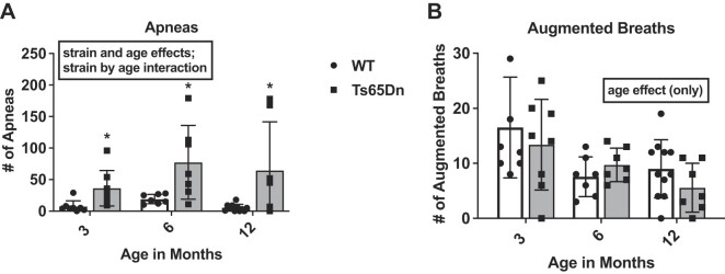
Individuals with Down syndrome (Ds) are at increased risk of respiratory infection, aspiration pneumonia, and apnea. The Ts65Dn mouse is a commonly used model of Ds, but there have been no formal investigations of awake breathing and respiratory muscle function in these mice. We hypothesized that breathing would be impaired in Ts65Dn vs. wild-type (WT), and would be mediated by both neural and muscular inputs. Baseline minute ventilation was not different at 3, 6, or 12 mo of age. However, VT/Ti, a marker of the neural drive to breathe, was lower in Ts65Dn vs. WT and central apneas were more prevalent. The response to breathing hypoxia was not different, but the response to hypercapnia was attenuated, revealing a difference in carbon dioxide sensing, and/or motor output in Ts65Dn. Oxygen desaturations were present in room air, demonstrating that ventilation may not be sufficient to maintain adequate oxygen saturation in Ts65Dn. We observed no differences in arterial PO2 or PCO2, but Ts65Dn had lower hemoglobin and hematocrit. A retrospective medical record review of 52,346 Ds and 52,346 controls confirmed an elevated relative risk of anemia in Ds. We also performed eupneic in-vivo electromyography and in-vitro muscle function and histological fiber typing of the diaphragm, and found no difference between strains. Overall, conscious respiration is impaired in Ts65Dn, is mediated by neural mechanisms, and results in reduced hemoglobin saturation. Oxygen carrying capacity is reduced in Ts65Dn vs. WT, and we demonstrate that individuals with Ds are also at increased risk of anemia.

Keywords: apnea; diaphragm; hypercapnia; hypoxemia; hypoxia; plethysmography; trisomy 21; ventilation.


Conflict of interest statement

None of the authors has any conflict of interest.

Figures

Graphical Abstract
Graphical Abstract
Figure 1.
Figure 1.
Minute ventilation during baseline and 10 min of hypoxia and hypercapnia. Average minute ventilation (mL/min) for baseline (room air: 20.93% O2; balanced N2) and minute by minute response to 10% hypoxia (10% O2; balanced N2) was measured in (A) 3 mo wild-type (WT) mice (n = 7) and Ts65Dn (Ts) mice (n = 8), (B) 6 mo WT mice (n = 7) and Ts mice (n = 7), and (C) 12 mo WT mice (n = 11) and Ts mice (n = 7). Following a 10-min recovery, minute by minute response to 5% hypercapnia (5% CO2; 20.93% O2; balanced N2) was measured in the same (D) 3 , (E) 6, and (F) 12 mo WT and Ts mice. Hypercapnic exposure revealed a strain difference, but only at 12 mo where Ts mice showed a lower MV response (*P = .03 raw/absolute values, P = .015 vs. baseline) compared to WT counterparts. No significant differences were detected in response to hypoxia (P > .05).
Figure 2.
Figure 2.
Apneas and augmented breaths in conscious mice. (A) Apneas (lack of flow ≥ 0.5 s) and (B) Augmented Breaths for WT mice (n = 26) and Ts65Dn mice (n = 22) over 30 min breathing air. Apneas and augmented breaths were determined over 30 min. Apneas were defined as periods of suspended breathing lasting > 0.5 s, and augmented breaths were indicated by a sharp rise in the breathing trace above 1.25 mL/s followed by a sharp decrease below −0.75 mL/s. Strain (P < .001), age (P < .001), and a strain by age interaction (P < .001) were detected for apneas, with fewer apneas in WT mice. Augmented breaths were not different between strains, although an age effect (P = .002) was observed with fewer augmented breaths detected in older mice.
Figure 3.
Figure 3.
Hypoxemias in conscious mice. The MouseOx pulse oximeter was used to detect oxygen saturation over 1 h of room air breathing. Oxygen saturation below 85% for up to 5 s was counted as 1 hypoxemia in WT (n = 34) mice and Ts65Dn (n = 30). A strain (P = .03) and age effect (P = .045) were detected.
Figure 4.
Figure 4.
Arterial blood sampling in conscious mice. (A) Partial pressure of oxygen (PaO2), (B) partial pressure of carbon dioxide (PaCO2), (C) hemoglobin, and (D) hematocrit for WT mice (n = 18) and Ts65Dn (n = 10). Mice were implanted with a femoral catheter under a plane of anesthesia and allowed to recover. Once mice were ambulating throughout the cage and eating, a sample was collected in awake mice breathing room air. There were no significant differences for PaO2 (P = .896) or PaCO2 (P = .516) between groups. Hemoglobin (Hb) and hematocrit (Hct) were different. One mouse was removed due to Hb and Hct levels > 3 SD from the mean.
Figure 5.
Figure 5.
Diaphragm EMG in anesthetized mice. (A) Root-mean-squared (RMS) EMG amplitude for WT mice (n = 15) and Ts65Dn (n = 6) and a raw signal for a (B) WT mouse and (C) Ts65Dn mouse. Mice were anesthestized with isoflurane and a fine wire was inserted into the diaphragm. Electromyography signals were amplified at a gain of 500, filtered between 10 Hz and 1 kHz using an analog amplifier (Model 3000 Differential AC/DC Amplifier, A–M Systems). Data were collected and averaged over 1 min. There were no significant differences for RMS EMG (P = .112). One mouse was removed due to values > 3 SD from the mean.
Figure 6.
Figure 6.
In-vitro diaphragm strip muscle function. (A) Twitch and tetanic peak-stress (N/cm2). (B) Force–frequency curves expressed as a function of specific force production (N/cm2) at simulation frequencies between 10 and 300 Hz in wild type (WT; n = 16) and Ts65Dn (Ts; n = 8) mice, (C) relative fatigue development of diaphragm strips from WT and Ts mice during a 30-min fatigue protocol where strips were stimulated submaximally every 2 s at 30 Hz, and (D) relative force recovery over 15 min immediately following the fatiguing protocol. No significant differences between groups were detected for contractile measures (P > .05).
Figure 7.
Figure 7.
Diaphragm muscle histology. (A) Mean CSA values for type I/IIa, type IIx, and type IIb (P > .05). B.) Type I/IIa, IIx, and IIb fiber were counted and expressed as a percentage of the total number of muscle fibers on the section (P > .05). (C) and (D) Representative images of diaphragm obtained from WT (C) and Ts65Dn (D) mice.

References

    1. Egan JF, Benn PA, Zelop CM, Bolnick A, Gianferrari E, Borgida AF. Down syndrome births in the United States from 1989 to 2001. Am J Obstet Gynecol. 2004;191(3):1044–1048. - PubMed
    1. Shin M, Besser LM, Kucik JEet al. Prevalence of Down syndrome among children and adolescents in 10 regions of the United States. Pediatrics. 2009;124(6):1565–1571. - PubMed
    1. Birth defects surveillance data from selected states, 1998–2002. Birth Defect Res A. 2005;73(10):758–853. - PubMed
    1. Survey of Income and Program Participation . 2006.
    1. Lane SBaDL, ed. The Development of Intelligence. New York, NY: Holt, Rinehart and Winston, 1985.

Publication types

LinkOut - more resources