. 2023 Dec 8;133(12):966-988.
doi: 10.1161/CIRCRESAHA.122.321836. Epub 2023 Nov 13.

Cyclin D-CDK4 Disulfide Bond Attenuates Pulmonary Vascular Cell Proliferation

Affiliations

Cyclin D-CDK4 Disulfide Bond Attenuates Pulmonary Vascular Cell Proliferation

Hannah Knight et al. Circ Res. .

Abstract

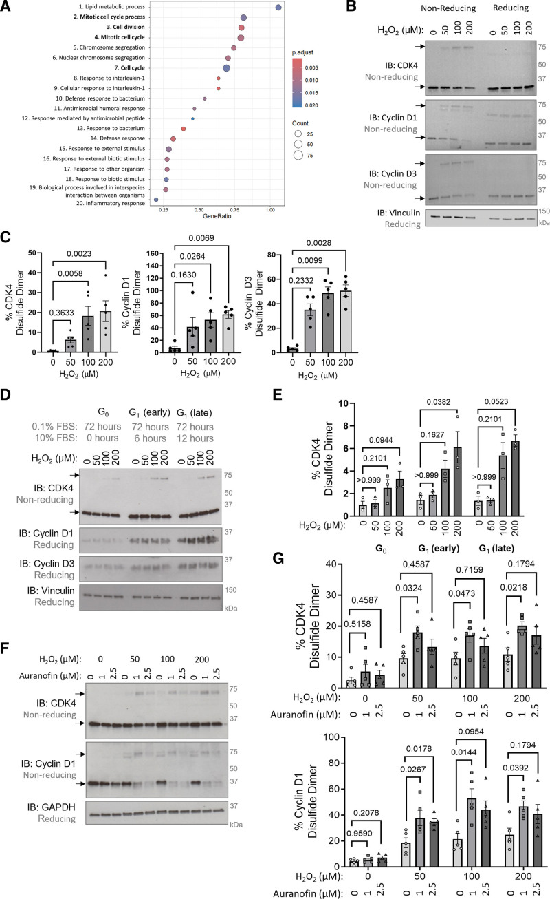
Background: Pulmonary hypertension (PH) is a chronic vascular disease characterized, among other abnormalities, by hyperproliferative smooth muscle cells and a perturbed cellular redox and metabolic balance. Oxidants induce cell cycle arrest to halt proliferation; however, little is known about the redox-regulated effector proteins that mediate these processes. Here, we report a novel kinase-inhibitory disulfide bond in cyclin D-CDK4 (cyclin-dependent kinase 4) and investigate its role in cell proliferation and PH.

Methods: Oxidative modifications of cyclin D-CDK4 were detected in human pulmonary arterial smooth muscle cells and human pulmonary arterial endothelial cells. Site-directed mutagenesis, tandem mass-spectrometry, cell-based experiments, in vitro kinase activity assays, in silico structural modeling, and a novel redox-dead constitutive knock-in mouse were utilized to investigate the nature and definitively establish the importance of CDK4 cysteine modification in pulmonary vascular cell proliferation. Furthermore, the cyclin D-CDK4 oxidation was assessed in vivo in the pulmonary arteries and isolated human pulmonary arterial smooth muscle cells of patients with pulmonary arterial hypertension and in 3 preclinical models of PH.

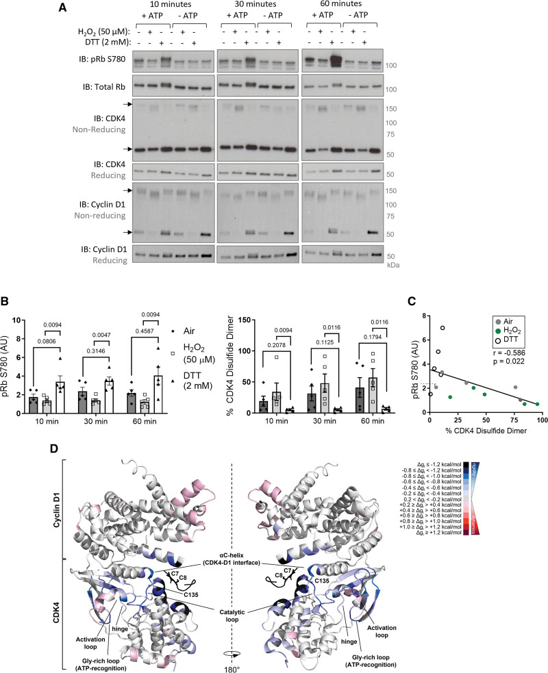
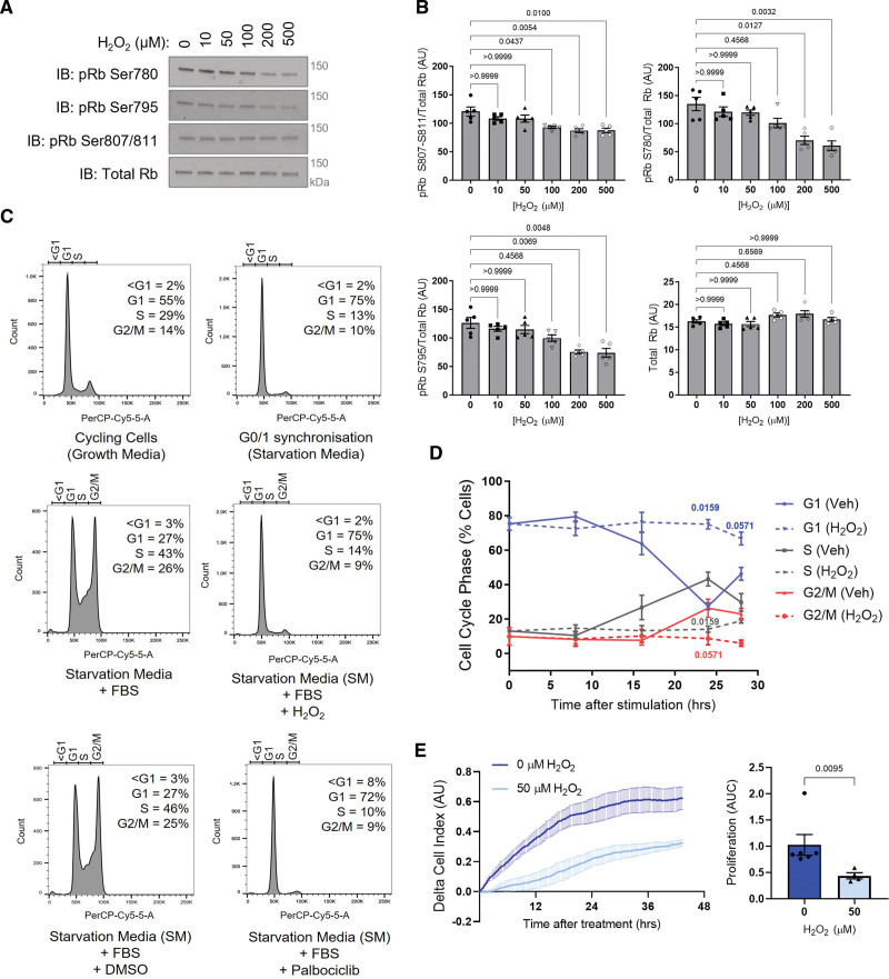
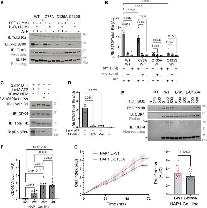
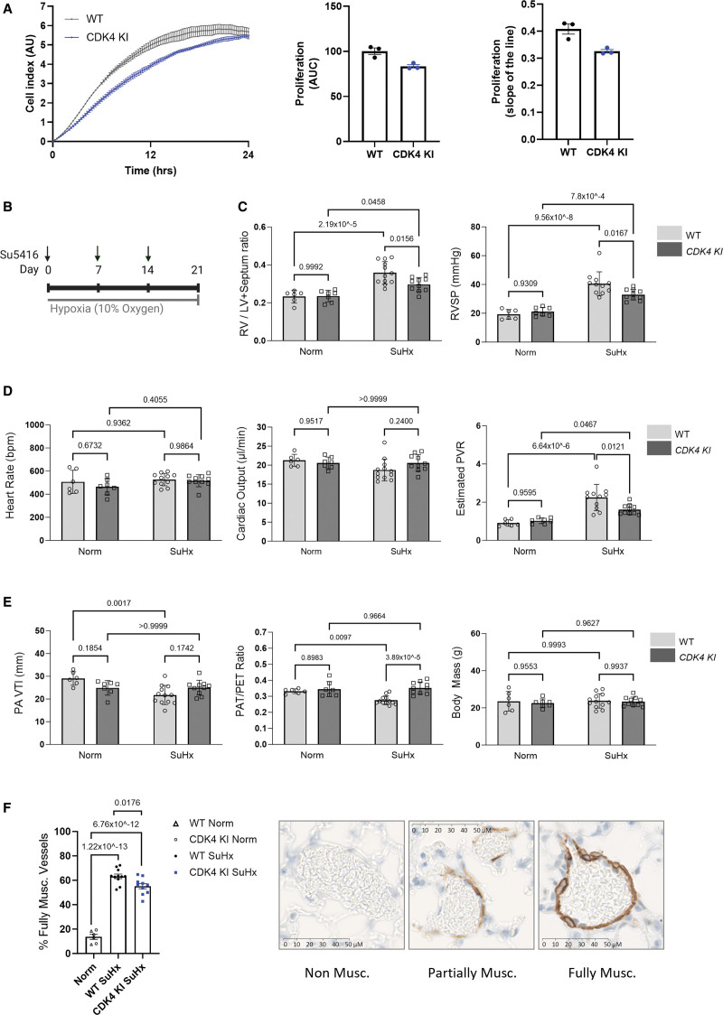
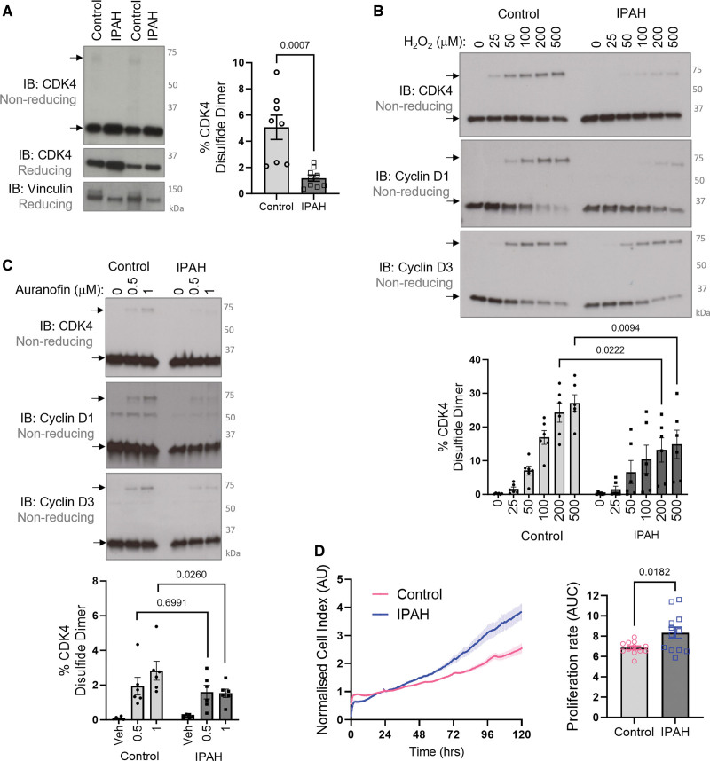
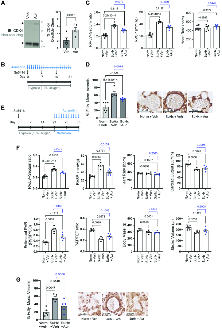
Results: Cyclin D-CDK4 forms a reversible oxidant-induced heterodimeric disulfide dimer between C7/8 and C135, respectively, in cells in vitro and in pulmonary arteries in vivo to inhibit cyclin D-CDK4 kinase activity, decrease Rb (retinoblastoma) protein phosphorylation, and induce cell cycle arrest. Mutation of CDK4 C135 causes a kinase-impaired phenotype, which decreases cell proliferation rate and alleviates disease phenotype in an experimental mouse PH model, suggesting this cysteine is indispensable for cyclin D-CDK4 kinase activity. Pulmonary arteries and human pulmonary arterial smooth muscle cells from patients with pulmonary arterial hypertension display a decreased level of CDK4 disulfide, consistent with CDK4 being hyperactive in human pulmonary arterial hypertension. Furthermore, auranofin treatment, which induces the cyclin D-CDK4 disulfide, attenuates disease severity in experimental PH models by mitigating pulmonary vascular remodeling.

Conclusions: A novel disulfide bond in cyclin D-CDK4 acts as a rapid switch to inhibit kinase activity and halt cell proliferation. This oxidative modification forms at a critical cysteine residue, which is unique to CDK4, offering the potential for the design of a selective covalent inhibitor predicted to be beneficial in PH.

Keywords: cell cycle; cell proliferation; hypertension, pulmonary; myocytes, smooth muscle; oxidation-reduction.


Conflict of interest statement

Disclosures None.

Figures

Figure 1.
Figure 1.
Cyclin D-CDK4 (cyclin-dependent kinase 4) forms an inducible intermolecular disulfide dimer. A, An Affymetrix GeneChip microarray with gene enrichment pathway analysis of approximately the top 1000 genes of defined biological function with an altered expression between mice exposed to chronic hypoxia (10% oxygen) or normoxia (21% oxygen) for 3 days, n=6 mice per group. The top 20 gene-enriched pathways are shown based on pathway analysis performed in R studio using the gseGO function of the clusterProfiler package. B, CDK4, cyclin D1, and cyclin D3 form oxidant-induced intermolecular disulfide bonds. Monomeric and disulfide dimeric (indicated by black arrows) CDK4, cyclin D1, or cyclin D3 were detected by nonreducing immunoblotting in human pulmonary arterial smooth muscle cells (HPASMCs) after treatment with H2O2. Vinculin was used as a loading control. C, The percentage of CDK4, cyclin D1, and cyclin D3 observed as a disulfide dimer in response to H2O2 treatment. As the data sample size with an n<6 cannot be reliably tested for normality, P values are calculated using a nonparametric Kruskal-Wallis test followed by Dunn multiple comparisons to compare H2O2-induced disulfide formation with 0 µmol/L control (n=5 independent experiments); the results are shown as means±SEM. D, Cyclin D expression is higher in G1 phase cells than quiescent cells, causing an increase in disulfide cyclin D-CDK4. Monomeric and disulfide dimeric CDK4 were detected by nonreducing immunoblotting of HPASMCs synchronized into the G0/1 phase by serum starvation (0.1% FBS) for 72 hours, followed by stimulation with 10% FBS for 6 or 12 hours. HPASMCs were then treated with H2O2 for 15 minutes. Cyclin D1, cyclin D3, and vinculin (loading control) were detected by immunoblotting under reducing conditions. E, Quantification of the percentage of CDK4 observed as a disulfide dimer. As the data sample size with an n<6 cannot be reliably tested for normality, P values are calculated using a nonparametric Kruskal-Wallis test followed by Dunn multiple comparisons to compare H2O2-induced disulfide formation with 0 µmol/L control within cell cycle phase (n=3 independent experiments); the results are shown as means±SEM. F, Auranofin potentiates accumulation of the oxidant-induced cyclin D1-CDK4 disulfide bond. Monomeric and disulfide dimeric (indicated by black arrows) CDK4 and cyclin D1 were detected by nonreducing immunoblotting in HPASMCs after pretreatment with the thioredoxin reductase inhibitor, auranofin, for 25 minutes followed by the addition of H2O2 for 15 minutes. GAPDH was used as a loading control. G, The proportion of CDK4 and cyclin D1 observed as a disulfide dimer after treatment with auranofin and H2O2. P values are calculated using a nonparametric Kruskal-Wallis test followed by Dunn multiple comparisons to compare disulfide formation in response to auranofin treatment within 0, 50, 100 or 200 µM H2O2-treatment group (n=5 independent experiments); the results are shown as means±SEM.
Figure 2.
Figure 2.
The intermolecular disulfide bond forms between CDK4 (cyclin-dependent kinase 4) C135 and cyclin D1 C7/8. A, The cyclin D1-CDK4 crystal structure (PDB 2W96) showing the locations of CDK4 C135 and cyclin D1 C7/8 in black. The distance (Å) between cysteine residues was measured using PyMOL. The cyclin D1 protein structure and CDK4 protein structure are shown in gray and green, respectively. B, C7/8A cyclin D1 and C135A CDK4 are redox-dead. Immunoblots show disulfide dimeric cyclin D1 and CDK4 (indicated by black arrows) in human pulmonary arterial smooth muscle cells (HPASMCs) overexpressed with wild-type (WT) or C135A CDK4-FLAG and WT or C7/8A cyclin D1-HA and treated with H2O2. Cyclin D1 (HA-tag) and CDK4 (FLAG-tag) were detected by nonreducing immunoblotting. Red arrows indicate additional artificial bands. C, The percentage of cyclin D1 and CDK4 observed as a disulfide dimer in overexpressed HPASMCs. As the data sample size with an n<6 cannot be reliably tested for normality, P values are calculated using unpaired 2-tailed nonparametric Mann-Whitney U test to compare between vehicle and H2O2 treatment for each mutant; nonparametric Kruskal-Wallis test followed by Dunn multiple comparisons was used to compare disulfide formation in response to 200 µM H2O2 treatment between each mutant and WT (n=5 independent experiments); the results are shown as means±SEM. D, BLAST alignments show CDK4 C135 is not conserved between cell cycle CDK proteins. E, BLAST alignments show cyclin D1 C7/8 is conserved between cyclin D subtypes. All sequences were obtained from the UniProt database with identification codes presented in brackets. F, C5/6A cyclin D3 and C135A CDK4 are redox-dead. Disulfide dimeric cyclin D3 and CDK4 (indicated by black arrows) in HPASMCs overexpressed with WT or C135A CDK4-FLAG and WT or C5/6A cyclin D3-HA. Cells were treated with H2O2 for 15 minutes. Cyclin D3 and CDK4 (FLAG-tag) were detected by nonreducing immunoblotting. Red arrows indicate additional artificial bands. G, The percentage of cyclin D3 and CDK4 observed as a disulfide dimer in overexpressed HPASMCs. P values are calculated using unpaired 2-tailed nonparametric Mann-Whitney U test to compare between H2O2 treatment and vehicle for each mutant; nonparametric Kruskal-Wallis test followed by Dunn multiple comparisons was used to compare disulfide formation in response to 200 µM H2O2 treatment between each mutant and WT (n=5 independent experiments); the results are shown as means±SEM. H and I, Analysis of an extracted ion chromatogram from an LC-MS/MS precursor ion survey scan identified an ion of (H) m/z 681.554+ and (I) m/z 656.824+, which correspond with the peptide 127GLDFLHANCIVHR139 in CDK4 bound through a disulfide bond to the peptide 1MELLCCEGTR10 in cyclin D3, as shown in the red boxes. The cyclin D3 peptide was identified with dioxidation of the free cysteine residue, and acetylation of the N-terminal, either with the N-terminal methionine (H) present, or (I) absent.
Figure 3.
Figure 3.
Oxidation of C135 inhibits cyclin D1-CDK4 (cyclin-dependent kinase 4) kinase activity. A, An in vitro kinase activity assay showing oxidation inhibits kinase activity of recombinant cyclin D1-CDK4 toward recombinant Rb substrate. Cyclin D1-CDK4 protein was oxidized by either air or H2O2 or reduced by DTT. Phosphorylation of Rb protein substrate was measured after incubation for 10, 30, or 60 minutes. pRb S780, total Rb, CDK4, and cyclin D1 were detected by immunoblotting under reducing conditions. Disulfide CDK4 and disulfide cyclin D1 was detected by immunoblotting under nonreducing conditions. B, Quantification of the proportion of CDK4 observed as a disulfide dimer, and pRb S780 normalized to control (untreated with no ATP). As the data sample size with an n<6 cannot be reliably tested for normality, P values are calculated using a nonparametric Kruskal-Wallis test followed by Dunn multiple comparisons to compare Air or H2O2 treatment to DTT for 10, 30, or 60-minute time points (n=5 independent experiments); the results are shown as means±SEM. C, Negative correlation between pRb S780 and CDK4 disulfide formation at the 60-minute time point. Analysis was performed using Simple Linear Regression model showing the line of best fit and Pearson correlation coefficient (r; n=5 independent experiments). D, AlloSigMA analysis of probing of CDK4 C135 and cyclin D1 C7/8 shows extensive rigidification of CDK4. The panel shows a cartoon model of the cyclin D1-CDK4 complex (PDB 2W96). The cysteine residues involved in the formation of the intermolecular disulfide bond, and the key secondary structure elements of interest are labelled. The per-residue allosteric free energy (Δgi) values obtained in the AlloSigMA calculation are mapped onto the structure: the darker the shade of red or blue, the greater the destabilization or gain in rigidity, respectively. DTT indicates dithiothreitol; and Rb, retinoblastoma.
Figure 4.
Figure 4.
Oxidation decreases cyclin D-CDK4 (cyclin-dependent kinase) substrate phosphorylation and induces G1 phase cell cycle arrest. A, Oxidation of human pulmonary arterial smooth muscle cells (HPASMCs), induced by acute H2O2 treatment, decreased. Rb (retinoblastoma) phosphorylation at S780, S795, or S807-811, and total Rb were detected by immunoblotting under reducing conditions. B, Quantification of Rb phosphorylation, normalized to total Rb. As the data sample size with an n<6 cannot be reliably tested for normality, P values are calculated using a nonparametric Kruskal-Wallis test followed by Dunn post hoc multiple comparisons to compare Rb phosphorylation in response to H2O2 with the 0 µmol/L control (n=5 independent experiments); the results are shown as means±SEM. C, Histograms show cell cycle analysis using flow cytometry of propidium iodide-stained HPASMCs. Continuously cycling cells were maintained in DMEM growth medium (10% FBS; upper left), while G0/1 phase synchronized cells were maintained in starvation media (0.1% FBS) for 40 hours (upper right). Synchronized cells were stimulated to enter the cell cycle with 10% FBS alone for 24 hours (middle left), FBS plus 50 µmol/L H2O2 for 24 hours (middle right), FBS plus DMSO vehicle (bottom left), or FBS plus 10 µmol/L palbociclib for 24 hours (bottom right). The proportion of cells in each phase of the cell cycle at this time point was analyzed by the Watson Pragmatic cell cycle model and an average from 4 to 5 independent experiments is provided. D, H2O2 causes G1 phase cell cycle arrest. Graph shows the proportion of synchronized cells in each cell cycle phase with time after stimulation with 10% FBS alone (solid lines) or 10% FBS plus 50 µmol/L H2O2 (dashed lines). P values are calculated using unpaired 2-tailed nonparametric Mann-Whitney U test to compare between vehicle and H2O2 treatment in cell cycle phases at each time point (0 and 8 hours, n=5 experimental sample per group; 16, 24, or 28 hours, n=4 experimental samples per group); the results are shown as means±SEM. E, Proliferation of HPASMCs treated with 50 µmol/L H2O2 or vehicle. Cells were seeded in xCELLigence real-time cell analysis (RTCA) E-plates at 3×10⁴ cells/well in starvation media (0.1% FBS). After 40 hours, cells were stimulated with 10% FBS with or without H2O2. Proliferation rate of HPASMCs after treatment was quantified using area under the curve. As the data sample size with an n<6 cannot be reliably tested for normality, the P value is calculated using an unpaired 2-tailed nonparametric Mann-Whitney U test to compare between area under the curve for 0 and 50 µmol/L (n=4 independent experiments/ biological replicates per group, including n=2 performed in technical replicates for 0 µmol/L, altogether comprising n=6 replicates for 0 µmol/L and n=4 replicates for 50 µmol/L); the results are shown as means±SEM.
Figure 5.
Figure 5.
C135A CDK4 (cyclin-dependent kinase) is kinase-impaired causing a decreased proliferation rate of cells. A, An in vitro kinase activity assay of coimmunoprecipitated WT or mutant cyclin D1-CDK4 shows C135A/S CDK4 is kinase-impaired. WT, C135A, C135S, or C78A CDK4-FLAG, and WT cyclin D1-HA were overexpressed and coimmunoprecipitated from HAP1 (near-haploid human cell line) CDK4 KO cells. Protein was incubated with 2 mmol/L DTT, 1 µmol/L H2O2 or vehicle (H2O) to reduce or oxidize, respectively. Rb (retinoblastoma) phosphorylation was measured by immunoblotting after 60-minute incubation with recombinant Rb protein substrate. B, Quantification of pRb S780 normalized to total Rb. As the data sample size with an n<6 cannot be reliably tested for normality, P values are calculated using a nonparametric Kruskal-Wallis test followed by Dunn post hoc multiple comparisons to compare H2O2 or no ATP-treated conditions to DTT+ATP control within each mutant group, and DTT-treated conditions between the WT, C78A, and C135A/S mutants (n=5 independent experiments); the results are shown as means±SEM. C, An in vitro kinase activity assay shows thiol alkylation inhibits cyclin D1-CDK4 kinase activity toward Rb. Cyclin D1-CDK4 was reduced by DTT and incubated with 10 mmol/L N-ethylmaleimide (NEM) or 10 mmol/L maleimide for 2 hours. Phosphorylation of recombinant Rb substrate was measured by immunoblotting after incubation for 60 minutes. CDK4, cyclin D1, total Rb, and pRb S780 were detected by immunoblotting under reducing conditions. D, Quantification of pRb S780 normalized to total Rb after incubation with or without NEM or maleimide. As the data sample size with an n<6 cannot be reliably tested for normality, P values are calculated using a nonparametric Kruskal-Wallis test followed by Dunn post hoc multiple comparisons to compare Rb phosphorylation in response to alkylating agents with no alkylating agent+ATP control (n=3 independent experiments); the results are shown as means±SEM. E, Reducing immunoblots showing CDK4 expression and nonreducing immunoblots showing monomeric and disulfide dimeric CDK4 in CDK4 KO HAP1 cells, WT HAP1 cells, and HAP1 lentiviral stable cell lines expressing WT (L-WT) or C135A (L-C135A) CDK4. Cells were treated with H2O2 for 15 minutes. Vinculin was used as a loading control. F, Quantification of CDK4 expression normalized to vinculin. The data passed the normality test performed by the Shapiro-Wilk test. P value is calculated using 1-way ANOVA followed by Tukey post hoc multiple comparisons to compare between the groups (n=7 independent experiments per group); the results are shown as means±SEM. G, A decreased proliferation rate of L-C135A HAP1 cells compared with L-WT HAP1 cells. Proliferation of L-WT and L-C135A HAP1 cells was measured by electrical impedance (cell index) for 72 hours. Cells were seeded in xCELLigence real-time cell analysis (RTCA) E-plates at 2×10⁴ cells/well. Proliferation rate was quantified by area under the curve (AUC) of L-WT and L-C135A cells measured by RTCA. As the data sample size with an n<6 cannot be reliably tested for normality, P value is calculated using unpaired 2-tailed nonparametric Mann-Whitney U test to compare AUC values between L-WT and L-C135A (n=5 independent biological replicates per group, in technical quadruplicates for the first n=4 and duplicates for the last n=1, altogether comprising 18 replicates); the results are shown as means±SEM. DTT indicates dithiothreitol.
Figure 6.
Figure 6.
Redox-dead C135A CDK4 (cyclin-dependent kinase) KI mice develop a less severe disease phenotype in Sugen/hypoxia experimental models of pulmonary hypertension (PH). A, Mouse lung endothelial cells (MLECs) isolated from C135A CDK4 KI mice demonstrate a decreased proliferation rate compared with MLECs isolated from wild-type (WT) mice. Proliferation of MLECs was measured by electrical impedance (cell index) for 24 hours. Cells were seeded in xCELLigence gelatin and fibronectin-coated real-time cell analysis (RTCA) E-plates at 5×10³ cells/well. Proliferation rate was analyzed by area under the curve of MLECs, and the slope is calculated from the linear portion of the graph (first 14 hours). Two WT or 2 CDK4 KI mouse lungs were pooled for one cell isolation procedure per genotype, and the proliferation experiment was run in technical triplicate. B, The Sugen (SU5146)/hypoxia mouse model of PH was induced by exposure to hypoxia (10% O2) for 3 weeks along with weekly subcutaneous injections of 20 mg/kg Sugen (Su5416). Control mice were maintained in normoxia (Norm, 21% oxygen). Male and female mice were used. C, Right ventricular (RV) hypertrophy (RV/LV+septum ratio) and RVSP (mm Hg), (D) heart rate (beats per minute), cardiac output (μL/minute) and estimated pulmonary vascular resistance (PVR), (E) pulmonary flow velocity time integral (PA VTI), pulmonary flow acceleration time (PAT)/pulmonary ejection time (PET) ratio and body mass were measured in both sexes of WT or KI mice to assess the severity of PH. C through E, An assessment of the data normality was performed by the Shapiro-Wilk test; all data parameters presented in C, D, and E of this figure passed the normality test. As the data passed the test for normality, P values were calculated using a parametric test 2-way ANOVA followed by Tukey multiple comparisons tests to compare between all groups (WT Norm, n=6; CDK4 KI Norm, n=7; WT SuHx, n=12; CDK4 KI SuHx, n=10), and the results are shown as means±SEM. F, Muscularization of small pulmonary arteries (10–100 µm) was measured using alpha smooth muscle actin (α-SMA) immunohistochemical staining of mouse lung sections after induction of the Sugen/hypoxia pulmonary hypertension model. The data passed the normality test performed by the Shapiro-Wilk test. P values were calculated using a 1-way ANOVA followed by Tukey multiple comparisons test to compare between all groups (Norm, n=6; WT SuHx, n=11; CDK4 KI SuHx, n=10); the results are shown as means±SEM. WT and CDK4 KI mice from normoxic control groups were combined into 1 group.
Figure 7.
Figure 7.
Idiopathic pulmonary arterial hypertension (PAH; IPAH) pulmonary arteries and human pulmonary arterial smooth muscle cells (HPASMCs) have less CDK4 (cyclin-dependent kinase 4) disulfide than donor controls. A, Formation of the CDK4 disulfide bond is decreased in pulmonary arteries of IPAH patients compared with healthy volunteer controls. Nonreducing immunoblots show monomeric and disulfide dimeric CDK4, while reducing immunoblots show CDK4 expression with vinculin used as a loading control. Quantification is provided for the percentage of CDK4 observed as a disulfide dimer. The data passed the normality test performed by the Shapiro-Wilk test. P values were calculated using the parametric unpaired 2-tailed t test to compare between 2 groups (control, n=8; IPAH, n=9), and the results are shown as means±SEM. B, IPAH HPASMCs have an attenuated response to oxidant treatment compared with donor control cells. HPASMCs were treated with H2O2 for 15 minutes in DMEM growth medium. The formation of disulfide cyclin D-CDK4 was detected by nonreducing immunoblotting probed for CDK4, cyclin D1, and cyclin D3. The graph shows quantification of the percentage of CDK4 that is detected as a disulfide dimer. The data passed the normality test performed by the Shapiro-Wilk test. P values were calculated using the parametric 2-way ANOVA followed by Sidak post hoc multiple comparisons test (n=6 per group); the results are shown as means±SEM. C, IPAH HPASMCs accumulate less disulfide CDK4 than donor control cells in response to auranofin treatment. HPASMCs were treated with auranofin for 1 hour in serum-free DMEM. Monomeric and disulfide dimeric CDK4, cyclin D1, and cyclin D3 were detected by nonreducing immunoblotting. The graph shows quantification of the proportion of CDK4 that was observed to form a disulfide (n=6 per group). The data did not pass the normality test performed by the Shapiro-Wilk test. P values are calculated using an unpaired 2-tailed nonparametric Mann-Whitney U test to compare 0.5 or 1 μM Aur-treatment between Control and IPAH groups (n=6 per group). The results are shown as means±SEM. D, HPASMCs isolated from patients with IPAH have an increased proliferation rate compared with donor control HPASMCs. Proliferation was measured by electrical impedance in real time using xCELLigence (representative of n=6 independent biological replicates per group, in technical duplicate). Proliferation rate was then analyzed using area under the curve (AUC). The data passed the normality test performed by the Shapiro-Wilk test. P value is calculated using the parametric unpaired 2-tailed t test to compare between 2 groups (n=6 independent biological replicates per group, in technical duplicate), and the results are shown as means±SEM.
Figure 8.
Figure 8.
Pro-oxidant treatment provides therapeutic benefits in experimental models of pulmonary hypertension (PH). A, Thioredoxin reductase inhibition by auranofin treatment potentiated the accumulation of disulfide CDK4 (cyclin-dependent kinase 4). Mice were treated, by intraperitoneal injection, with auranofin (10 mg/kg) or vehicle (10% DMSO/saline) for 6 hours before rapid isolation and snap-freezing of pulmonary arteries. Mouse pulmonary arteries were analyzed by nonreducing immunoblotting to assess monomeric and disulfide dimeric CDK4 (representative of n=5/group). Graphs show quantification of the proportion of CDK4 that was observed as a disulfide dimer. As the data sample size with an n<6 cannot be reliably tested for normality, a P value is calculated using an unpaired 2-tailed nonparametric Mann-Whitney U test to compare between Vehicle and Aur-treatment (n=5 per group), and the results are shown as means±SEM. B, The Sugen (SU5146)/hypoxia mouse model of PH was induced by exposure to hypoxia (10% O2) for 3 weeks along with weekly subcutaneous injections of 20 mg/kg Sugen (Su5416). Control mice were maintained in normoxia (Norm, 21% oxygen). Auranofin (Aur, 10 mg/kg per day) or vehicle (Veh, 10% DMSO/saline) was injected daily for the duration of the model. C, Right ventricular (RV) systolic pressure (RVSP, mm Hg), RV hypertrophy (RV/LV+septum ratio), and heart rate (beats per minute) were measured to assess the severity of PH. As the data sample size with an n<6 cannot be reliably tested for normality, P values are calculated using nonparametric Kruskal-Wallis test followed by Dunn multiple comparisons to compare Vehicle and Aur-treated SuHx groups to Norm+Veh; unpaired 2-tailed nonparametric Mann-Whitney U test was used to compare between Vehicle and Aur-treatment SuHx groups (Norm+Veh, n=6; SuHx+Veh, SuHx+Aur, n=5), and the results are shown as means±SEM. D, Muscularization of small pulmonary arteries (10–100 µm) was measured using α-SMA immunohistochemical staining, of mouse lung sections after induction of the Sugen/hypoxia pulmonary hypertension model for 3 weeks along with daily injections of 10 mg/kg auranofin or vehicle. P values are calculated using nonparametric Kruskal-Wallis test followed by Dunn multiple comparisons to compare the proportion of fully muscularized vessels in Vehicle or Aur-treated SuHx groups to Norm+Veh; unpaired 2-tailed nonparametric Mann-Whitney U test was used to compare between Vehicle and Aur-treatment SuHx groups (Norm+Veh, n=6; SuHx+Veh, SuHx+Aur, n=5), the results are shown as means±SEM. Representative images of each treatment group are provided, along with a 50 µm scale bar. E, Pulmonary hypertension was modeled using Sugen (SU5416)/hypoxia (SuHx) in Wistar rats by a single subcutaneous injection of 20 mg/kg Sugen followed by exposure to hypoxia (10% oxygen) for 3 weeks. Rats were then returned to normoxia (21% oxygen) and treated daily with 8 mg/kg Auranofin (Aur) or Vehicle (Veh, 10% saline/DMSO). F, RV hypertrophy, RVSP, heart rate, cardiac output, pulmonary vascular resistance (PVR), pulmonary acceleration time (PAT)/pulmonary ejection time (PET) ratio, body mass, and stroke volume were analyzed to assess disease severity. As the data sample size with an n<6 cannot be reliably tested for normality, P values are calculated using a nonparametric Kruskal-Wallis test followed by Dunn multiple comparisons to compare parameters in Vehicle and Aur-treated SuHx groups to Norm+Veh; unpaired 2-tailed nonparametric Mann-Whitney U test compare between Vehicle and Aur-treatment SuHx groups (n=5 per group), the results are shown as means±SEM. G, Muscularization of small pulmonary arteries (10–100 µm) was measured using alpha smooth muscle actin (α-SMA) immunohistochemical staining of rat lung sections after induction of the Sugen/hypoxia pulmonary hypertension model followed by treatment with auranofin or vehicle. P values are calculated using a nonparametric Kruskal-Wallis test followed by Dunn multiple comparisons to compare the proportion of fully muscularized vessels in Vehicle and Aur-treated SuHx groups to Norm+Veh; unpaired 2-tailed nonparametric Mann-Whitney U test was used to compare between Vehicle and Aur-treatment SuHx groups (n=5 per group), the results are shown as means±SEM. Representative images of each treatment group are provided, along with a 50 µm scale bar.

References

    1. Schermuly RT, Ghofrani HA, Wilkins MR, Grimminger F. Mechanisms of disease: pulmonary arterial hypertension. Nat Rev Cardiol. 2011;8:443–455. doi: 10.1038/nrcardio.2011.87 - PMC - PubMed
    1. Shimoda LA, Laurie SS. Vascular remodeling in pulmonary hypertension. J Mol Med (Berl). 2013;91:297–309. doi: 10.1007/s00109-013-0998-0 - PMC - PubMed
    1. Wilkins MR, Ghofrani HA, Weissmann N, Aldashev A, Zhao L. Pathophysiology and treatment of high-altitude pulmonary vascular disease. Circulation. 2015;131:582–590. doi: 10.1161/CIRCULATIONAHA.114.006977 - PubMed
    1. Guignabert C, Tu L, Le Hiress M, Ricard N, Sattler C, Seferian A, Huertas A, Humbert M, Montani D. Pathogenesis of pulmonary arterial hypertension: lessons from cancer. Eur Respir Rev. 2013;22:543–551. doi: 10.1183/09059180.00007513 - PMC - PubMed
    1. Masri FA, Xu W, Comhair SA, Asosingh K, Koo M, Vasanji A, Drazba J, Anand-Apte B, Erzurum SC. Hyperproliferative apoptosis-resistant endothelial cells in idiopathic pulmonary arterial hypertension. Am J Physiol Lung Cell Mol Physiol. 2007;293:L548–L554. doi: 10.1152/ajplung.00428.2006 - PubMed

Publication types