. 2024 Feb 7;14(2):jkad258.
doi: 10.1093/g3journal/jkad258.

A nematode model to evaluate microdeletion phenotype expression

Affiliations

A nematode model to evaluate microdeletion phenotype expression

Katianna R Antkowiak et al. G3 (Bethesda). .

Abstract

Microdeletion syndromes are genetic diseases caused by multilocus chromosomal deletions too small to be detected by karyotyping. They are typified by complex pleiotropic developmental phenotypes that depend both on the extent of the deletion and variations in genetic background. Microdeletion alleles cause a wide array of consequences involving multiple pathways. How simultaneous haploinsufficiency of numerous adjacent genes leads to complex and variable pleiotropic phenotypes is not well understood. CRISPR/Cas9 genome editing has been shown to induce microdeletion-like alleles at a meaningful rate. Here, we describe a microdeletion allele in Caenorhabditis elegans recovered during a CRISPR/Cas9 genome editing experiment. We mapped the allele to chromosome V, balanced it with a reciprocal translocation crossover suppressor, and precisely defined the breakpoint junction. The allele simultaneously removes 32 protein-coding genes, yet animals homozygous for this mutation are viable as adults. Homozygous animals display a complex phenotype including maternal effect lethality, producing polynucleated embryos that grow into uterine tumors, vulva morphogenesis defects, body wall distensions, uncoordinated movement, and a shortened life span typified by death by bursting. Our work provides an opportunity to explore the complexity and penetrance of microdeletion phenotypes in a simple genetic model system.

Keywords: cytokinesis; genome instability; microdeletion; nematode; phenotype; pleiotropy; sterility.


Conflict of interest statement

Conflicts of interest The author(s) declare no conflicts of interest.

Figures

Fig. 1.
Fig. 1.
The sprDf1 microdeletion induces a complex pleiotropic phenotype. The WRM31 strain is a heterozygous animal that contains a microdeletion allele (sprDf1) on chromosome V and an nT1[qIs51] balancer chromosome that suppresses crossover repair of the deficiency. Only 2 viable genotypes are produced by self-fertilization of this strain: GFP− homozygotes (sprDf1/sprDf1 V) and GFP+ heterozygotes (sprDf1/nT1[qIs51]). a and b) Adult GFP− sprDf1 homozygous worms are shorter and thicker (see also Supplementary Fig. 3) than their GFP+ heterozygous siblings and form a large uterine tumor c) that causes a protruding vulva (white arrow) and frequently leads to body wall distention (arrow to the right of the image). The phenotype is easy to follow using standard light microscopy. The scale bar in panels a and b represents a distance of 50 microns, and in panel c it represents 20 microns.
Fig. 2.
Fig. 2.
Characterization of the sprDf1 microdeletion allele. a) Recombination analysis following a cross of WRM31 with the Hawaiin strain (CB4856). The number of recombination events at each SNP is represented by the numerator, while the denominator represents the number of phenotypic cross progeny assessed at each SNP. The mutation lies to the left of SNP snp_Y69A9L (V-5) on chromosome V. This region is shown below with vertical lines indicating the position of WRM31-specific SNPs and Indels detected by whole genome sequencing, and rectangles indicating WRM31 specific SV. b) The sprDf1 microdeletion allele. The identities and positions of the protein coding genes eliminated in the microdeletion are shown below the map. c) Single worm PCR of multiple GFP+ and GFP− siblings (labeled) to evaluate the presence or absence of 3 genes predicted to be eliminated by the microdeletion (trm-2A, unc-34, and rnst-2), one gene that lies just outside of the microdeletion allele (ptr-16), and with primers that flank the breakpoint predicted in whole genome sequencing of WRM31. In all cases, the expected amplicon product is marked with an asterisk. d) Sequencing trace revealing the precise junction of the microdeletion allele (dashed line).
Fig. 3.
Fig. 3.
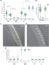
The sprDf1 allele induces a maternal effect lethal phenotype with polynucleated misshapen embryos. a) The total brood size was determined by counting the number of embryos and animals deposited on a plate by a single hermaphrodite adult throughout its fertile life span. The total viable brood was determined by counting the number of embryos that hatched into animals that survived to at least the L2 stage. The data are represented as violin plots. The genotype is given below the graph. Open circles correspond to the total brood produced by a given animal. Closed circles correspond to the viable brood. The bar denotes the mean. Statistical significance was assessed using a 1-way ANOVA comparing all samples, total and viable, with Bonferroni correction for multiple hypothesis testing. The abbreviation nT1 is used in place of nT1[qIs51] throughout the figure. b) The hatch rate was calculated by dividing the number of viable animals by the total brood. The box represents the quartiles 1–3, and the whiskers denote the full range of the data. Open circles represent the hatch rate per adult hermaphrodite. Stars represent far outliers, and circles with an X represent near outliers as calculated by the Tukey method. The black bar represents the median. A single asterisk denotes Padj < 8.3e-3 comparing all data sets to +/+, and a double asterisk represents a significant decrease with Padj < 8.3e-3 comparing all data sets to sprDf1/nT1[qIs51] in a 1 way ANOVA with Bonferroni correction for multiple hypothesis testing. c) Representative micrographs of young adult sprDf1/sprDf1 homozygotes and sprDf1/nT1[qIs51] heterozygotes collected 4 days post hatching. The yellow arrows indicate polynucleated embryos, while the white arrows represent embryos that appear normal. The scale bars represent a distance of 20 microns. d) The fraction of polynucleated embryos retained in utero per animal was determined at day 4, 5, or 6 post hatching. The mean is denoted by a horizontal black bar. The P-values were calculated by 1-way ANOVA with post hoc Bonferroni correction for multiple hypothesis testing across all genotypes and all days measured. A single asterisk represents a statistically significant difference with adjusted P-value < 1.1e-3 with reference to the day 4 +/+ control.
Fig. 4.
Fig. 4.
Microdeletion sprDf1 mutants are prone to death by bursting. DIC micrographs of a GFP− sprDf1/sprDf1 homozygous worm observed while bursting are shown. The full movie is available as Supplementary Movie 1. a) This frame corresponds to time zero, the beginning of the movie. b) This frame corresponds to the same animal, 4 minutes and 57 seconds later. The scale bars in both panels represent a distance of 20 microns. c) The fraction of animals that died by bursting plotted as a function of days posthatching is shown with GFP+ heterozygotes and GFP− homozygous siblings. The left panel compares sprDf1/spDf1 homozygotes to sprDf1/nT1[qIs51] heterozygotes, while the right panel shows both +/+ and +/nT1[qIs51] genotypes. The data shown represent the sum of multiple synchronizations, with N ranging from 28 to 166 animals per genotype per day. The abbreviation nT1 is used in place of nT1[qIs51] throughout the figure. The error bars represent the standard error. Statistical significance was determined by Kaplan Meier estimation using the logrank method with StatPlus software and Bonferroni correction for multiple hypothesis testing. d) The fraction of animals surviving is represented as in panel c for all 4 genotypes.
Fig. 5.
Fig. 5.
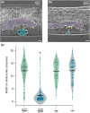
sprDf1 homozygotes have a squashed vulva and are shorter and wider than their heterozygous siblings. a) DIC micrograph of an L4 sprDf1/nT1[qIs51] heterozygote showing the developing vulva. The vulval lumen is indicated by the lower cyan shape, and the uterus is indicated by the upper purple shape. The UTSE valve which separates the uterus from the vulva is indicated by asterisks. The arrow denotes where the measurement is made to define the width of the vulval lumen b) Equivalently aged L4 homozygous sprDf1/sprDf1 worms display a dramatic reduction in the width of the vulval lumen (a sqv or squashed vulva phenotype), and the UTSE valve does not form properly (asterisks). In panels a and b, the scale bar represents 5 microns. c) The distribution of luminal widths of the vulva is shown for homozygous animals vs heterozygous siblings for sprDf1/sprDf1, sprDf1/nT1[qIs51], +/+, and +/nT1[qIs51] genotypes. Statistical significance was calculated from a 1-way ANOVA with Bonferonni correction for multiple hypothesis testing. An asterisk indicates an adjusted P-value of <8.3e-. Each point represents the luminal of from a single animal, and bars represent the mean. The abbreviation nT1 is used in place of nT1[qIs51] throughout the figure.
Fig. 6.
Fig. 6.
Homozygous microdeletion mutant animals are uncoordinated. a and b). Still frames from movies collected for GFP+ sprDf1/nT1[qIs51] heterozygous worms (top row) and GFP− sprDf1/sprDf1 homozygous worms (bottom row). The full movie can be seen in Supplementary Movie 2. The time stamps indicate time post initiation of filming, and the scale bar represents 200 microns. c and d). The total length of the path traveled (c) and the rate of travel is shown. Each point represents the distance traveled by an individual animal and bars represent the mean. The genotypes and animals’ ages are shown below. Statistical significance between genotypes was calculated with a 1-way ANOVA with Bonferroni post hoc correction for multiple hypothesis testing, where a single asterisk denotes Padj < 8.3e-5. The abbreviation nT1 is used in place of nT1[qIs51] throughout the figure.

References

    1. Abyzov A, Urban AE, Snyder M, Gerstein M. 2011. CNVnator: an approach to discover, genotype, and characterize typical and atypical CNVs from family and population genome sequencing. Genome Res. 21(6):974–984. doi:10.1101/gr.114876.110. - DOI - PMC - PubMed
    1. Albarqi MMY, Ryder SP. 2021. The endogenous mex-3 3´UTR is required for germline repression and contributes to optimal fecundity in C. elegans. PLoS Genet. 17(8):e1009775. doi:10.1371/journal.pgen.1009775. - DOI - PMC - PubMed
    1. Almedom RB, Liewald JF, Hernando G, Schultheis C, Rayes D, Pan J, Schedletzky T, Hutter H, Bouzat C, Gottschalk A. 2009. An ER-resident membrane protein complex regulates nicotinic acetylcholine receptor subunit composition at the synapse. EMBO J. 28(17):2636–2649. doi:10.1038/emboj.2009.204. - DOI - PMC - PubMed
    1. Arribere JA, Bell RT, Fu BX, Artiles KL, Hartman PS, Fire AZ. 2014. Efficient marker-free recovery of custom genetic modifications with CRISPR/Cas9 in Caenorhabditis elegans. Genetics. 198(3):837–846. doi:10.1534/genetics.114.169730. - DOI - PMC - PubMed
    1. Carelle-Calmels N, Saugier-Veber P, Girard-Lemaire F, Rudolf G, Doray B, Guérin E, Kuhn P, Arrivé M, Gilch C, Schmitt E, et al. . 2009. Genetic compensation in a human genomic disorder. N Engl J Med. 360(12):1211–1216. doi:10.1056/NEJMoa0806544. - DOI - PubMed

Publication types

LinkOut - more resources