. 2023 Nov 13;6(20):CASE23418.
doi: 10.3171/CASE23418. Print 2023 Nov 13.

Bilateral infraoptic origin of the anterior cerebral artery: illustrative case

Affiliations

Bilateral infraoptic origin of the anterior cerebral artery: illustrative case

Neil Majmundar et al. J Neurosurg Case Lessons. .

Abstract

Background: A bilateral infraoptic origin of the anterior cerebral arteries (ACAs) is a rare anatomical variant that can be encountered during anterior skull base surgery. The ACAs arise from the internal carotid artery (ICA) at the level of the ophthalmic artery and course medially, traveling inferior to the ipsilateral optic nerves. Herein, the authors discuss the different configurations of the anatomical variant, its prevalence, and hypotheses leading to the variable configuration of this anomaly.

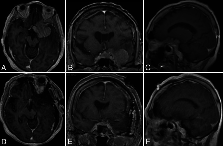
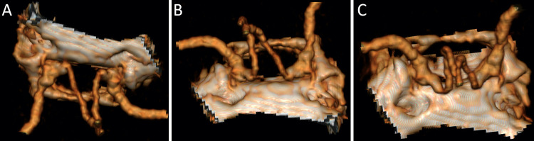
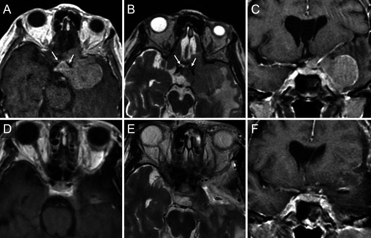
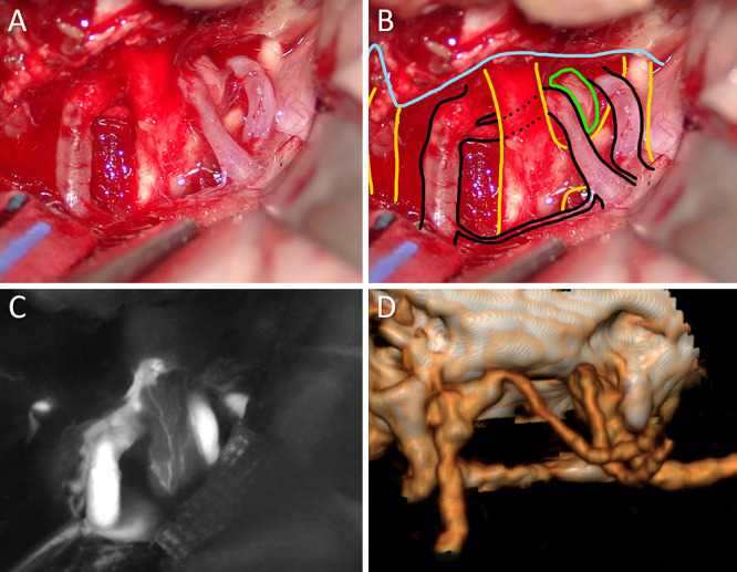
Observations: A 67-year-old woman presented with worsening dizziness over a week-long period and was found to have a large left sphenocavernous meningioma with optic, cavernous, and suprasellar extension. The tumor incorporated the left supraclinoid ICA and its branches. She underwent a left modified orbitozygomatic craniotomy for tumor resection. Early identification of the aberrant ACA anatomy was crucial in avoiding vascular injury.

Lessons: While this variant is typically encountered during the treatment of vascular pathologies-namely, intracranial aneurysms-its existence should be kept in mind during the treatment of any anterior skull base pathology. Failure to account for the presence of this variant may lead to potential intraoperative complications.

Keywords: anterior cerebral artery; cerebrovascular; meningioma; skull base surgery; skull base tumor.


Conflict of interest statement

Disclosures The authors report no conflict of interest concerning the materials or methods used in this study or the findings specified in this paper.

Figures

FIG. 1
FIG. 1
Preoperative (A–C) and postoperative (D–F) T1-weighted post-gadolinium magnetic resonance imaging (MRI) in the axial, coronal, and sagittal planes. Preoperative images demonstrate the large left sphenocavernous meningioma, while the postoperative images demonstrate gross-total resection of the sphenocavernous meningioma.
FIG. 2
FIG. 2
Different rotated views of the three-dimensional (3D) reconstruction of the patient’s postoperative computed tomography angiography (CTA). A: Anteroposterior view along the anterior skull base as if one were approaching this anatomical area from a transbasal approach. This clearly demonstrates the bilateral infraoptic loops of the A1 segments originating from the supraclinoidal internal carotid artery (ICA). B: Anteroposterior coronal view along the anterior skull base, again demonstrating the course of the infraoptic A1 segments. C: View from posterior to anterior demonstrating the dramatic inferior and then posterior loops of the infraoptic A1 segments.
FIG. 3
FIG. 3
Preoperative (A–C) and postoperative (D–F) MRI. A: Axial T1-weighted post-gadolinium sequence demonstrates the large left-sided sphenocavernous meningioma. At the medial aspect of the lesion, the white arrows mark the flow voids of the medially coursing bilateral infraoptic anterior cerebral arteries (ACAs). B: Axial T2-weighted sequence further highlights some of the detail of the flow voids of the bilateral infraoptic A1 ACAs. C: Coronal T1-weighted post-gadolinium image demonstrates the position of the optic chiasm in relation to the infraoptic A1 ACA segment. D: Axial T1-weighted post-gadolinium image shows the extent of tumor resection from the sphenoid wing and lateral cavernous sinus. Additionally, the supraclinoid ICA origin of these bilateral A1 ACA segments is illustrated in cross-section. E: Axial T2-weighted image demonstrates the flow voids of the infraoptic ACA origin arising from the ICA. F: Coronal T1-weighted post-gadolinium image demonstrates the extent of the left sphenocavernous meningioma resection as well as the natural repositioning of the optic chiasm previously displaced cranially and medially by the tumor.
FIG. 4
FIG. 4
Intraoperative microsurgical anatomy findings. A: After resection of the tumor at the sphenoid wing and up to the lateral cavernous sinus, a panoramic view of the proximal ICA, optic nerves, and ACA complex is afforded. B: The same photograph as in panel A with the addition of the graphic overlay of important anatomical elements. The blue line is the dura of the middle and anterior cranial fossae. The slit in the dura on the far left of the panel is decompressing cranial nerve (CN) III as it enters the posterior cavernous sinus. The left anterior clinoid process was removed extradurally prior to intradural exposure. The arteries of the left supraclinoid region are noted in black, with the infraoptic origin of the A1 segment of the ACA. The dotted lines estimate its course inferior to the left optic nerve. The optic nerves and optic chiasm are outlined in yellow. Note the hypoplastic ipsilateral left A1 segment in the expected position superior to the optic tract. The pituitary gland is outlined in green. The contralateral optic nerve and contralateral infraoptic course of the right A1 segment can be appreciated in this view. C: Indocyanine green angiography illustrates the arterial course and good perfusion of the optic nerve following tumor resection. D: A 3D reconstruction of the patient’s postoperative CTA rotated into a surgical view to match with panels A and B, demonstrating the supraclinoid ICA origin of these bilateral infraoptic A1 segments. Note the absence of the left anterior clinoid process.

References

    1. Robinson LR. An unusual human anterior cerebral artery. J Anat. 1959;93(1):131–133. - PMC - PubMed
    1. Osborn AG, Jacobs JM, Osborn AG. Diagnostic Cerebral Angiography. 2nd ed. Lippincott-Raven; 1999.
    1. Uchino A. Infraoptic anterior cerebral artery (ACA) versus carotid-ACA anastomosis. Surg Radiol Anat. 2017;39(8):937. - PubMed
    1. Kochar PS, Soin P, Elfatairy K. Infraoptic anterior cerebral artery or carotid-anterior cerebral artery anastomosis: a very rare embryological variation. Case series and review of literature. Clin Imaging. 2021;79:8–11. - PubMed
    1. Tanaka K, Ishida F, Tanioka S, Suzuki H. A ruptured anterior communicating artery aneurysm with infra-optic course of the anterior cerebral artery: a case report and a short review. NMC Case Rep J. 2021;8(1):465–472. - PMC - PubMed