. 2023 Nov 13;13(1):19773.
doi: 10.1038/s41598-023-47127-3.

Chia seeds and coenzyme Q10 alleviate iron overload induced hepatorenal toxicity in mice via iron chelation and oxidative stress modulation

Affiliations

Chia seeds and coenzyme Q10 alleviate iron overload induced hepatorenal toxicity in mice via iron chelation and oxidative stress modulation

Shimaa A Sadek et al. Sci Rep. .

Abstract

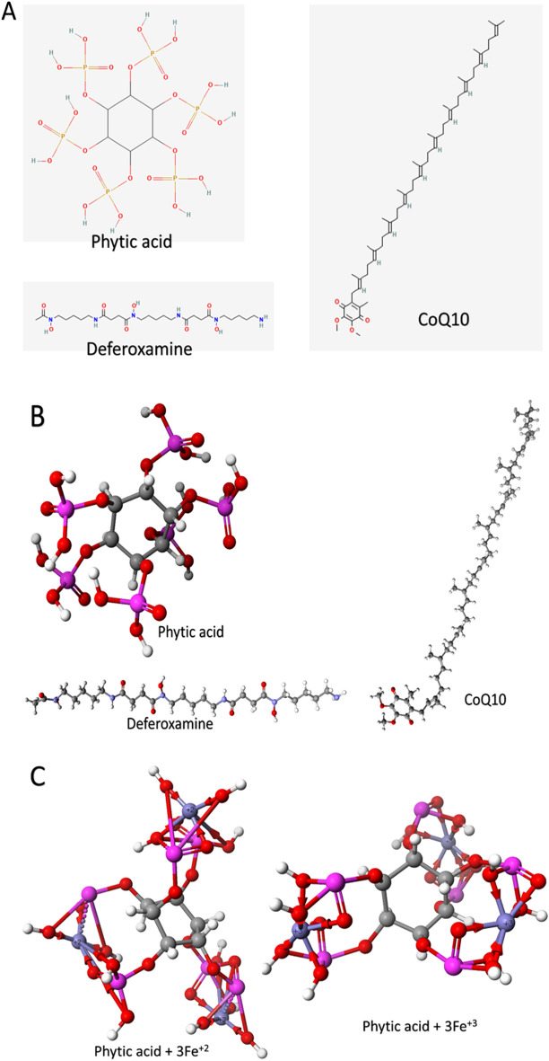
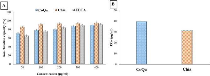
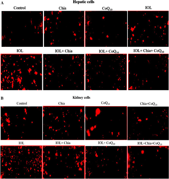
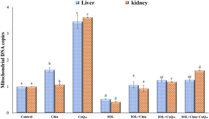
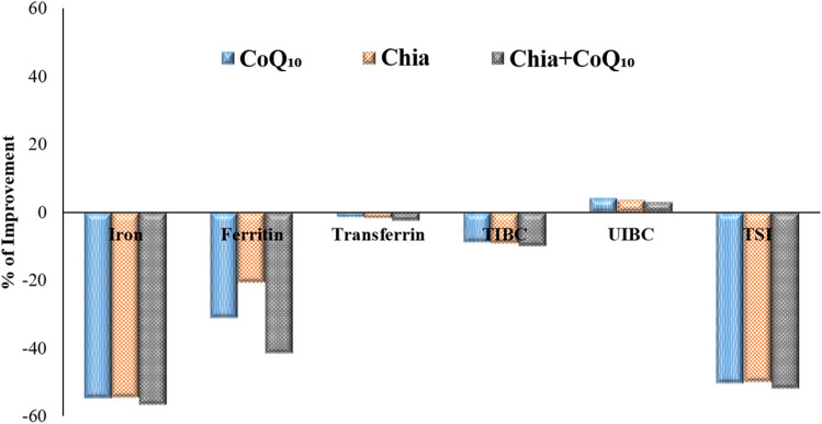
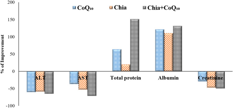
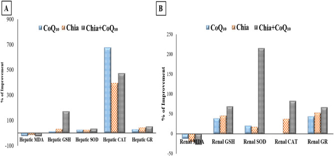
Iron overload (IOL) can cause hepatorenal damage due to iron-mediated oxidative and mitochondrial damage. Remarkably, combining a natural iron chelator with an antioxidant can exert greater efficacy than monotherapy. Thus, the present study aimed to evaluate the efficacy of Chia and CoQ10 to chelate excess iron and prevent hepatorenal oxidative damage in IOL mice. Male Swiss albino mice (n = 49) were randomly assigned to seven groups: control, dietary Chia, CoQ10, IOL, IOL + Chia, IOL + CoQ10, and IOL + Chia + CoQ10. Computational chemistry indicates that the phytic acid found in the Chia seeds is stable, reactive, and able to bind to up to three iron ions (both Fe2+ and Fe3+). IOL induced a significant (P < 0.05) increase in serum iron, ferritin, transferrin, TIBC, TSI, RBCs, Hb, MCV, MCH, WBCs, AST, ALT, creatinine, and MDA. IOL causes a significant (P < 0.05) decrease in UIBC, platelets, and antioxidant molecules (GSH, SOD, CAT, and GR). Also, IOL elicits mitochondrial membrane change depolarization, and DNA fragmentation and suppresses mitochondrial DNA copies. Furthermore, substantial changes in hepatic and renal tissue, including hepatocellular necrosis and apoptosis, glomerular degeneration, glomerular basement membrane thickening, and tubular degeneration, were observed in the IOL group. Dietary Chia and CoQ10 induced significant (P < 0.05) amelioration in all the mentioned parameters. They can mostly repair the abnormal architecture of hepatic and renal tissues induced by IOL, as signified by normal sinusoids, normal central veins, and neither glomerular damage nor degenerated tubules. In conclusion, the combined treatment with Chia + CoQ10 exerts more pronounced efficacy than monotherapy in hepatorenal protection via chelating excess iron and improved cellular antioxidant status and hepatorenal mitochondrial function in IOL mice.


Conflict of interest statement

The authors declare no competing interests.

Figures

Figure 1
Figure 1
The structure of phytic acid, CoQ10, and deferoxamine in 2D (A) and 3D (B). The optimized formed complexes of phytic acid with Fe2+ and Fe3+ calculated at the PM6 level of theory (C). Atoms are colored according to this scheme; Carbon: gray, Oxygen: red, Nitrogen: violet, Phosphorus: pink, Iron: purple, and Hydrogen: white.
Figure 2
Figure 2
In vitro iron chelation potency of CoQ10 and Chia seeds. (A) Iron chelation capacity percentage of CoQ10 and Chia seeds. (B) Half-maximal effective concentration (EC50) of the CoQ10 and Chia seeds.
Figure 3
Figure 3
(A) The modulating efficacies of dietary chia and CoQ10 on the hepatic mitochondrial membrane potential (Δψ) of iron-overloaded mice. CoQ10: Coenzyme Q10 (300 mg/kg body weight); Diet containing Chia seeds (362 g/kg diet); IOL: Iron overload. (B) The modulating efficacies of dietary chia and CoQ10 on the renal mitochondrial membrane potential (Δψ) of iron-overloaded mice. CoQ10: Coenzyme Q10 (300 mg/kg body weight); Diet containing Chia seeds (362 g/kg diet); IOL: Iron overload.
Figure 4
Figure 4
Modulating efficacies of dietary chia and CoQ10 on mitochondrial DNA copies of iron-overloaded mice. Values are mean ± SEM (n = 7). Values with different superscript letters in the same raw are significantly different at P < 0.05. CoQ10: Coenzyme Q10 (300 mg/kg body weight); Diet containing Chia seeds (362 g/kg diet); IOL: Iron overload.
Figure 5
Figure 5
Photomicrographs of the mice's liver architecture of different experimental groups stained with hematoxylin and eosin. (A) control group showing well-preserved liver architecture with round polygonal hepatocytes, normal distribution of sinusoidal spaces and Kupffer cells. (B,C) Dietary Chia and CoQ10 liver sections demonstrate distinct and relatively normal hepatic architecture. (D,E) The iron overload group showed severe hepatic injury with inflammatory infiltration in the sinusoidal spaces, enlarged kupffer cells and degenerated hepatocytes. (F,G) Iron-overloaded mice treated with dietary Chia or CoQ10 showed slight amelioration in hepatic structure with few degenerated hepatocytes. (H) Iron-overloaded mice treated with combined therapy (Chia and CoQ10) showed more pronounced improvement of the liver structure, confirmed by normal sinusoids and central vein. H: Hepatocyte, S: Sinusoidal space, K: Kupffer cell, CV: Central vein, DH: Degenerated hepatocyte. The red arrow refers to inflammatory infiltration.
Figure 6
Figure 6
Photomicrographs of the mice's kidney architecture of different experimental groups stained with hematoxylin and eosin. (A) The control group showed normal renal structure with renal corpuscles surrounded by Bowman’s capsule and intact proximal and distal convoluted tubules. (B,C) Dietary Chia and CoQ10 kidney sections show distinct and relatively normal kidney architecture. (D,E) The iron overload group showed severe renal injury with glomerular degeneration (DG), glomerular atrophy, tubular degeneration and lymphocyte distribution (red arrow). (F,G) Iron-overloaded mice treated with dietary Chia and CoQ10 showed more or less normal appearance of the control group with slight lymphocyte distribution (red arrow). (H) Iron-overloaded mice treated with combined dietary Chia and CoQ10 showed a marked recovery of the renal structure, as indicated by neither destruction of tubular structure nor degenerated proximal tubule. G: glomeruli, BS: Bowman’s space, BC: Bowman’s capsule, CD: collecting duct, DT: degenerated tubule.
Figure 7
Figure 7
Improvement percentage of monotherapy (Chia or CoQ10) and combined therapy (Chia + CoQ10) on hematological parameters.
Figure 8
Figure 8
Improvement percentage of monotherapy (Chia or CoQ10) and combined therapy (Chia + CoQ10) on iron indices.
Figure 9
Figure 9
Improvement percentage of monotherapy (Chia or CoQ10) and combined therapy (Chia + CoQ10) on hepatorenal function markers.
Figure 10
Figure 10
Improvement percentage of monotherapy (Chia or CoQ10) and combined therapy (Chia + CoQ10) on oxidative/antioxidative stress markers.
Figure 11
Figure 11
Improvement percentage of monotherapy (Chia or CoQ10) and combined therapy (Chia + CoQ10) on DNA fragmentation.

References

    1. Brittenham GM. Iron-chelating therapy for transfusional iron overload. N. Engl. J. Med. 2011;364:146–156. doi: 10.1056/NEJMct1004810. - DOI - PMC - PubMed
    1. Entezari S, et al. Iron chelators in treatment of iron overload. J. Toxicol. 2022;2022:4911205. doi: 10.1155/2022/4911205. - DOI - PMC - PubMed
    1. Valko M, Morris H, Cronin TDM. Metals, toxicity and oxidative stress. Curr. Med. Chem. 2005;12:1161–1208. doi: 10.2174/0929867053764635. - DOI - PubMed
    1. Scaramellini N, et al. Interpreting iron homeostasis in congenital and acquired disorders. Pharmaceuticals. 2023;16:329. doi: 10.3390/ph16030329. - DOI - PMC - PubMed
    1. Wallace DF. The regulation of iron absorption and homeostasis. Clin. Biochem. Rev. 2016;37:51–62. - PMC - PubMed

Publication types