. 2023 Dec;5(12):2131-2147.
doi: 10.1038/s42255-023-00919-3. Epub 2023 Nov 13.

Inhibition of the proline metabolism rate-limiting enzyme P5CS allows proliferation of glutamine-restricted cancer cells

Affiliations

Inhibition of the proline metabolism rate-limiting enzyme P5CS allows proliferation of glutamine-restricted cancer cells

Samantha J Linder et al. Nat Metab. 2023 Dec.

Abstract

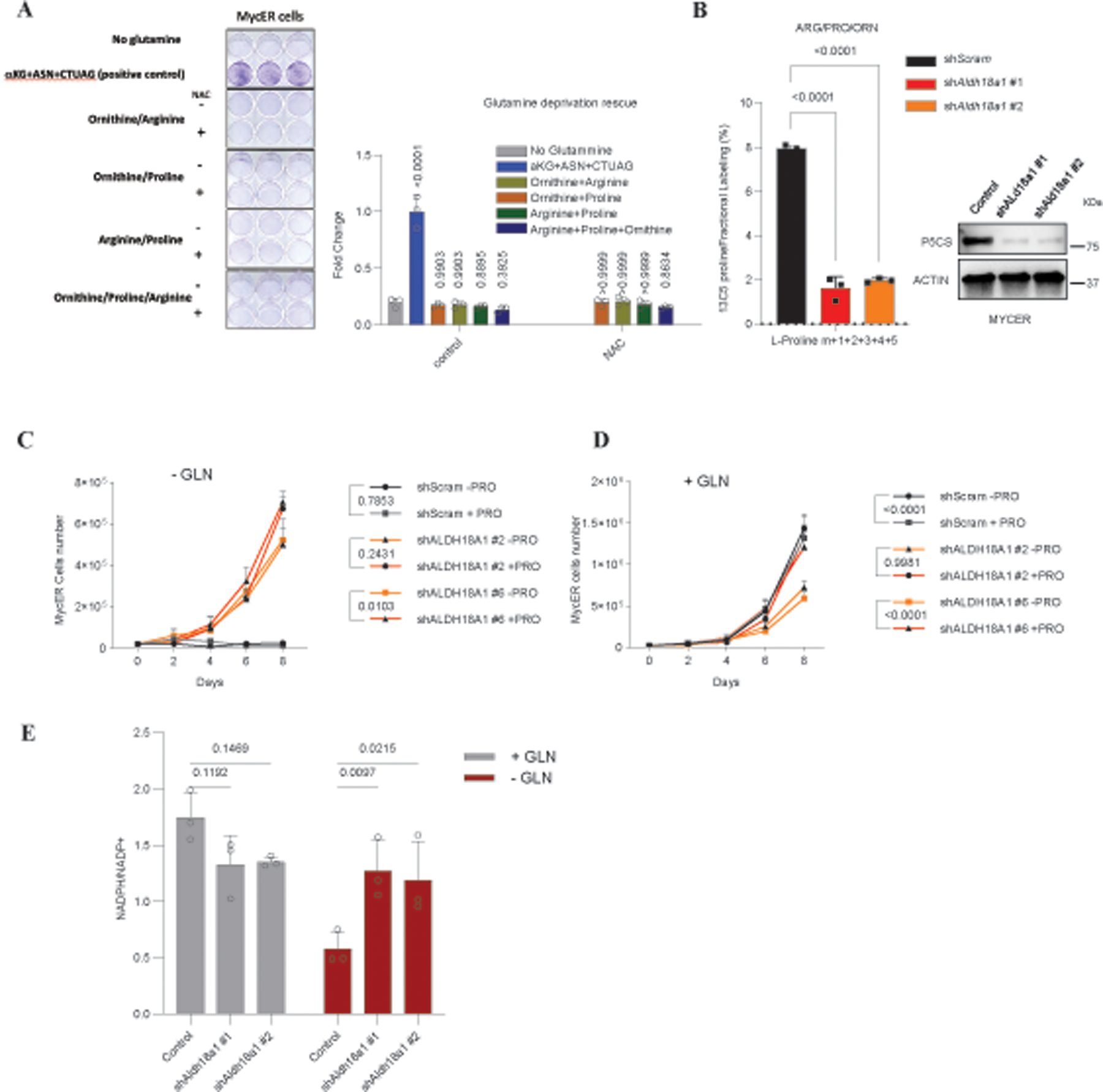
Glutamine is a critical metabolite for rapidly proliferating cells as it is used for the synthesis of key metabolites necessary for cell growth and proliferation. Glutamine metabolism has been proposed as a therapeutic target in cancer and several chemical inhibitors are in development or in clinical trials. How cells subsist when glutamine is limiting is poorly understood. Here, using an unbiased screen, we identify ALDH18A1, which encodes P5CS, the rate-limiting enzyme in the proline biosynthetic pathway, as a gene that cells can downregulate in response to glutamine starvation. Notably, P5CS downregulation promotes de novo glutamine synthesis, highlighting a previously unrecognized metabolic plasticity of cancer cells. The glutamate conserved from reducing proline synthesis allows cells to produce the key metabolites necessary for cell survival and proliferation under glutamine-restricted conditions. Our findings reveal an adaptive pathway that cancer cells acquire under nutrient stress, identifying proline biosynthesis as a previously unrecognized major consumer of glutamate, a pathway that could be exploited for developing effective metabolism-driven anticancer therapies.


Conflict of interest statement

Competing interests:

Authors declare no competing interests.

Figures

Extended Data Figure 1.
Extended Data Figure 1.. Quality controls of the genome-wide shRNA-library, GLN deprivation screen.
A) Western blot of MycER and Myc transcriptional target Ldha in whole cell lysate (WCL) and chromatin isolated from MycER MEFs exposed to 200nM 4-OHT for 24 and 48 hours. B) Phase contrast microscopy of MycER cells + 200nM 4-OHT grown in normal media for 3 days or without GLN for 3 and 11 days. C) Heatmap showing pairwise sample-sample Euclidean distances, arranged by treatment group. Dendrograms show hierarchical clustering to highlight similarities between samples. D) MA plot of log2(mean counts-per-million) against log2(median fold-change), with points colored by density to highlight data trend(s). E) Levels of Glutamine (GLN), Glutamate (GLU), and Proline (PRO) in Fetal bovine serum. Data are represented as mean ± SD of three independent experiments F) WT and two clonal P5CS CRISPR KO mouse kidney epithelial cell lines grown in the presence of 2mM GLN for 1 week, or absence of GLN for 1 month, with corresponding quantification of growth. Statistically significant differences were evaluated using a paired, one-tailed T-Test, with the error bars representing standard deviation (SD). G) Growth curves of control and Aldh18a1 knockdown cells under +GLN and -GLN conditions. Data are represented as mean ± SD of three independent experiments. Statistical significance was determined using two-way ANOVA. H) Repeats of western blot against P5CS in short-term (S) or long-term (L) culture of shScram and shAldh18a1 MycER MEFs in the presence or absence of 2mM glutamine. * p ≤ 0.05, ** p ≤ 0.01, *** p ≤ 0.01, **** p ≤ 0.0001.
Extended Data Figure 2.
Extended Data Figure 2.. Proline depletion upon P5CS/Aldh18a1 downregulation.
A) Relative abundance of intracellular PRO in WT mouse kidney epithelial cells from two different thaws (WT-1 and WT-2) and two P5CS KO subclones (C2 and C9) infected either with empty vector (+Vec) or reconstituted with P5CS (+P5CS). Cells were grown for 24 hours in proline-free high-glucose DMEM medium with 0.65mM GLN and 2.5%FBS, and metabolite levels normalized by total protein (n=3) Statistical significance was determined using an unpaired, T-Test, with error bars indicating SD. B) Metabolomic profiling of amino acid consumption and secretion. This panel of plots illustrates the relative consumption and secretion rates of various amino acids by control and P5CS KD cells, under low-glutamine conditions. The lower bar graphs show the consumption rates of amino acids from the media, while the upper ones depict the secretion rates of amino acids into the media. Data are represented as mean ± SD of three independent experiments. The statistical analysis was performed using two-way ANOVA. * p ≤ 0.05, ** p ≤ 0.01, *** p ≤ 0.01, **** p ≤ 0.0001.
Extended Data Figure 3.
Extended Data Figure 3.. Evaluation of Cell Death in Response to Glutamine Deprivation
A) Gating strategy for sorting MycER control cells and shALDH18A1 cells under different conditions: normal medium, glutamine-deprived medium, and medium supplemented with 7 mM alpha-ketoglutarate (aKG). B) Propidium iodide staining of NUGC2 control and shALDH18A1 cells quantified by Flow cytometry. Data are presented as mean ± SD from three independent experiments. Statistical analysis was performed using two-way ANOVA. On the right, the gating strategy for sorting in control and shALDH18A1 NUGC2 cells under different conditions: normal medium, glutamine-deprived medium, and medium supplemented with 7 mM alpha-ketoglutarate (aKG). * p ≤ 0.05, ** p ≤ 0.01, *** p ≤ 0.01, **** p ≤ 0.0001.
Extended Data Figure 4.
Extended Data Figure 4.. Proline is not a major source of carbon and/or nitrogen in GLN-starved cells.
A) Graphical representation of the 13C5 glutamate labeling pathway, illustrating its incorporation into intermediates of the TCA cycle, amino acids, and pyrimidines. B) Tracer experiment using 13C5 labeled glutamate in Control and P5CS Knockdown Cells. The bar chart illustrates the proportion of proline derived from glutamine in both control and P5CS knockdown cells, as well as the corresponding increase in the labeling of amino acids, nucleosides, and TCA cycle intermediates. Data are represented as mean ± SD of three independent experiments. The statistical analysis was performed using two-way ANOVA. C) Crystal violet staining and corresponding quantification of MycER shScram cells cultured in the presence or absence of GLN for 5 days. Cells were cultured in DMEM supplemented with 7mM dimethyl-α-ketoglutarate (α-KG), 2mM ASN, 15mM GlcNAc, or 250μM each cytidine (C), thymidine (T), uridine (U), adenosine (A), and guanosine (G) in indicated combinations. Statistical significance was determined using an unpaired, Two-tailed, T-Test, with error bars indicating SD. D) Crystal violet staining and corresponding quantification of MycER shScram cells cultured in the presence or absence of GLN for 5 days. Cells were cultured in DMEM supplemented with 7mM dimethyl-α-ketoglutarate (α-KG), 2mM ASN, 15mM GlcNAc, or 250μM each cytidine (C), thymidine (T), uridine (U), adenosine (A), and guanosine (G) in presence or absence of L-Methionine sulfoximine (MSO). Statistical significance was determined using ordinary one-way ANOVA followed by Tukey’s post hoc test for multiple comparisons. Error bars represent standard deviation (SD). E) Cell proliferation of Aldh18a1 KD cells transfected with either siScram or two independent siP5CDH grown in the absence of GLN for 5 days (n=3) (Top). Corresponding western blot verifying P5CS and P5CDH knockdown (Bottom). Statistical significance was determined using ordinary one-way ANOVA followed by Tukey’s post hoc test for multiple comparisons. Error bars represent standard deviation (SD). F) Crystal violet staining and corresponding quantification of MycER shScram cells cultured in the presence or absence of GLN for 5 days. Cells were cultured in DMEM supplemented with 0.5mM Proline, 2mM ASN, or 250uM each C, T, U, A, and G, in indicated combinations. All error bars are SD. Grey square: Presence, white square: Absence * p ≤ 0.05, ** p ≤ 0.01, *** p ≤ 0.01, **** p ≤ 0.0001.
Extended Data Figure 5.
Extended Data Figure 5.. Downregulation of P5CS maintains NADPH/ NADP+ ratio in low GLN
A) Crystal violet staining and corresponding quantification of MycER cells cultured in the absence of GLN or in the presence of aKG (7mM) + ASN (2mM) + C,T,U,A, and G (250uM each) or Ornithine/arginine 2mM or Ornithine/Proline 2mM or Arginine/Proline 2mM, or Ornithine/proline/Arginine 2mM. Each condition with or without N-Acetyl Cysteine (NAC) 5 days. Statistical significance was determined using ordinary one-way ANOVA followed by Dunnett’s post hoc test for multiple comparisons. Error bars represent standard deviation (SD). B) This represents the percentage of incorporation of 13C5-labeled glutamine into L-Proline in MycER shScram, shAldh18a1 #1, and shAldh18a1 #2 cells. These cells were cultured in DMEM supplemented with dialyzed FBS and 2mM each of labeled Glutamine (13C5), Arginine, Proline, and Ornithine (n=3). The fraction that was not labeled was designated as Proline M+0. Corresponding western blot verifying P5CS knockdown (right side). Statistical significance was determined using ordinary one-way ANOVA followed by Dunnett’s post hoc test for multiple comparisons. Error bars represent standard deviation (SD). C and D) Left panel: Growth curves of control and Aldh18a1 knockdown cells under -GLN and +GLN conditions, in the presence of 0.5 mM Proline. Data are represented as mean ± SD of three independent experiments. Statistical significance was determined using a two-way ANOVA. Right panel: Growth curves of control and Aldh18a1 knockdown cells under +Proline and -Proline conditions, in the context of +GLN. Data are represented as mean ± SD of three independent experiments. The statistical analysis was performed using a two-way ANOVA. E) bar graph representing the NADPH/NADP+ ratio in MycER cells, comparing the control group with two separate shALDH18A1 knockdown groups, under low glutamine conditions for 48 hours. Data are represented as mean ± SD of three independent experiments. The statistical analysis was performed using two-way ANOVA. * p ≤ 0.05, ** p ≤ 0.01, *** p ≤ 0.01, **** p ≤ 0.0001.
Extended Data Figure 6.
Extended Data Figure 6.. Sensitivity of human cancer cell lines to GLN restriction.
A) Kaplan-Meier survival curves analyzing the The Cancer Genome Atlas (TCGA) human uterine/endometrial or stomach cancer patient samples based on Aldh18a1 mRNA expression divided in quartiles. B) Bar plot illustrating the distribution of Uterine Endometrioid Carcinoma, Uterine Mixed Endometrial Carcinoma, and Uterine Serous Carcinoma cases, classified by low or high expression levels of ALDH18A1. (TCGA) C) Scatter plot demonstrating the ALDH18A1 mRNA expression levels across Uterine Endometrioid Carcinoma and Uterine Serous Carcinoma cases, based on TCGA data. Statistical analysis was conducted using an unpaired two-tailed t-test, and data are presented as mean ± SD. D) Bar plot demonstrating the distribution of Uterine cancer cases by histological grade, categorized by low or high ALDH18A1 RNA expression, as per TCGA data. E) Representative images and quantification of sensitivity to glutamine depletion of human cancer cell lines grown in the presence or absence of GLN for 4 days, represented by percent survival. Survival was calculated by dividing the average number of cells counted on Day 4 in wells treated with no GLN by the average number of cells in wells with GLN (n = 3 wells). F) Western blot analysis of NUGC2 and PC3 cell lines using scramble shRNA or two independent shRNAs targeting the 3’UTR of ALDH18A1.
Extended Data Figure 7.
Extended Data Figure 7.. Variation in P5CS Expression Levels among Human Cancer Cell Lines under Glutamine Deprivation
A) Bar plot illustrating the weights of subcutaneous xenograft tumors from NUGC2 cells (PLKO, shALDH18A1, and rescue with dox-inducible NUGC2). Statistical analysis was conducted using a one-way ANOVA followed by Tukey’s multiple comparison test. Error bars represent standard deviation (SD). B) 15N-labeled ammonia (15NH4+) in NUGC2 gastric cancer cells with genetically downregulated P5CS. Data are represented as mean ± SD of three independent experiments. Statistical significance was determined using two-way ANOVA. C) Western blot of P5CS in human cancer cell lines that do not endogenously downregulate P5CS upon acute GLN starvation. Cells were cultured in the presence or absence of 2mM GLN for 24 hrs. D) Comparison of P5CS levels in xenograft tumors derived from MKN45 cells. On the left, a western blot illustrates the relative P5CS protein levels in tumor extracts, while on the right, a bar graph represents the quantification of these levels. Statistical significance was determined using a one-tailed unpaired t-test with Welch’s correction for unequal variances, with error bars indicating standard deviation (SD). E) Additional representative immunofluorescence (IF) of a tumor section of uterine serous carcinoma, depicting staining by IF for P5CS (red), GS (green), and Dapi (blue). On the right, a selected area of the tumors demonstrates an inverse correlation between P5CS and GS expression. F) Total number of cells with partner P5CSlow/GShigh or vice-versa detected in five tumor sections of uterine serous carcinoma (USC). Statistical analysis was performed using an unpaired two-tailed Mann-Whitney test, and the standard deviation (SD) is shown. p ≤ 0.05, ** p ≤ 0.01, *** p ≤ 0.01, **** p ≤ 0.0001.
Fig. 1.
Fig. 1.. A genome-wide glutamine deprivation screen identified Aldh18a1 as a modulator of glutamine metabolism.
A) Graphical representation of glutamine depleted screen, where cells are infected with the TRC2 lentiviral mouse shRNA library, selected, and subjected to glutamine deprivation to select for glutamine independent clones. Created with BioRender.com. B) Representative actively proliferating clones arising from the glutamine-deprived KD screen after 22 days of glutamine depletion (n=3). C) MA plot of log2(mean counts-per-million) against log2(median fold-change) for each shRNA, with points colored red for genes with at least two shRNAs passing thresholds (≥ 5 cpm in control or treatment and fold-change of ≥ 2). Points for all other shRNAs are in grey. Labels indicate positions of shRNAs targeting Aldh18a1. D) Plot showing distribution of log2(median fold-change) for all shRNAs (blu) with finite fold-change values, with points horizontally jittered according to density. shRNAs targeting Aldh18a1 are in red. E) In the upper part, western blot of P5CS in MycER MEFs stably expressing a scrambled control short hairpin (shScram) or two independent short hairpins targeting Aldh18a1. In the lower part, cell proliferation of control or Aldh18a1 knockdown MycER MEFs while grown in glutamine-deprived media for ten days (n=3). P-values were calculated using an unpaired t-test with FDR correction using the Benjamini-Hochberg method. Data are presented as mean values ± SD. F) Phase-contrast microscopy of control or Aldh18a1 knockdown MycER MEFs after 2, 10, and 16 days glutamine deprivation. G) representative western blot of P5CS in short-term (S) vs. long-term (LT) culture of shScram and shAldh18a1 MycER MEFs in complete or glutamine deprived media, and corresponding quantification (n=3) Statistical significance was determined using an unpaired, Two-tailed T-Test. Data are presented as mean values ± SD, (n=3).
Fig. 2.
Fig. 2.. Decreased Aldh18a1 expression re-wires cellular metabolism.
A) Graphical representation of the proline biosynthetic pathway, whereby proline is synthesized via glutamate by P5CS, the encoded protein of Aldh18a1. B) Graphical representation of 15NH4 labeling of newly synthesized glutamine, catalyzed by GS, and its utilization for downstream metabolites. C) Fractional labeling of select metabolites from samples labeled with 15NH4Cl for 24 hours: glutamine; ASN; ASP; NADH; hexosamines UDP-GlcNac and UDP-galactose; and nucleotides, UMP, CMP, IMP, and AMP. Statistical significance was determined using 2way ANOVA. Data are presented as mean values ± SD. N=3 biological independent samples. D) Western blot of P5CS and ASNS proteins in shScram and shAldh18a1 MycER MEFs. E) Crystal violet staining and corresponding quantification of MycER shScram cells cultured in complete or glutamine-deprived media, with or without supplementation of 2mM ASN for 5 days. Statistical significance was determined using a paired, Two-tailed T-Test. Data are presented as mean values ± SD. N=3 biological independent samples. F) Bar plot of control or Aldh18a1 knockdown MycER MEFs while grown in complete or glutamine deprived media, + or – 1mM MSO on + or – 10 µM of CB-839 for 5 days. Statistical significance was determined using two-way ANOVA followed by Tukey’s post hoc test for multiple comparisons. Data are presented as mean values ± SD, N=6 biological independent samples.
Fig.3
Fig.3. Glutamine biosynthesis allows α-KG dependent survival and nucleotide/asparagine-dependent proliferation in the absence of exogenous glutamine.
A) Crystal violet staining and corresponding colorimetric quantification of MycER shScram cells cultured in complete or deprived, while grown in complete or glutamine deprived media, + or - 2mM ASN, 15mM GlcNAc, or 250μM each cytidine (C), thymidine (T), uridine (U), adenosine (A), and guanosine (G) in indicated combinations for 5 days with corresponding quantification. Data are presented as mean values ± SD. N=3 biological independent samples. B) Propidium iodide staining of MycER cells quantified by Flow cytometry. Data are presented as mean ± SD from three independent experiments. Statistical analysis was performed using two-way ANOVA. C) Graphical representation of the 13C5 glutamine labeling pathway, illustrating its incorporation into intermediates of the TCA cycle, amino acids, and pyrimidines D) Tracer experiment using 13C5 labeled glutamine in Control and P5CS Knockdown Cells. The bar chart illustrates the proportion of proline derived from glutamine in both control and P5CS knockdown cells, as well as the corresponding increase in the labeling of amino acids, nucleosides, and TCA cycle intermediates. Data are represented as mean ± SD of three independent experiments. The statistical analysis was performed using two-way ANOVA. E) Crystal violet staining and corresponding quantification of MycER shScram cells cultured in complete or deprived media for 5 days. Cells were cultured in DMEM supplemented with 7mM dimethyl-α-ketoglutarate (α-KG), 2mM ASN, 15mM GlcNAc, or 250μM each cytidine (C), thymidine (T), uridine (U), adenosine (A), and guanosine (G) in indicated combinations. Data are presented as mean values ± SD. N=3 biological independent samples. F) Model of working hypothesis: Aldh18a1 deficient cells overcome glutamine dependency by re-routing glutamate away from proline biosynthesis and towards α-KG, ASN, and nucleotide production. Together, these carbon and nitrogen sources support cell survival and proliferation in the context of glutamine-deprived condition. Grey square: Presence ; white square: Absence
Fig. 4
Fig. 4. Downregulation of P5CS in human cancer cells leads to glutamine resistance and increased sensitivity to MSO inhibitor.
A) Kaplan-Meier survival curves analyzing The Cancer Genome Atlas (TCGA) human uterine/endometrial or stomach cancer patient samples based on Aldh18a1 mRNA expression with log-rank p-value (Mantel-Haenszel test) calculated in R with survdiff. B) Cell proliferation of control or ALDH18A1 knockdown NUGC2 cells while grown in glutamine-deprived media for eight days (n=3). Statistical significance was determined using two-way ANOVA followed by Tukey’s post hoc test for multiple comparisons. Data are presented as mean values ± SD. C) Cell proliferation of control or ALDH18A1 knockdown PC3 cells while grown in normal or glutamine-deprived media for eight days (n=3). Statistical significance was determined using ordinary one-way ANOVA followed by Tukey’s post hoc test for multiple comparisons. Data are presented as mean values ± SD. D) Bar plot experiment of NUGC2 cells cultured in complete or glutamine-deprived media for 5 days. Cells were cultured in DMEM supplemented with 3.5mM α-KG, 2mM ASN, and 125μM each C, T, U, A, and G. Statistical significance was determined using 2way ANOVA, multiple test Turkey. Data are presented as mean values ± SD. N=3 biological independent samples. E) Dose-response to MSO inhibitor in various cancer cell lines. From left to right the panel shows the response of PC3, NUGC2, and MFE-280 cell lines to varying concentrations of MSO. Statistical significance was determined using two-way ANOVA followed by Sidak’s post hoc test for multiple comparisons. Data are presented as mean values ± SD. N=3 biological independent samples. F) Dose-response to CB-839 inhibitor in various cancer cell lines. From left to right the panel shows the response of PC3, NUGC2, and MFE-280 cell lines to varying concentrations of MSO. Statistical significance was determined using two-way ANOVA followed by Sidak’s post hoc test for multiple comparisons. Data are presented as mean values ± SD. N=3 biological independent samples. G) Western blot analysis of the NUGC2 cell line, comparison between control, shALDH18A1, and shALDH18A1 rescued with exogenous expression of P5CS, (n=3). H) line graph representing the progression of tumor volume over time in control (8), shALDH18A1 (8), and in the inducible ALDH18A1 (8) tumors. Statistical significance was determined using two-way ANOVA followed by Tukey’s post hoc test for multiple comparisons. Data are presented as mean values ± SD. N=8 independent animals.
Fig. 5
Fig. 5. Human cancer cells reduce P5CS expression as an adaptation to glutamine restriction
A) Representative western blots of P5CS in human cancer cell lines found to endogenously downregulate P5CS upon acute glutamine starvation, with corresponding quantification. EN, MKN45, OCUM1, and VCAP cells were cultured in complete or glutamine-deprived media for 24 hrs (n=3). Statistical significance was determined using an unpaired t-test with Welch’s correction for unequal variances, applying individual variance for each group. The False Discovery Rate (FDR) was controlled using the two-stage step-up method by Benjamini, Krieger, and Yekutieli Data are presented as mean values ± SD. B) Graphical representation of experimental design: MKN45 or EN cells were cultured in complete or glutamine-deprived condition long term (~ 3 weeks), then injected into matching flanks of SCID mice. Created with BioRender.com. C) Representative xenograft tumors removed from SCID mice injected with MKN45 (1×10⁶ cells + 20% Matrigel; n=11) or EN (2×10⁶+20% Matrigel; n=7) cells cultured in complete or glutamine deprived media (Left). Tumors were removed at 5 weeks (MKN45) or 6–8 weeks (EN). Fold tumor size, relative to matching +glutamine flank control (Right). Statistical significance was determined using a two-tailed unpaired t-test with Welch’s correction for unequal variances. Data are presented as mean values ± SD. D) EN and EN_LG (Long-term low glutamine) fractional labeling of select metabolites from samples labeled with 15NH4Cl for 24 hours: GLN (glutamine); ASN (asparagine); ASP (aspartate); NADH; hexosamines UDP-GlcNac and UDP-galactose; and nucleotides, UMP, CMP, IMP, and AMP. Right: immunoblot showing representative image of P5CS levels in control and low glutamine EN cells. Statistical significance was determined using 2way ANOVA. Data are presented as mean values ± SD. N=3 biological independent samples. E) Representative immunofluorescence (IF) of a tumor section of uterine serous carcinoma, depicting staining by IF for P5CS (red), GS (green), and Dapi (blue). On the right, a selected area of the tumors demonstrates an inverse correlation between P5CS and GS expression (n=3). F) Quantitative analysis of the cytoplasmic intensity of P5CS and GS in the same cells, based on immunofluorescence measurements in five independent tumors. Statistical analysis was performed using a paired, Two-tailed t-test. Data are presented as mean values ± SD. N=5 independent tumors for P5CSlow/GShigh and respectively N=4 independent tumors for P5CShigh/GSlow. G) Examples of immunostaining against P5CS and GS in gastric cancer sections. Note that areas of high P5CS exhibit low GS staining (left image) and vice versa (right image). H) Quantitative analysis of the samples indicated in J. Data is presented as average optical density (from at least three independent regions) for each marker (n=3). Statistical significance was determined using an unpaired, Two-tailed t-test with a Mann-Whitney test for non-normally distributed data. Data are presented as mean values ± SD, (n=1).

References

    1. Altman ABJ, Stine ZE, Dang CV, From Krebs to clinic: glutamine metabolism to cancer therapy. Nat Rev Cancer. 16, 619–34 (2016). - PMC - PubMed
    1. Zhang J, Pavlova NN, Thompson CB, Cancer cell metabolism: the essential role of the nonessential amino acid, glutamine. Embo J 36, 1302–1315 (2017). - PMC - PubMed
    1. Krebs HA, Metabolism of amino-acids: The synthesis of glutamine from glutamic acid and ammonia, and the enzymic hydrolysis of glutamine in animal tissues. Biochem J 29, 1951–69 (1935). - PMC - PubMed
    1. Eagle H, The minimum vitamin requirements of the L and HeLa cells in tissue culture, the production of specific vitamin deficiencies, and their cure. J Exp Medicine. 102, 595–600 (1955). - PMC - PubMed
    1. Bott J, Peng I-C, Fan Y, Faubert B, Zhao L, Li J, Neidler S, Sun Y, Jaber N, Krokowski D, Lu W, Pan J-A, Powers S, Rabinowitz J, Hatzoglou M, Murphy DJ, Jones R, Wu S, Girnun G, Zong W-X, Oncogenic Myc Induces Expression of Glutamine Synthetase through Promoter Demethylation. Cell Metab 22, 1068–77 (2015). - PMC - PubMed