. 2023 Nov 13;22(1):312.
doi: 10.1186/s12933-023-02057-2.

Treatment with recombinant Sirt1 rewires the cardiac lipidome and rescues diabetes-related metabolic cardiomyopathy

Affiliations

Treatment with recombinant Sirt1 rewires the cardiac lipidome and rescues diabetes-related metabolic cardiomyopathy

Sarah Costantino et al. Cardiovasc Diabetol. .

Abstract

Background: Metabolic cardiomyopathy (MCM), characterized by intramyocardial lipid accumulation, drives the progression to heart failure with preserved ejection fraction (HFpEF). Although evidence suggests that the mammalian silent information regulator 1 (Sirt1) orchestrates myocardial lipid metabolism, it is unknown whether its exogenous administration could avoid MCM onset. We investigated whether chronic treatment with recombinant Sirt1 (rSirt1) could halt MCM progression.

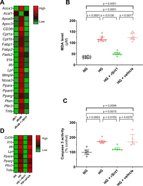
Methods: db/db mice, an established model of MCM, were supplemented with intraperitoneal rSirt1 or vehicle for 4 weeks and compared with their db/ + heterozygous littermates. At the end of treatment, cardiac function was assessed by cardiac ultrasound and left ventricular samples were collected and processed for molecular analysis. Transcriptional changes were evaluated using a custom PCR array. Lipidomic analysis was performed by mass spectrometry. H9c2 cardiomyocytes exposed to hyperglycaemia and treated with rSirt1 were used as in vitro model of MCM to investigate the ability of rSirt1 to directly target cardiomyocytes and modulate malondialdehyde levels and caspase 3 activity. Myocardial samples from diabetic and nondiabetic patients were analysed to explore Sirt1 expression levels and signaling pathways.

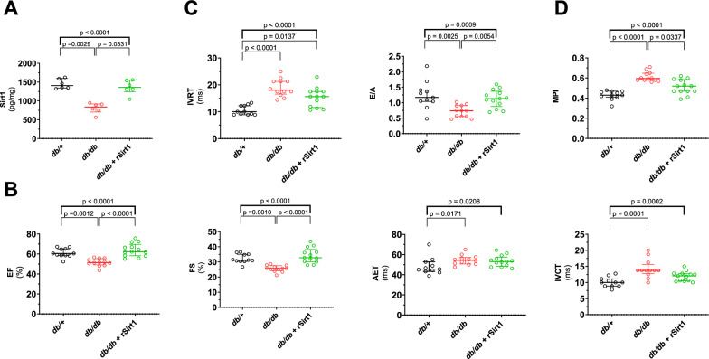
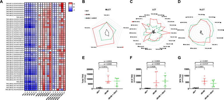
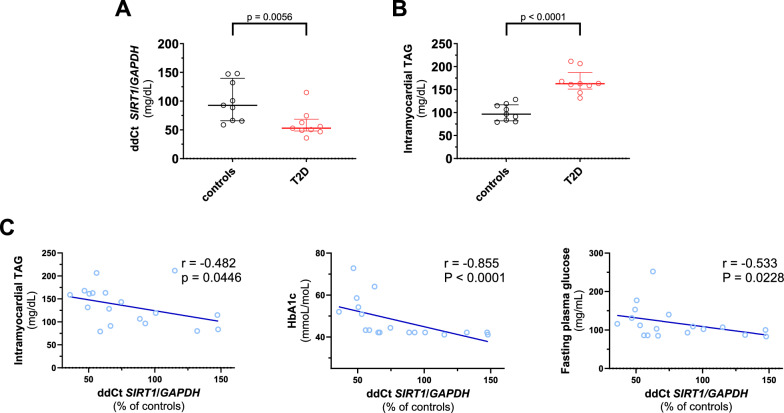
Results: rSirt1 treatment restored cardiac Sirt1 levels and preserved cardiac performance by improving left ventricular ejection fraction, fractional shortening and diastolic function (E/A ratio). In left ventricular samples from rSirt1-treated db/db mice, rSirt1 modulated the cardiac lipidome: medium and long-chain triacylglycerols, long-chain triacylglycerols, and triacylglycerols containing only saturated fatty acids were reduced, while those containing docosahexaenoic acid were increased. Mechanistically, several genes involved in lipid trafficking, metabolism and inflammation, such as Cd36, Acox3, Pparg, Ncoa3, and Ppara were downregulated by rSirt1 both in vitro and in vivo. In humans, reduced cardiac expression levels of Sirt1 were associated with higher intramyocardial triacylglycerols and PPARG-related genes.

Conclusions: In the db/db mouse model of MCM, chronic exogenous rSirt1 supplementation rescued cardiac function. This was associated with a modulation of the myocardial lipidome and a downregulation of genes involved in lipid metabolism, trafficking, inflammation, and PPARG signaling. These findings were confirmed in the human diabetic myocardium. Treatments that increase Sirt1 levels may represent a promising strategy to prevent myocardial lipid abnormalities and MCM development.

Keywords: Cardiometabolic; Diabetes; Lipidome; Metabolic cardiomyopathy; Sirt1; Therapy.


Conflict of interest statement

T.F.L. has outside this work received research and educational grants to the institution Abbott, Amgen, AstraZeneca, Boehringer-Ingelheim, Daichi-Sankyo, Menarini Foundation, Novartis, Novo Nordisk, Roche Diagnostics, Sanofi and Vifor and honoraria from Amgen, Dacadoo, Daichi-Sankyo, Menarini Foundation, Novartis, Novo Nordisk, Philips and Pfizer. F.R. has not received personal payments from pharmaceutical companies or device manufacturers in the last three years (remuneration for the time spent in activities, such as participation as steering committee member of clinical trials and member of the Pfizer Research Award selection committee in Switzerland, were made directly to the University of Zurich). The Department of Cardiology (University Hospital of Zurich/University of Zurich) reports research-, educational- and/or travel grants from Abbott, Amgen, Astra Zeneca, Bayer, Berlin Heart, B. Braun, Biosense Webster, Biosensors Europe AG, Biotronik, BMS, Boehringer Ingelheim, Boston Scientific, Bracco, Cardinal Health Switzerland, Corteria, Daiichi, Diatools AG, Edwards Lifesciences, Guidant Europe NV (BS), Hamilton Health Sciences, Kaneka Corporation, Kantar, Labormedizinisches Zentrum, Medtronic, MSD, Mundipharma Medical Company, Novartis, Novo Nordisk, Orion, Pfizer, Quintiles Switzerland Sarl, Sahajanand IN, Sanofi, Sarstedt AG, Servier, SIS Medical, SSS International Clinical Research, Terumo Deutschland, Trama Solutions, V-Wave, Vascular Medical, Vifor, Wissens Plus, ZOLL. The research and educational grants do not impact F.R.’s personal remuneration. F.P. has received personal fees from Novo Nordisk and is a scientific consultant for Vectura Pharma.

Figures

Fig. 1
Fig. 1
rSirt1 treatment rescues cardiac function in a mouse model of MCM. A Myocardial levels of rSirt1 in the three experimental groups. B Indices of systolic function (ejection fraction, fractional shortening, aortic ejection time and isovolumic contraction time) across the three experimental groups. C Diastolic function assessed as isovolumic relaxation time and E/A in the three experimental groups. D Myocardial performance index in the three experimental groups. Data are presented as box plots showing median [IQR] and compared by the Kruskal–Wallis test (upper bold bar) with Dunn post hoc test. *p < 0.05, **p < 0.01. AET aortic ejection time, EF ejection fraction, FS fractional shortening IVCT isovolumic contraction time, IVRT isovolumic relaxation time MPI Myocardial performance index rSirt1 recombinant Sirt1
Fig. 2
Fig. 2
Cardiac lipidomic signature in MCM is modulated by rSirt1 treatment. A Heat map showing levels of different lipid species across the three experimental groups (n = 6 for each group). B-D Radar plots describing Medium and long-chain triacylglycerols, long-chain triacylglycerols, and very long-chain triacylglycerols in the three experimental groups. Red dots: statistically significant difference between db/db mice treated with rSirt1 and db/db mice. Black dots: statistically significant difference between db/db mice treated with rSirt1 and db/ + mice; E–G Total triacylglycerols content, total 22:6- containing triacylglycerols content, total triacylglycerols containing only saturated fatty acids content in the three experimental groups. Data are presented as median and internal normalised within each species to provide a standardised measurement A-D; in radar plots, data are expressed in log10 changes and each grey line represent a 0.2 fold change) or as box plots E–G showing median [IQR] and compared by the Kruskal–Wallis test (upper bold bar) and corrected for multiple testing by the two stage step-up Benjamini, Krieger and Yekutieli false discovery rate method at an alpha level of 0.05. *p < 0.05, **p < 0.01. Full data are reported in Supplementary Table 1. LCT Long-chain triacylglycerols MLCT medium and long-chain triacylglycerols, rSirt1 recombinant Sirt1, SFA saturated fatty acid, TAG triacylglycerols VLCT very long-chain triacylglycerols
Fig. 3
Fig. 3
Mechanisms underpinning rSirt1-induced preservation of cardiac function in MCM. A Heat map showing differential mean relative expression levels of genes involved in lipid metabolism, trafficking and inflammation in the three experimental groups (n = 4 for each group). B–C In vitro assays showing levels of oxidative stress and apoptosis in H9c2 cardiomyocytes exposed to normal glucose (black dots and plots; n = 6), high glucose (red dots and plots; n = 6), high glucose and rSirt1 (green dots and plots; n = 6), high glucose and vehicle (pink dots and plots; n = 6). Data are presented as box plots showing median [IQR] and compared by the Kruskal–Wallis test (upper bold bar) with Dunn post hoc test. *p < 0.05, **p < 0.01. D Heat map showing differential mean relative expression levels of genes involved in lipid metabolism, trafficking and inflammation in the four experimental groups (n = 5 for each group). HG high glucose MDA malondialdehyde; NG normal glucose; rSirt1 recombinant Sirt1
Fig. 4
Fig. 4
Sirt1 signalling is related to MCM features in human myocardial samples from diabetic and nondiabetic patients. A Expression levels of Sirt1 assessed by real-time PCR in myocardial samples from diabetic (n = 9) and nondiabetic (n = 9) patients. B Intramyocardial triacylglycerol levels in myocardial samples from diabetic (n = 9) and nondiabetic (n = 9) patients. C Scatterplot showing the correlation of myocardial Sirt1 levels with myocardial triacylglycerols, plasma HbA1c and fasting plasma glucose in the study population (n = 18). Data are presented as box plots showing median [IQR] and compared by the Mann–Whitney U test. Correlations between variables were assessed by Spearman’s test. *p < 0.05, **p < 0.01. T2D type 2 diabetes TAG triacylglycerols
Fig. 5
Fig. 5
PPARG and PPARG-related genes in human myocardial specimens. Expression levels of PPARG and PPARG-downstream genes assessed by real-time PCR in myocardial samples from diabetic (n = 9) and nondiabetic (n = 9) patients. Data are presented as violin plots showing median [IQR] and compared by the Mann–Whitney U test. Correlations between variables were assessed by Spearman’s test. *p < 0.05, **p < 0.01. CD36 cluster of differentiation 36, FAS fas cell Surface death receptor, GAPDH Glyceraldehyde-3-phosphate dehydrogenase. LPL lipoprotein lipase PLIN5 perilipin 5, PPARA peroxisome proliferator-activated receptor alpha, PPARG peroxisome proliferator-activated receptor gamma, T2D type 2 diabetes

References

    1. Chen Z, Jin ZX, Cai J, Li R, Deng KQ, Ji YX, et al. Energy substrate metabolism and oxidative stress in metabolic cardiomyopathy. J Mol Med (Berl) 2022;100(12):1721–1739. doi: 10.1007/s00109-022-02269-1. - DOI - PubMed
    1. Schiattarella GG, Altamirano F, Tong D, French KM, Villalobos E, Kim SY, et al. Nitrosative stress drives heart failure with preserved ejection fraction. Nature. 2019;568(7752):351–356. doi: 10.1038/s41586-019-1100-z. - DOI - PMC - PubMed
    1. Costantino S, Paneni F, Cosentino F. Ageing, metabolism and cardiovascular disease. J Physiol. 2016;594(8):2061–2073. doi: 10.1113/JP270538. - DOI - PMC - PubMed
    1. Chew NWS, Ng CH, Tan DJH, Kong G, Lin C, Chin YH, et al. The global burden of metabolic disease: data from 2000 to 2019. Cell Metab. 2023;35(3):414–428.e413. doi: 10.1016/j.cmet.2023.02.003. - DOI - PubMed
    1. Nishida K, Otsu K. Inflammation and metabolic cardiomyopathy. Cardiovasc Res. 2017;113(4):389–398. doi: 10.1093/cvr/cvx012. - DOI - PubMed

Publication types