. 2023 Nov 13;11(1):180.
doi: 10.1186/s40478-023-01670-2.

Heterogeneity of cortical pTDP-43 inclusion morphologies in amyotrophic lateral sclerosis

Affiliations

Heterogeneity of cortical pTDP-43 inclusion morphologies in amyotrophic lateral sclerosis

Rachel H Tan et al. Acta Neuropathol Commun. .

Abstract

Background: Despite the presence of significant cortical pTDP-43 inclusions of heterogeneous morphologies in patients diagnosed with amyotrophic lateral sclerosis (ALS), pathological subclassification is routinely performed in the minority of patients with concomitant frontotemporal dementia (FTD).

Objective: In order to improve current understanding of the presence and relevance of pathological pTDP-43 subtypes in ALS, the present study examined the pattern of cortical pTDP-43 aggregates in 61 ALS cases without FTD.

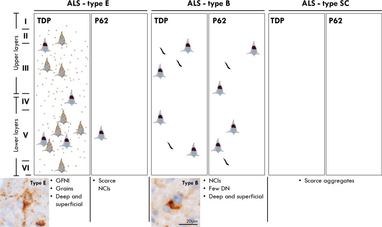
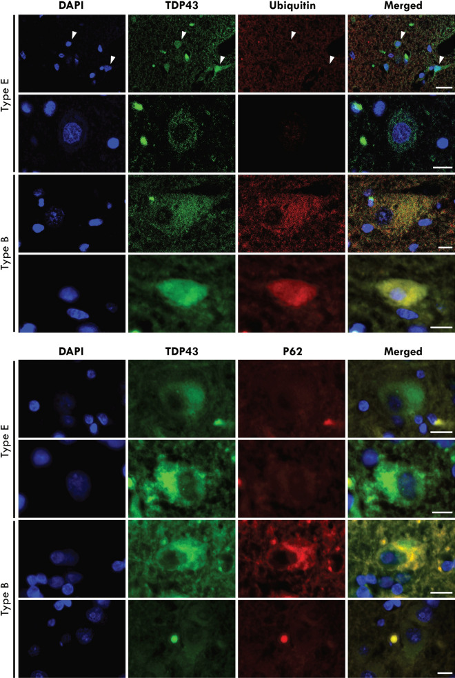
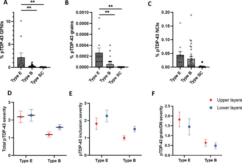
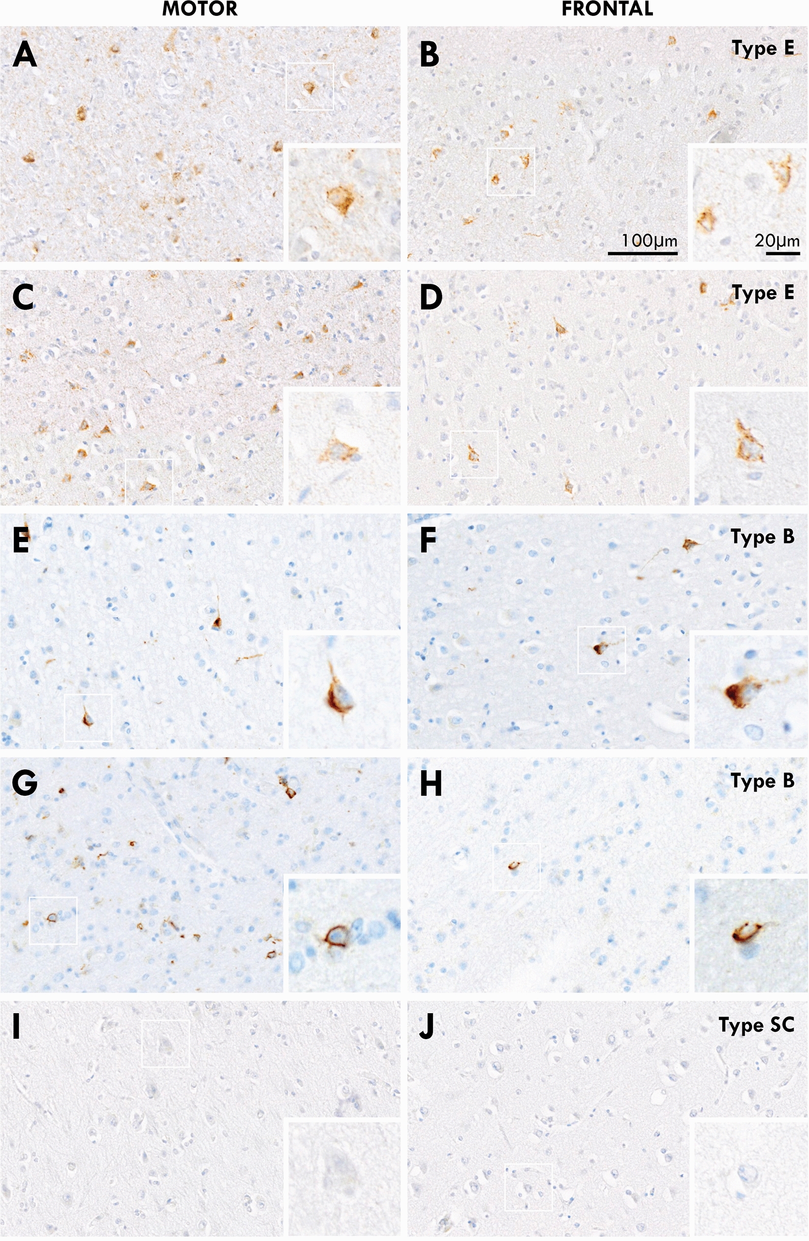
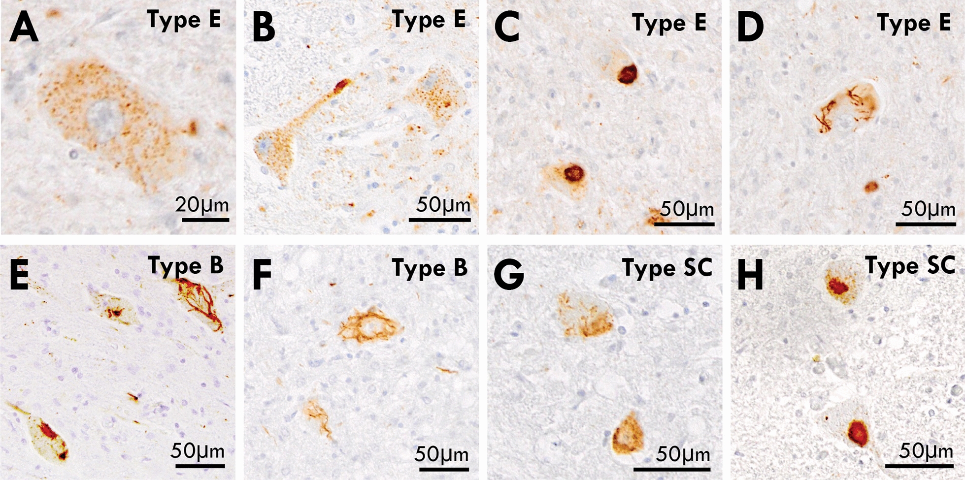
Results: Based on the presence, morphology and composition of pTDP-43 pathology, three distinct ALS-TDP subtypes were delineated: (1) A predominant pattern of pTDP-43 granulofilamentous neuronal inclusions (GFNIs) and grains that were immuno-negative for p62 was identified in 18% of cases designated ALS-TDP type E; (2) neuronal cytoplasmic inclusions (NCIs) that were immuno-positive for both pTDP-43 and p62 were observed in 67% of cases assigned ALS-TDP type B; and (3) scarce cortical pTDP-43 and p62 aggregates were identified in 15% of cases coined ALS-TDP type SC (scarce cortical). Quantitative analyses revealed a significantly greater burden of pTDP-43 GFNI and grains in ALS-TDP type E. Principal component analysis demonstrated significant relationships between GFNIs, grains and ALS-TDP subtypes to support the distinction of subtypes E and B. No significant difference in age at death or disease duration was found between ALS-TDP subgroups to suggest that these subtypes represent earlier or later stages of the same disease process. Instead, a significantly higher ALS-TDP stage, indicating greater topographical spread of pTDP-43, was identified in ALS-TDP type E. Alzheimer's disease neuropathological change (ABC score ≥ intermediate) and Lewy body disease (Braak stage ≥ IV) was more prevalent in the ALS-TDP type SC cohort, which also demonstrated a significantly lower overall cognitive score.

Conclusion: In summary, the present study demonstrates that ALS-TDP does not represent a single homogenous neuropathology. We propose the subclassification of ALS-TDP into three distinct subtypes using standard immuno-stains for pTDP-43 and p62 in the motor cortex, which is routinely sampled and evaluated for diagnostic neuropathological characterisation of ALS. We propose that future studies specify both clinicopathological group and pTDP-43 subtype to advance current understanding of the pathogenesis of clinical phenotypes in pTDP-43 proteinopathies, which will have significant relevance to the development of targeted therapies for this heterogeneous disorder.

Keywords: Amyotrophic lateral sclerosis; Frontotemporal lobar degeneration; pTDP-43.


Conflict of interest statement

The authors declare that they have no competing interests.

Figures

Fig. 1
Fig. 1
A schematic illustration of the motor cortex for the classification of ALS-TDP subtypes. Cases with a predominant pattern of pTDP-positive granulofilamentous neuronal inclusions (GFNIs) and grains not identified on corresponding p62-immunostained sections were assigned a type E; cases with round neuronal cytoplasmic inclusions ± few dystrophic neurites that were observed throughout the cortical layers of both pTDP-43 and p62 immunostained sections were assigned a type B; cases with scarce pTDP-43 and p62 aggregates that were insufficient for subclassification were assigned a type SC (scarce cortical)
Fig. 2
Fig. 2
Micrographs of the characteristic pathologies observed in the motor cortex of ALS-TDP subtypes. Cases with ALS-TDP type E demonstrated a predominant pattern of pTDP-positive granulofilamentous neuronal inclusions (GFNIs, asterix) and grains (white arrowhead) not identified on corresponding p62-immunostained sections in both C9ORF72-carriers and non-carriers. Cases with ALS-TDP type B had a predominant pattern of round neuronal cytoplasmic inclusions (black arrowhead) and occasional dystrophic neurites (arrow) that were observed throughout the cortical layers of both pTDP-43 and p62 immunostained sections in both C9ORF72-carriers and non-carriers. Cases with ALS-TDP type SC (scarce cortical) had scarce pTDP-43 and p62 aggregates that could not be subtyped
Fig. 3
Fig. 3
Cortical inclusions were immunopositive for pTDP-43 but not ubiquitin or p62 in ALS-TDP type E. In contrast to pTDP-43 aggregates that were ubiquitin-negative in ALS-TDP type E, pTDP-43 aggregates were ubiquitin-positive in ALS-TDP type B cases. Similarly, pTDP-43 aggregates were p62-negative in ALS-TDP type E but p62-positive in ALS-TDP type B cases
Fig. 4
Fig. 4
Quantitative analyses of motor cortical pTDP-43 morphologies in ALS-TDP subgroups. A significantly greater burden of pTDP-43 granulofilamentous neuronal inclusions (GFNIs) and grains were found in ALS-TDP type E (n = 11) compared to ALS-TDP type B (n = 35) and ALS-TDP type SC (n = 8) cases, even when when the two ALS-TDP type E cases with the most severe pathology were excluded from analysis (p ≤ 0.005) ( A- B). No significant difference in pTDP-43 neuronal cytoplasmic inclusions (NCIs) were found between groups ( C). Semi-quantitative analysis of pTDP-43 burden revealed a similar burden of pathological pTDP-43 in the superficial and deep layers within subtypes ( D- F). **p ≤ 0.005
Fig. 5
Fig. 5
A side-by-side comparison of pTDP-43 in the motor and frontal cortex of the same patients with ALS-TDP type E (A-D), ALS-TDP type B (E- H) and ALS-TDP type SC (I, J). Regional differences in pathological subtypes were not identified within cases
Fig. 6
Fig. 6
pTDP-43 inclusions in the spinal cord of ALS-TDP subtypes. A predominance of granulofilamentous neuronal inclusions and grains were observed in the anterior horn of the spinal cord of ALS-TDP type E cases (A, B) and these were accompanied by occasional dense compact and skein-like inclusions (C, D) similar to that observed in ALS-TDP type B (E, F) and ALS-TDP type SC cases (G, H)

References

    1. Kiernan MC, Vucic S, Cheah BC, Turner MR, Eisen A, Hardiman O, et al. Amyotrophic lateral sclerosis. Lancet. 2011;377(9769):942–955. doi: 10.1016/S0140-6736(10)61156-7. - DOI - PubMed
    1. Kiernan MC, Vucic S, Talbot K, McDermott CJ, Hardiman O, Shefner JM, et al. Improving clinical trial outcomes in amyotrophic lateral sclerosis. Nat Rev Neurol. 2021;17(2):104–118. doi: 10.1038/s41582-020-00434-z. - DOI - PMC - PubMed
    1. Tan RH, Ke YD, Ittner LM, Halliday GM. ALS/FTLD: experimental models and reality. Acta Neuropathol. 2017;133(2):177–196. doi: 10.1007/s00401-016-1666-6. - DOI - PubMed
    1. Braak H, Ludolph AC, Neumann M, Ravits J, Del Tredici K. Pathological TDP-43 changes in Betz cells differ from those in bulbar and spinal alpha-motoneurons in sporadic amyotrophic lateral sclerosis. Acta Neuropathol. 2017;133(1):79–90. doi: 10.1007/s00401-016-1633-2. - DOI - PMC - PubMed
    1. Mackenzie IR, Neumann M, Baborie A, Sampathu DM, Du Plessis D, Jaros E, et al. A harmonized classification system for FTLD-TDP pathology. Acta Neuropathol. 2011;122(1):111–113. doi: 10.1007/s00401-011-0845-8. - DOI - PMC - PubMed

Publication types

Substances