. 2023 Oct 24;24(21):15544.
doi: 10.3390/ijms242115544.

Mesenchymal Stromal Cells from Perinatal Tissues as an Alternative for Ex Vivo Expansion of Hematopoietic Progenitor and Stem Cells from Umbilical Cord Blood

Affiliations

Mesenchymal Stromal Cells from Perinatal Tissues as an Alternative for Ex Vivo Expansion of Hematopoietic Progenitor and Stem Cells from Umbilical Cord Blood

Ximena Bonilla et al. Int J Mol Sci. .

Abstract

Umbilical cord blood (UCB) serves as a source of hematopoietic stem and progenitor cells (HSPCs) utilized in the regeneration of hematopoietic and immune systems, forming a crucial part of the treatment for various benign and malignant hematological diseases. UCB has been utilized as an alternative HSPC source to bone marrow (BM). Although the use of UCB has extended transplantation access to many individuals, it still encounters significant challenges in selecting a histocompatible UCB unit with an adequate cell dose for a substantial proportion of adults with malignant hematological diseases. Consequently, recent research has focused on developing ex vivo expansion strategies for UCB HSPCs. Our results demonstrate that co-cultures with the investigated mesenchymal stromal cells (MSCs) enable a 10- to 15-fold increase in the cellular dose of UCB HSPCs while partially regulating the proliferation capacity when compared to HSPCs expanded with early acting cytokines. Furthermore, the secretory profile of UCB-derived MSCs closely resembles that of BM-derived MSCs. Moreover, both co-cultures exhibit alterations in cytokine secretion, which could potentially impact HSPC proliferation during the expansion process. This study underscores the fact that UCB-derived MSCs possess a remarkably similar supportive capacity to BM-derived MSCs, implying their potential use as feeder layers in the ex vivo expansion process of HSPCs.

Keywords: hematopoietic stem and progenitor cells ex vivo expansion; hematopoietic stem and progenitor cells transplantation; mesenchymal stromal cells; perinatal tissues; umbilical cord blood.


Conflict of interest statement

The authors declare no conflict of interest.

Figures

Figure 1
Figure 1
Proliferation capacity of umbilical cord blood hematopoietic stem and progenitor cells (HSPCs) co-cultured with mesenchymal stromal cells (7-day culture). A comparison is made with HSPCs in monoculture (HSPC+Cytokines, depicted in green) treated with early acting cytokines (SCF, TPO, FLT3L). Co-culture with bone-marrow-derived MSCs (HSPC+BM-MSC, depicted in red). Co-culture with Wharton’s-jelly-derived MSCs (HSPC+WJ-MSC, depicted in blue). Co-culture with umbilical-cord-blood-derived MSCs (HSPC+UCB-MSC, depicted in orange). (A) Paired analysis of donors in mono- and co-culture. (B) Proliferation analysis of different donors under mono- and co-culture conditions. (C) Proliferation analysis of CFSE-labeled HSPCs and Mean Fluorescence Intensity (MFI) analysis via flow cytometry. Red: Unlabeled cells. Blue: Cells synchronized via 24 h starvation. Orange: HSPC+Cytokines. Light Green: HSPC+BM-MSC. Dark Green: HSPC+WJ-MSC. Violet: HSPC+UCB-MSC (Representative image). (D) Analysis of the MFI of CFSE-labeled HSPCs under different culture conditions. Statistical analysis performed using the Wilcoxon paired test (non-significant) and Mann–Whitney test (p < 0.05). * Data statistically significant. Values are presented as the number of times HSPCs are doubled.
Figure 2
Figure 2
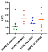
Migration capacity of umbilical cord blood hematopoietic stem progenitor cells (HSPCs) in co-culture with mesenchymal stromal cells (7-day culture). The migratory behavior of HSPCs co-cultured with mesenchymal stromal cells (MSCs) is assessed using a transwell system and chemotactic/adhesive stimuli for a duration of three hours, followed by flow-cytometry-based cell counting. The migration capacity of the co-cultured HSPCs is compared with that of HSPCs cultured alone with early acting cytokines (HSPC+Cytokines, depicted in green). Additionally, co-cultures are performed with bone-marrow-derived MSCs (HSPC+BM-MSC, shown in red), Wharton’s-jelly-derived MSCs (HSPC+WJ-MSC, depicted in blue), and umbilical-cord-blood-derived MSCs (HSPC+UCB-MSC, represented in orange). Statistical analysis is conducted using the Kruskal–Wallis test on unpaired data, and no significant differences in migration capacity are observed among the different co-culture conditions.
Figure 3
Figure 3
Evaluation of the expression of primitive markers in umbilical cord blood hematopoietic stem progenitor cells (HSPCs) in co-culture with mesenchymal stromal cells (MSC) (7-day culture). The flow-cytometry-based assessment of primitive and differentiation markers in HSPCs cultivated with MSCs is depicted. Expression levels of these markers in freshly isolated HSPCs are quantified and juxtaposed with HSPCs in monoculture (HSPC+Cytokines, green) treated with early acting cytokines (SCF, TPO, FLT3). Co-culture with bone-marrow-derived MSCs (HSPC+BM-MSC, red). Co-culture with Wharton’s-jelly-derived MSCs (HSPC+WJ-MSC, blue). Co-culture with umbilical-cord-blood-derived MSCs (HSPC+UCB-MSC, Orange). (A) Quantification of Mean Fluorescence Intensity (MFI) for CD34. (B) Median Fluorescence Intensity (MFI) for CD38. Statistical analysis was performed using the Wilcoxon test for paired data (p < 0.05). * Data statistically significant.
Figure 4
Figure 4
Evaluation of the clonogenic potential of umbilical cord blood hematopoietic stem progenitor cells (HSPC) in co-culture with mesenchymal stromal cells (MSC) (14-day culture). The clonogenic potential of CD34+ cells expanded in co-cultured with mesenchymal stromal cells (MSCs) is assessed by counting Colony-Forming Units (CFUs) formed in methylcellulose culture from HSPCs expanded in co-cultured with MSCs. The clonogenic capacity of the co-cultured HSPCs is compared with that of HSPCs cultured alone with early acting cytokines (HSPC+Cytokines, depicted in green). Additionally, co-cultures are performed with bone-marrow-derived MSCs (HSPC+BM-MSC, shown in red), Wharton’s-jelly-derived MSCs (HSPC+WJ-MSC, depicted in blue), and umbilical-cord-blood-derived MSCs (HSPC+UCB-MSC, represented in orange). Statistical analysis was conducted using the Kruskal–Wallis test for unpaired data (p < 0.05).
Figure 5
Figure 5
Cytokine secretion profile of HSPCs + MSCs co-cultures. The change in secretion levels of eight cytokines is presented as the Log2 of the fold change between hematopoietic progenitor cell and mesenchymal stromal cell co-cultures from each tissue. (A) Bone marrow. (B) Wharton’s jelly. (C) Umbilical cord blood. Samples are grouped according to their cytokine expression profile. Each row represents a sample, and each column represents a cytokine. Both samples and cytokines were grouped based on their similarity. (D) Sample grouping by treatment. (E) Sample grouping by source. Exp: HSPC+Cytokines w/o MSC culture.
Figure 6
Figure 6
Principal component analysis (PCA) according to HSPCs properties. A two-dimensional PCA was conducted with donors of HSPCs who underwent comprehensive functionality assays (proliferation, migration, differentiation, and secretory profile). (A) Correlation circle of evaluated variables according to contribution scale, where variables with a higher contribution are shown in red and those with lower contribution are shown in green. (B) Two-dimensional PCA based on MSC expansion strategy (co-cultures with MSCs from different sources versus HSPCs expanded with early action cytokines), displaying the contribution of these variables to the principal components and allowing the clear grouping and discrimination of Wharton’s jelly data (red) from other groups (blue).

References

    1. Emiloju O.E., Potdar R., Jorge V., Gupta S., Varadi G. Clinical Advancement and Challenges of Ex Vivo Expansion of Human Cord Blood Cells. Clin. Hematol. Int. 2019;2:18–26. doi: 10.2991/chi.d.191121.001. - DOI - PMC - PubMed
    1. Lund T.C., Boitano A.E., Delaney C.S., Shpall E.J., Wagner J.E. Advances in Umbilical Cord Blood Manipulation—From Niche to Bedside. Nat. Rev. Clin. Oncol. 2015;12:163–174. doi: 10.1038/nrclinonc.2014.215. - DOI - PMC - PubMed
    1. Pinho S., Frenette P.S. Haematopoietic Stem Cell Activity and Interactions with the Niche. Nat. Rev. Mol. Cell Biol. 2019;20:303–320. doi: 10.1038/s41580-019-0103-9. - DOI - PMC - PubMed
    1. Gluckman E. Current Status of Umbilical Cord Blood Hematopoietic Stem Cell Transplantation. Exp. Hematol. 2000;28:1197–1205. doi: 10.1016/S0301-472X(00)00540-3. - DOI - PubMed
    1. MacMillan M.L., Robin M., Harris A.C., DeFor T.E., Martin P.J., Alousi A., Ho V.T., Bolaños-Meade J., Ferrara J.L.M., Jones R., et al. A Refined Risk Score for Acute Graft-versus-Host Disease That Predicts Response to Initial Therapy, Survival, and Transplant-Related Mortality. Biol. Blood Marrow Transpl. 2015;21:761–767. doi: 10.1016/j.bbmt.2015.01.001. - DOI - PMC - PubMed