. 2023 Oct 26;24(21):15619.
doi: 10.3390/ijms242115619.

Vimentin Localization in the Zebrafish Oral Cavity: A Potential Role in Taste Buds Regeneration

Affiliations

Vimentin Localization in the Zebrafish Oral Cavity: A Potential Role in Taste Buds Regeneration

Marialuisa Aragona et al. Int J Mol Sci. .

Abstract

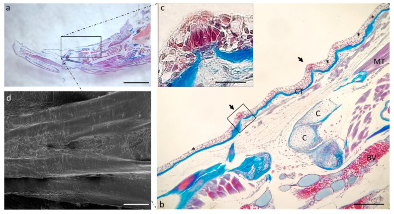
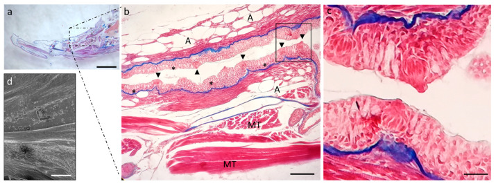
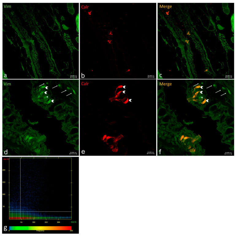
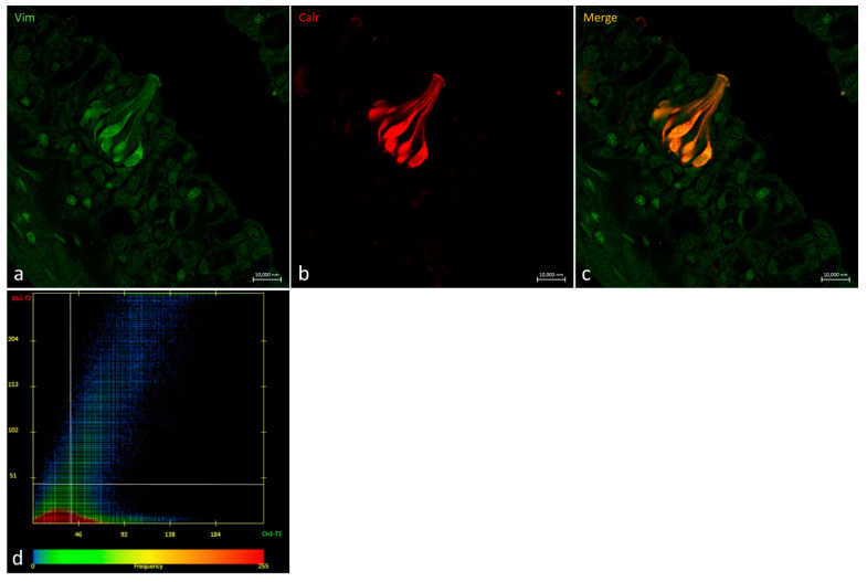
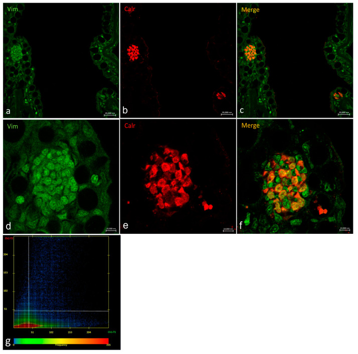
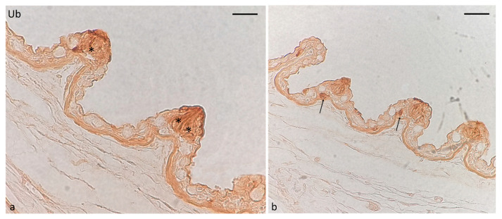
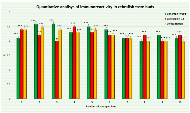
The morphology of the oral cavity of fish is related to their feeding habits. In this context, taste buds are studied for their ability to catch chemical stimuli and their cell renewal capacity. Vimentin RV202 is a protein employed as a marker for mesenchymal cells that can differentiate along different lineages and to self-renew, while Calretinin N-18 is employed as a marker of sensory cells, and ubiquitin is a protein crucial for guiding the fate of stem cells throughout development. In this study, a surface morphology investigation and an immunohistochemical analysis have been conducted. The results of the present study reveal, for the first time, the presence of Vimentin RV202 in a taste bud cell population of zebrafish. Some taste bud cells are just Vimentin RV202-immunoreactive, while in other cells Vimentin RV202 and Calretinin N-18 colocalize. Some taste buds are just reactive to Calretinin N-18. Vimentin RV202-immunoreactive cells have been observed in the connective layer and in the basal portion of the taste buds. The immunoreactivity of ubiquitin was restricted to sensory cells. Further studies are needed to elucidate the role of Vimentin RV202 in the maturation of taste bud cells, its potential involvement in the regeneration of these chemosensory organs, and its eventual synergic work with ubiquitin.

Keywords: Calretinin N-18; SEM; Vimentin RV202; oral cavity; taste buds; zebrafish.


Conflict of interest statement

The authors declare no conflict of interest.

Figures

Figure 1
Figure 1
(a) Scanning electron microscope micrograph of a sagittal section of a zebrafish head. (b) Scanning electron microscope of two taste buds with different shapes. Higher magnification of onion-shaped taste bud (c) and pear-shaped taste bud (d). Scale bar: 1 mm (a) 10 μm (b) 20 μm (c) 10 μm (d).
Figure 2
Figure 2
(a) Scanning electron microscope micrograph of a median section of a zebrafish head. Well-developed lips (asterisks), semilunar valve (arrow), taste buds (arrowheads), mucosal folds (#), muscular tissue (MT). (b) Light micrograph (Masson’s trichrome with aniline blue staining): taste buds (inset), semilunar valve (black arrow). Stratified epithelium (E); connective tissue (ct); mucous cells (stars); adipocytes (A); bone (BT); cartilage (C); muscular tissue (MT). (c) High magnification of taste buds (arrowheads) of inset in image. Scale bar: 1 mm (a). Magnification 10×, scale bar 100 µm (b). Magnification 63× oil, scale bar 10 µm (c).
Figure 3
Figure 3
Scanning electron microscope micrograph of the dorsal surface of the tongue: (a) apex, (b) body, (c) root. Scale bar: 1 mm (ac).
Figure 4
Figure 4
(a) Stereomicrograph of a sagittal section of the tongue (Masson trichrome with aniline blue staining): apex of the tongue (circular insert). (b) Scanning electron microscope micrograph of the tongue apex. Magnification 4×, scale bar 1 mm (a). Scale bar: 1 mm (b). Magnification 10×, scale bar 200 µm (insert).
Figure 5
Figure 5
(a) Stereomicrograph of a sagittal section of the tongue: dorsal surface of the tongue body (insert). (b) Light micrographs of the dorsal surface of the tongue with taste buds (arrows). Connective tissue (CT); cartilage (C); muscular tissue (MT); blood vessel (BV); goblet cells (asterisks). (c) high magnification of taste bud in image. (b) Masson trichrome with aniline blue staining. (d) Scanning electron microscope micrograph of the tongue body. (a) Magnification 4×, scale bar 1 mm. (b) Magnification 10×, scale bar 200 µm. (c) Magnification 63× oil, scale bar 10 µm. (d) Scale bar: 1 mm.
Figure 6
Figure 6
(a) Stereomicrograph of a sagittal section of the tongue: dorsal surface of the tongue root (insert). (b) Light micrographs of the dorsal surface of the tongue root showing the lingual mucosa with several goblet cells (asterisks) and taste buds (arrowheads). Muscular tissue (MT), adipocites (A); Masson trichrome with aniline blue staining. (c) high magnification of taste buds in (b). (d) Scanning electron microscope micrograph of the tongue body. (a) Magnification 4×, scale bar 1 mm. (b) Magnification 10×, scale bar 200 µm. (c) Magnification 63× oil, scale bar 10 µm. (d) Scale bar: 1 mm.
Figure 7
Figure 7
Oropharyngeal taste buds. Immunohistochemical detection (immunofluorescence method) of Vimentin RV202 (a,d) and Calretinin N-18 (b,e). Immunohistochemical detection of Vimentin RV202 and Calretinin N-18 in colocalization view (c,f). Vimentin RV202-immunoreactive cells in the connective layer (arrows). Vimentin RV202-immunoreactive cells interspersed between taste bud sensory cells (asterisks). Taste bud sensory cells (gallon arrows). (ac) Magnification 10×; scale bar 50 µm. (df) Magnification 63×; scale bar 10 µm. (g) Scatter plot of Vimentin RV202/ Calretinin N-18 colocalization in zebrafish taste buds. The X-axis and Y-axis indicate the immunofluorescent signal, respectively, for Calretinin N-18 and Vimentin RV202. The intersection point between two lines indicates colocalization, which means the localization of Calretinin N-18 and Vimentin RV202 in the same cell. Zen 2011 (LSM 700 Zeiss software ZEN 3.7).
Figure 8
Figure 8
Oropharyngeal taste bud. (a) Immunohistochemical detection (immunofluorescence method) of Vimentin RV202 and (b) Calretinin N-18. (c) Immunohistochemical detection of Vimentin RV202 and Calretinin N-18 in colocalization view. (ac) Magnification 63×; scale bar 10 µm. (d) Scatter plot of Vimentin RV202/ Calretinin N-18 colocalization in zebrafish taste buds. The X-axis and Y-axis indicate the immunofluorescent signal, respectively, for Calretinin N-18 and Vimentin RV202. The intersection point between two lines indicates colocalization, which means the localization of Calretinin N-18 and Vimentin RV202 in the same cell. Zen 2011 (LSM 700 Zeiss software ZEN 3.7).
Figure 9
Figure 9
Transversal section of the basal part of a taste bud. (a,d) Immunohistochemical detection (immunofluorescence method) of Vimentin RV202 and (b,e) Calretinin N-18. (c,f) Immunohistochemical detection of Vimentin RV202 and Calretinin N-18 in colocalization view. (ac) Magnification10×; scale bar 20 µm. (df). Magnification 63×; scale bar 10 µm. (g) Scatter plot of Vimentin RV202/ Calretinin N-18 colocalization in zebrafish taste buds. The X-axis and Y-axis indicate the immunofluorescent signal, respectively, for Calretinin N-18 and Vimentin RV202. The intersection point between two lines indicates colocalization, which means the localization of Calretinin N-18 and Vimentin RV202 in the same cell. Zen 2011 (LSM 700 Zeiss software ZEN 3.7).
Figure 10
Figure 10
Immunohistochemical localization of ubiquitin immunoreactivity in oral taste buds of adult zebrafish. (a) Sensory cells (asterisks). (b) Isolated cells (arrows) near taste buds. Magnification: 60× (a); 40× (b).
Figure 11
Figure 11
Graphical representation of adult zebrafish taste bud cells immunoreactive to Vimentin RV202, Calretinin N-18, and cells with two proteins colocalized. The statistical analysis shows a distribution pattern of the two proteins. Data represent the average of measurements from random fields of ten sections. N°: mean of taste bud cells immunopositive to Vimentin RV202, Calretinin N-18, and colocalized. Statistical significance: *** p < 0.001.

Similar articles

Cited by

References

    1. Buddington R.K., Kuz’mina V. Chapter 23—Digestive System. In: Ostrander G.K., editor. The Laboratory Fish. Academic Press; London, UK: 2000. pp. 379–384. - DOI
    1. Abbate F., Germana G., De Carlos F., Montalbano G., Laura R., Levanti M., Germana A. The oral cavity of the adult zebrafish (Danio rerio) Anat. Histol. Embryol. 2006;35:299–304. doi: 10.1111/j.1439-0264.2006.00682.x. - DOI - PubMed
    1. Abbate F., Guerrera M.C., Levanti M., Laurà R., Aragona M., Mhalhel K., Montalbano G., Germanà A. Anatomical, histological and immunohistochemical study of the tongue in the rainbow trout (Oncorhynchus mykiss) Anat. Histol. Embryol. 2020;49:848–858. doi: 10.1111/ahe.12593. - DOI - PubMed
    1. Abbate F., Guerrera M.C., Levanti M., Laurà R., Aragona M., Mhalhel K., Montalbano G., Germanà A. Morphological characteristics of the blackspot seabream (Pagellus bogaraveo) tongue: A structural and immunohistochemical study. Anat. Histol. Embryol. 2022;51:103–111. doi: 10.1111/ahe.12769. - DOI - PMC - PubMed
    1. Germanà A., Paruta S., Germanà G.P., Ochoa-Erena F.J., Montalbano G., Cobo J., Vega J.A. Differential distribution of S100 protein and calretinin in mechanosensory and chemosensory cells of adult zebrafish (Danio rerio) Brain Res. 2007;1162:48–55. doi: 10.1016/j.brainres.2007.05.070. - DOI - PubMed

LinkOut - more resources