. 2023 Oct 29;24(21):15725.
doi: 10.3390/ijms242115725.

In Vitro Assessment of Cisplatin/Hyaluronan Complex for Loco-Regional Chemotherapy

Affiliations

In Vitro Assessment of Cisplatin/Hyaluronan Complex for Loco-Regional Chemotherapy

Sabrina Banella et al. Int J Mol Sci. .

Abstract

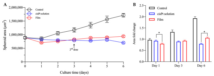
Loco-regional chemotherapy is a strategy used to achieve more precise anticancer drug effect directly on tumor mass, while decreasing whole body exposure, which can lead to undesirable side effects. Thus, the loco-regional chemotherapy is conceptually similar to the targeted drug delivery systems for delivering chemotherapeutics to cancer cells in a certain location of the body. Recently, it has been demonstrated that a novel polymeric film containing the complex between cisplatin (cisPt) and hyaluronan (sodium salt of hyaluronic acid; NaHA) enhanced in vivo efficacy and safety of cisplatin (cisPt) by loco-regional delivery in pleural mesothelioma. Biologically, hyaluronic acid (HA) binds with the CD44 receptor, which is a transmembrane glycoprotein overexpressed by other cancer cells. Thus, administering both cisPt and hyaluronan together as a complex loco-regionally to the tumor site could target cancer cells locally and enhance treatment safety. A slight excess of hyaluronan was required to have more than 85% cisPt complexation. In cell monolayers (2D model) the cisPt/NaHA complex in solution demonstrated dose- and time-dependent cytotoxic effect by decreasing the viability of pancreatic, melanoma, and lung cell lines (they all express CD44). At the same concentration in solution, the complex was as effective as cisPt alone. However, when applied as film to melanoma spheroids (3D model), the complex was superior because it prevented the tumor spheroid growth and, more importantly, the formation of new cell colonies. Hence, cisPt/NaHA complex could work in preventing metastases loco-regionally and potentially avoiding systemic relapses.

Keywords: CD44 receptor; chemotherapy; cisplatin; coordination complex; drug delivery system; hyaluronan; loco-regional therapy; melanoma; pleural mesothelioma.


Conflict of interest statement

Emeritus Professor Paolo Colombo is the administrator of PlumeStars s.r.l., the sponsor of the application for EMA orphan medicinal product designation of the cisplatin-loaded hyaluronan film.

Figures

Figure 1
Figure 1
Fraction of cisPt coordinated by NaHA as a function of (monomer:drug) molar ratio for: high (HMW; black) and low molecular weight (LMW; blue).
Figure 2
Figure 2
Concentration-dependent cytotoxicity of cisPt alone (black), cisPt/NaHA HMW (red), and cisPt + VA (blue) in: (A) BxPC3, (B) MIA PaCa-2, (C) A549, and (D) VEM-responsive A375 cell lines. Cell viability was determined by MTT assay after 48 h exposure to treatment. Data are reported as mean ± SEM (n = 6). Asterisks indicate a statistically significant difference (p < 0.05).
Figure 3
Figure 3
Cytotoxic effect of cisPt alone (black bar), cisPt/NaHA HMW (empty red bar) and cisPt/NaHA LMW (filled red bar), and cisPt + VA (blue bar) in MIA PaCa-2 (top plots), VEM-responsive A375 (middle plots), and VEM-resistant A375 (bottom plots). Cell viability was determined by MTT assay after 4 and 24 h exposure to 2 µM (left plots) and 10 µM (right plots) cisPt. Data are reported as mean ± SEM (n = 6). Asterisks indicate a statistically significant difference (p < 0.05).
Figure 4
Figure 4
Area of VEM-responsive (A) and VEM-resistant (B) A375 spheroids as a function of culture time with various treatment groups: untreated control (black), cisPt alone (red), cisPt/NaHA HMW complex (orange), valproic acid (VA, green), cisPt + VA (blue), and cisPt/NaHA HMW + VA (purple). Data are reported as mean ± SEM (n ≥ 3). Asterisks indicate a statistically significant difference (p < 0.05).
Figure 5
Figure 5
Representative images of (A) VEM-responsive A375 spheroids and (B) VEM-resistant A375 spheroids treated with no drug (control), cisPt alone, cisPt/NaHA HMW complex, cisPt + VA, VA alone and cisPt/NaHA HMW + VA at days 0–5 of treatment. The cisPt concentration was 1 µM in all cases, while the VA concentration was 100 µM. The last to the right series of fluorescence images depict cellular death of the spheroids in the various treatment groups. They are composite images of 4′,6-diamidino-2-phenylindole (DAPI) (blue), calcein acetoxymethyl (calcein AM) (green) and ethidium homodimer-1 (EthD-1) (red). Scale bar: 400 μm.
Figure 6
Figure 6
Area of VEM-responsive (A) and VEM-resistant (B) A375 spheroids as a function of time with various treatment groups: untreated control (black), cisPt alone (red), cisPt/NaHA HMW complex (orange), valproic acid (VA, green), cisPt + VA (blue), and cisPt/NaHA HMW + VA (purple). Data are reported as mean ± SEM (n ≥ 3). Asterisks indicate a statistically significant difference (p < 0.05).
Figure 7
Figure 7
Representative images of (A) VEM-responsive A375 spheroids and (B) VEM-resistant A375 spheroids treated with no drug (control), cisPt alone, cisPt/NaHA HMW complex, cisPt + VA, VA alone and cisPt/NaHA HMW + VA at days 0–2 of treatment. The cisPt concentration was 2.5 µM in all cases, while the NaHA and VA concentrations were 9 and 100 µM, respectively. Scale bar: 400 µm.
Figure 8
Figure 8
Optical microscopy image of the spheroid leaning on the film. The dashed line and the arrow indicate the outer edge of the film.
Figure 9
Figure 9
(A) Area of VEM-resistant spheroids as a function of culture time with various treatment groups: untreated control (black), cisPt solution (blue) and film (red). (B) Fold change in the area of VEM-resistant A375 spheroids treated during culture time vs. Day 0. Data are reported as mean ± SEM (n ≥ 6). Asterisks indicate a statistically significant difference (p < 0.05).
Figure 10
Figure 10
Representative images of VEM-resistant A375 spheroids treated with cisPt solution (25 µM) and film (cisPt 100 µM) following 6 days of treatment. Until Day 5 the magnification was 10× (scale bar: 400 µm), then it was 4x on the last day (scale bar: 1000 µm).
Figure 11
Figure 11
Crystal violet-stained images for control (A) and cisPt solution (B) groups.

References

    1. Agnihotri T.G., Gomte S.S., Jain A. Emerging theranostics to combat cancer: A perspective on metal-based nanomaterials. Drug Dev. Ind. Pharm. 2022;48:48585–48601. doi: 10.1080/03639045.2022.2153862. - DOI - PubMed
    1. Ghosh S. Cisplatin: The first metal based anticancer drug. Bioorg. Chem. 2019;88:102925. doi: 10.1016/j.bioorg.2019.102925. - DOI - PubMed
    1. Riley R.S., June C.H., Langer R., Mitchell M.J. Delivery technologies for cancer immunotherapy. Nat. Rev. Drug Discov. 2019;18:175–196. doi: 10.1038/s41573-018-0006-z. - DOI - PMC - PubMed
    1. Dang Y., Guan J. Nanoparticle-based drug delivery systems for cancer therapy. Smart Mater. Med. 2020;1:10–19. doi: 10.1016/j.smaim.2020.04.001. - DOI - PMC - PubMed
    1. Mu W., Chu Q., Liu Y., Zhang N. A Review on Nano-Based Drug Delivery System for Cancer Chemoimmunotherapy. Nanomicro. Lett. 2020;12:142. doi: 10.1007/s40820-020-00482-6. - DOI - PMC - PubMed

MeSH terms