. 2023 Oct 24;28(21):7240.
doi: 10.3390/molecules28217240.

Analysis of Skin Regeneration and Barrier-Improvement Efficacy of Polydeoxyribonucleotide Isolated from Panax Ginseng (C.A. Mey.) Adventitious Root

Affiliations

Analysis of Skin Regeneration and Barrier-Improvement Efficacy of Polydeoxyribonucleotide Isolated from Panax Ginseng (C.A. Mey.) Adventitious Root

Kwang-Soo Lee et al. Molecules. .

Abstract

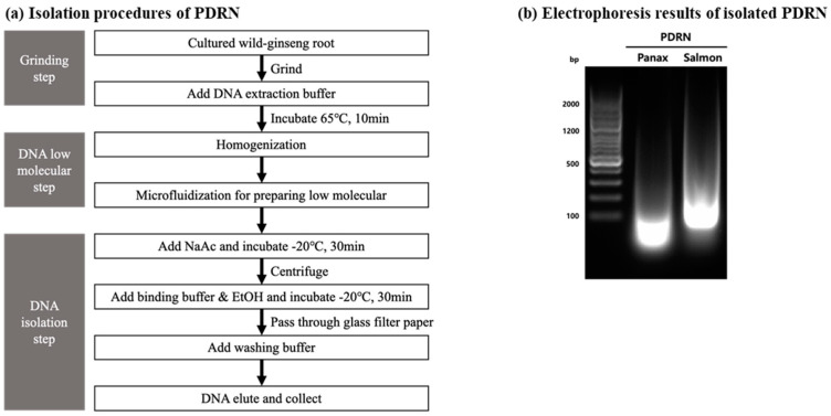
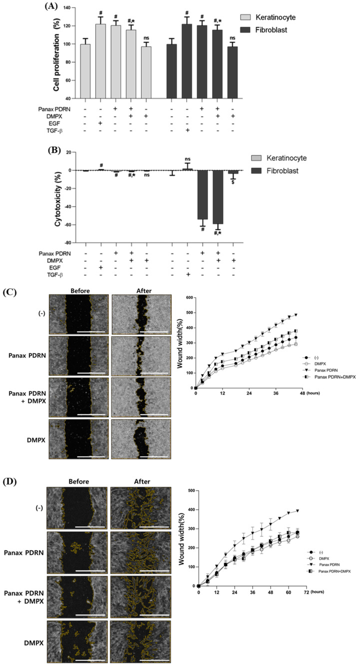
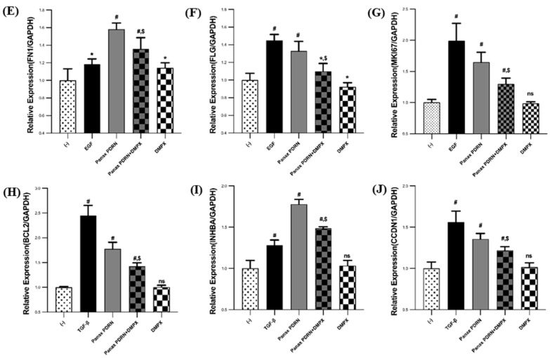
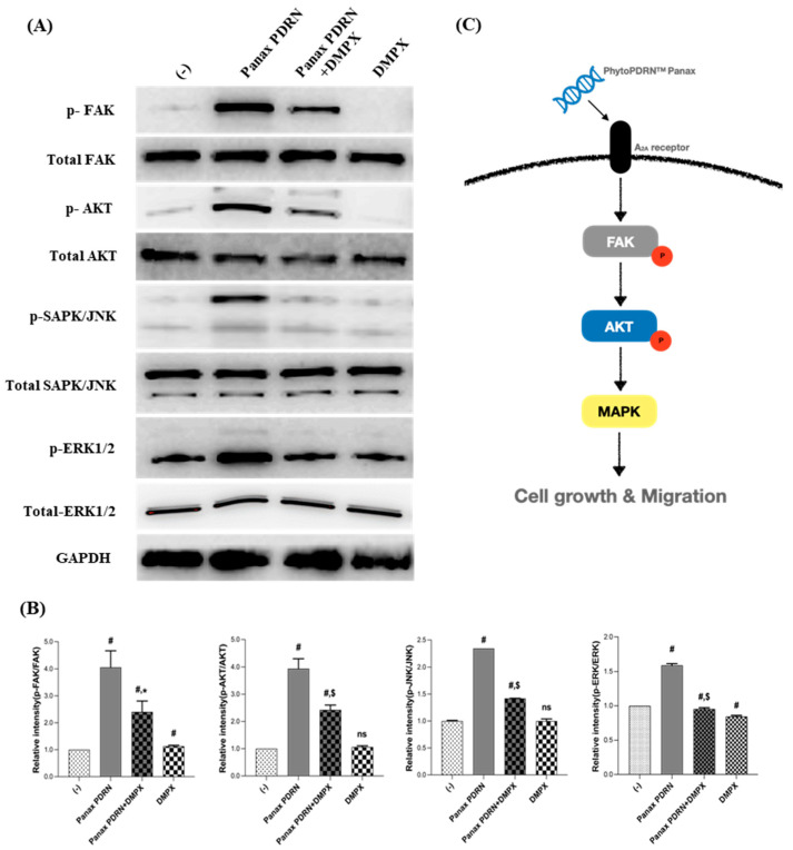
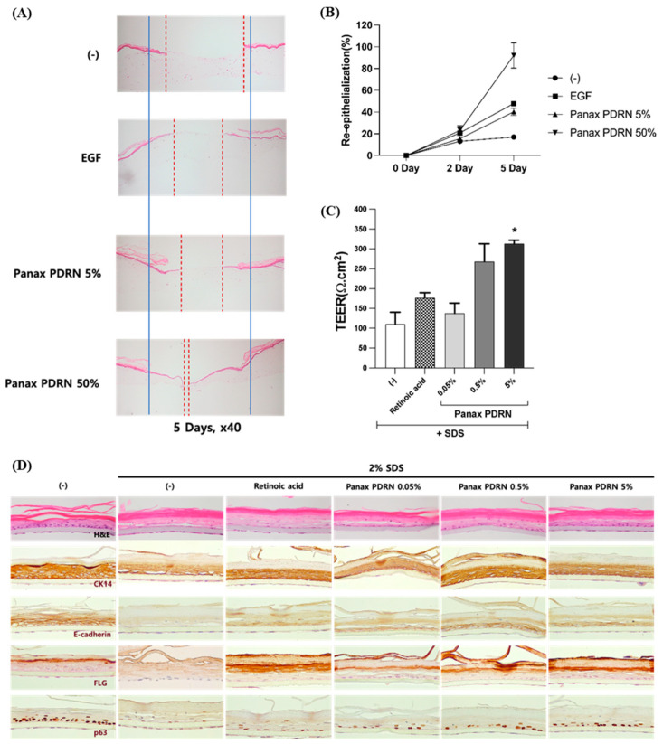
Polydeoxyribonucleotide (PDRN) has the ability to regenerate skin cells and improve the skin barrier and wound healing. This study investigated the possibility of replacing animal-derived PDRN with plant-derived PDRN. To test this, the adventitious roots of Korean ginseng (Panax ginseng C.A. Meyer), which is commonly used to treat various diseases, were suspension-cultivated through tissue culture; subsequently, PDRN was purified using microfluidization, an ultra-high-pressure physical grinding method. The results showed that purified Panax PDRN was effective in healing skin wounds and enhancing the skin barrier. Panax PDRN promoted the proliferation of keratinocytes and fibroblasts by increasing the expression of fibronectin, filaggrin, Ki-67, Bcl-2, inhibin beta A, and Cyclin D1. It also acted as an agonist of the adenosine A2A receptor and induced the phosphorylation of focal adhesion kinase, adenosine triphosphate-dependent tyrosine kinase, and mitogen-activated protein kinase. This activated signal transduction, thereby regenerating skin cells and strengthening the barrier. These results were not only observed in skin cells but also in an artificial skin model (KeraSkinTM). The use of plant-derived PDRN instead of animal-derived PDRN can promote animal welfare and environmental sustainability. Furthermore, Panax PDRN can potentially be a new plant-derived PDRN (PhytoPDRN) that may be utilized in the treatment of various skin diseases.

Keywords: Panax ginseng; plant-derived polydeoxyribonucleotide; polydeoxyribonucleotide; skin barrier; skin regeneration; wound healing.


Conflict of interest statement

The authors declare no conflict of interest.

Figures

Figure 1
Figure 1
Isolation and purification of Panax PDRN from adventitious roots of Panax ginseng using microfluidization. (a) Panax PDRN was isolated and purified from cultured cells mass-produced by plant tissue culture technology using an ultra-high-pressure extraction method. Low-molecular Panax PDRN can be obtained via hydrolysis, excluding chemicals. (b) The DNA size of purified Panax PDRN was compared with that of salmon PDRN through gel electrophoresis.
Figure 2
Figure 2
Analysis of cell proliferation and regeneration-promoting effects of Panax PDRN on keratinocytes and fibroblasts. (A) Cell proliferation and (B) cytotoxicity were analyzed for Panax PDRN in keratinocytes (HaCaT) and fibroblasts (HDF). (C,D) A wound-healing assay was performed to confirm cell proliferation and regeneration ability in HaCaT and HDF (scale bar represents 1000 μm). (EJ) The expression of each gene related to cell proliferation and regeneration (fibronectin, filaggrin, Ki-67, Bcl-2, inhibin A, and cyclin D1) was confirmed via qRT-PCR in HaCaT and HDF. mRNA levels were normalized to GAPDH expression. Expression levels for each gene are indicated as fold-changes compared with the control levels. # p < 0.01 compared with basal levels; * p < 0.05; $ p < 0.01 compared with Panax PDRN levels; ns means statistically not significant; DMPX: 3,7-Dimethyl-1-propargylxanthine.
Figure 2
Figure 2
Analysis of cell proliferation and regeneration-promoting effects of Panax PDRN on keratinocytes and fibroblasts. (A) Cell proliferation and (B) cytotoxicity were analyzed for Panax PDRN in keratinocytes (HaCaT) and fibroblasts (HDF). (C,D) A wound-healing assay was performed to confirm cell proliferation and regeneration ability in HaCaT and HDF (scale bar represents 1000 μm). (EJ) The expression of each gene related to cell proliferation and regeneration (fibronectin, filaggrin, Ki-67, Bcl-2, inhibin A, and cyclin D1) was confirmed via qRT-PCR in HaCaT and HDF. mRNA levels were normalized to GAPDH expression. Expression levels for each gene are indicated as fold-changes compared with the control levels. # p < 0.01 compared with basal levels; * p < 0.05; $ p < 0.01 compared with Panax PDRN levels; ns means statistically not significant; DMPX: 3,7-Dimethyl-1-propargylxanthine.
Figure 3
Figure 3
Effect of Panax PDRN on FAK-KT-MAPK signaling in keratinocytes. (A) FAK and AKT phosphorylation and MAPK activation were assessed via Western blot analysis, and (B) the quantitative results are shown. (C) Panax PDRN can activate a signaling pathway that induces cell proliferation through the A2A receptor. # p < 0.01 compared with basal levels; * p < 0.05; $ p < 0.01 compared with Panax PDRN levels; ns means statistically not significant.
Figure 4
Figure 4
Skin tissue regeneration and barrier function improvement effect of Panax PDRN in a 3D skin model. (A,B) Analysis of the skin regeneration ability in the 3D skin model using the wound-healing assay when treated with 5% or 50% Panax PDRN. (C) After treatment with Panax PDRN, the recovery of the damaged skin barrier was confirmed through TEER measurement and (D) immunostaining in the tissue. * p < 0.05 compared with basal levels. Red dash line: non epithelialized area; blue solid line: initial wound area.

References

    1. Ko I.G., Jin J.J., Hwang L., Kim S.H., Kim C.J., Jeon J.W., Chung J.Y., Han J.H. Adenosine A2A receptor agonist polydeoxyribonucleotide ameliorates short-term memory impairment by suppressing cerebral ischemia-induced inflammation via MAPK pathway. PLoS ONE. 2021;16:e0248689. doi: 10.1371/journal.pone.0248689. - DOI - PMC - PubMed
    1. Tonello G., Daglio M., Zaccarelli N., Sottofattori E., Mazzei M., Balbi A. Characterization and quantitation of the active polynucleotide fraction (PDRN) from human placenta, a tissue repair stimulating agent. J. Pharm. Biomed. Anal. 1996;14:1555–1560. doi: 10.1016/0731-7085(96)01788-8. - DOI - PubMed
    1. Zhou Y., Zeng X., Li G., Yang Q., Xu J., Zhang M., Mao X., Cao Y., Wang L., Xu Y., et al. Inactivation of endothelial adenosine A(2A) receptors protects mice from cerebral ischaemia-induced brain injury. Br. J. Pharmacol. 2019;176:2250–2263. doi: 10.1111/bph.14673. - DOI - PMC - PubMed
    1. Hwang K.H., Kim J.H., Park E.Y., Cha S.K. An effective range of polydeoxyribonucleotides is critical for wound healing quality. Mol. Med. Rep. 2018;18:5166–5172. doi: 10.3892/mmr.2018.9539. - DOI - PubMed
    1. Colangelo M.T., Galli C., Guizzardi S. The effects of polydeoxyribonucleotide on wound healing and tissue regeneration: A systematic review of the literature. Regen. Med. 2020;15:1801–1821. doi: 10.2217/rme-2019-0118. - DOI - PubMed

Substances