

Propolis Has an Anticancer Effect on Early Stage Colorectal Cancer by Affecting Epithelial Differentiation and Gut Immunity in the Tumor Microenvironment
- PMID: 37960147
- PMCID: PMC10648826
- DOI: 10.3390/nu15214494
Propolis Has an Anticancer Effect on Early Stage Colorectal Cancer by Affecting Epithelial Differentiation and Gut Immunity in the Tumor Microenvironment
Abstract
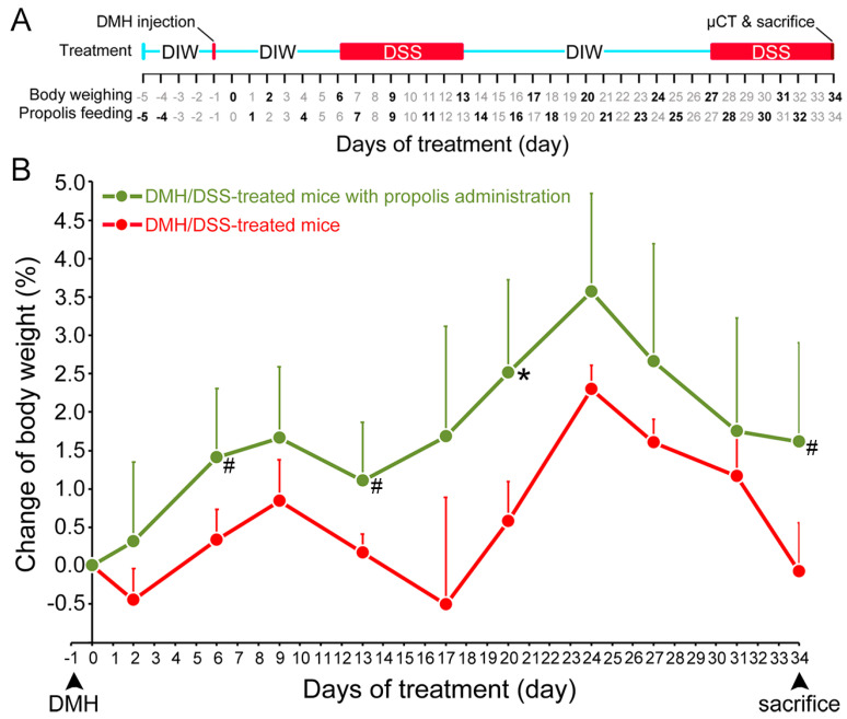
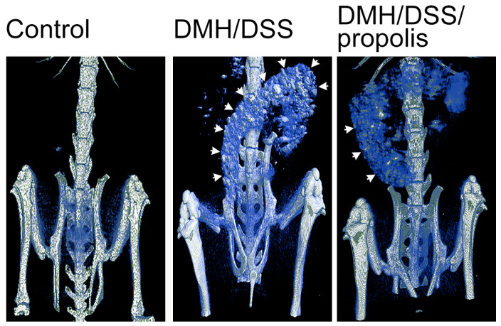
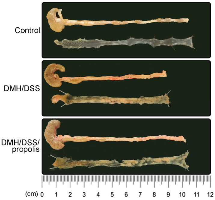
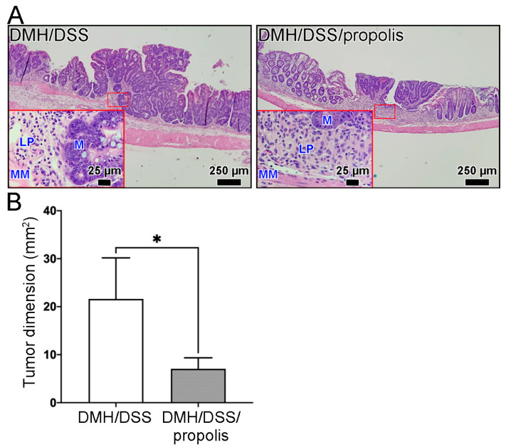
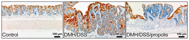
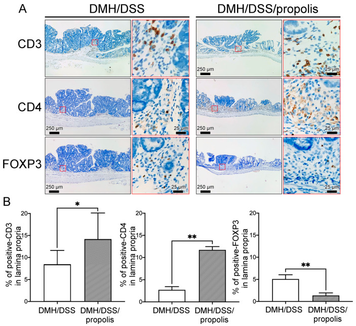
Colorectal cancer (CRC) is one of the most common cancers and is the second leading cause of cancer-related death in the world. Due to the westernization of diets, young patients with CRC are often diagnosed at advanced stages with an associated poor prognosis. Improved lifestyle choices are one way to minimize CRC risk. Among diet choices is the inclusion of bee propolis, long recognized as a health supplement with anticancer activities. Understanding the effect of propolis on the gut environment is worth exploring, and especially its associated intratumoral immune changes and its anticancer effect on the occurrence and development of CRC. In this study, early stage CRC was induced with 1,2-dimethylhydrazine (DMH) and dextran sulfate sodium (DSS) for one month in an animal model, without and with propolis administration. The phenotypes of early stage CRC were evaluated by X-ray microcomputed tomography and histologic examination. The gut immunity of the tumor microenvironment was assessed by immunohistochemical staining for tumor-infiltrating lymphocytes (TILs) and further comparative quantification. We found that the characteristics of the CRC mice, including the body weight, tumor loading, and tumor dimensions, were significantly changed due to propolis administration. With further propolis administration, the CRC tissues of DMH/DSS-treated mice showed decreased cytokeratin 20 levels, a marker for intestinal epithelium differentiation. Additionally, the signal intensity and density of CD3+ and CD4+ TILs were significantly increased and fewer forkhead box protein P3 (FOXP3) lymphocytes were observed in the lamina propria. In conclusion, we found that propolis, a natural supplement, potentially prevented CRC progression by increasing CD3+ and CD4+ TILs and reducing FOXP3 lymphocytes in the tumor microenvironment of early stage CRC. Our study could suggest a promising role for propolis in complementary medicine as a food supplement to decrease or prevent CRC progression.
Keywords: CD4 protein; cytokeratin 20; early stage colorectal cancer; forkhead box protein P3; gut immunity; propolis; tumor microenvironment.
Conflict of interest statement
The authors declare that they have no competing interest.
Figures
Similar articles
-
Increased number of forkhead box P3+ tumor-infiltrating lymphocytes correlates with high preoperative albumin level and better survival in patients with stage II or III colorectal cancer.Tumour Biol. 2015 Jul;36(7):5407-14. doi: 10.1007/s13277-015-3206-8. Epub 2015 Feb 20. Tumour Biol. 2015. PMID: 25697896
-
Tumor-infiltrating lymphocyte subsets and tertiary lymphoid structures in pulmonary metastases from colorectal cancer.Clin Exp Metastasis. 2016 Oct;33(7):727-39. doi: 10.1007/s10585-016-9813-y. Epub 2016 Jul 23. Clin Exp Metastasis. 2016. PMID: 27449756 Free PMC article.
-
A comparison of the local immune status between the primary and metastatic tumor in colorectal cancer: a retrospective study.BMC Cancer. 2018 Apr 3;18(1):371. doi: 10.1186/s12885-018-4276-y. BMC Cancer. 2018. PMID: 29614981 Free PMC article.
-
FOXP3+ Tregs: heterogeneous phenotypes and conflicting impacts on survival outcomes in patients with colorectal cancer.Immunol Res. 2015 Mar;61(3):338-47. doi: 10.1007/s12026-014-8616-y. Immunol Res. 2015. PMID: 25608795 Review.
-
Colorectal Cancer-Infiltrating Regulatory T Cells: Functional Heterogeneity, Metabolic Adaptation, and Therapeutic Targeting.Front Immunol. 2022 Jul 8;13:903564. doi: 10.3389/fimmu.2022.903564. eCollection 2022. Front Immunol. 2022. PMID: 35874729 Free PMC article. Review.
Cited by
-
Antitumor Effects and the Potential Mechanism of 10-HDA against SU-DHL-2 Cells.Pharmaceuticals (Basel). 2024 Aug 20;17(8):1088. doi: 10.3390/ph17081088. Pharmaceuticals (Basel). 2024. PMID: 39204193 Free PMC article.
-
Exploring the Prospective Role of Propolis in Modifying Aging Hallmarks.Cells. 2024 Feb 24;13(5):390. doi: 10.3390/cells13050390. Cells. 2024. PMID: 38474354 Free PMC article. Review.
References
-
- Baidoun F., Elshiwy K., Elkeraie Y., Merjaneh Z., Khoudari G., Sarmini M.T., Gad M., Al-Husseini M., Saad A. Colorectal Cancer Epidemiology: Recent Trends and Impact on Outcomes. Curr. Drug Targets. 2021;22:998–1009. - PubMed
-
- Hofseth L.J., Hebert J.R., Chanda A., Chen H., Love B.L., Pena M.M., Murphy E.A., Sajish M., Sheth A., Buckhaults P.J., et al. Early-onset colorectal cancer: Initial clues and current views. Nat. Rev. Gastroenterol. Hepatol. 2020;17:352–364. doi: 10.1038/s41575-019-0253-4. Correction in Nat. Rev. Gastroenterol. Hepatol. 2020, 17, 517. - DOI - PMC - PubMed
MeSH terms
Substances
Grants and funding
LinkOut - more resources
Full Text Sources
Medical
Research Materials
