. 2023 Oct 27;15(21):4574.
doi: 10.3390/nu15214574.

Intermittent Fasting Attenuates Metabolic-Dysfunction-Associated Steatohepatitis by Enhancing the Hepatic Autophagy-Lysosome Pathway

Affiliations

Intermittent Fasting Attenuates Metabolic-Dysfunction-Associated Steatohepatitis by Enhancing the Hepatic Autophagy-Lysosome Pathway

Kyung Eun Kim et al. Nutrients. .

Abstract

An intermittent fasting (IF) regimen has been shown to protect against metabolic dysfunction-associated steatohepatitis (MASH). However, the precise mechanism remains unclear. Here, we explored how IF reduced hepatic lipid accumulation, inflammation, and fibrosis in mice with MASH. The mice were fed a high-fat diet (HFD) for 30 weeks and either continued on the HFD or were subjected to IF for the final 22 weeks. IF reduced body weight, insulin resistance, and hepatic lipid accumulation in HFD-fed mice. Lipidome analysis revealed that IF modified HFD-induced hepatic lipid composition. In particular, HFD-induced impaired autophagic flux was reversed by IF. The decreased hepatic lysosome-associated membrane protein 1 level in HFD-fed mice was upregulated in HFD+IF-fed mice. However, increased hepatic lysosomal acid lipase protein levels in HFD-fed mice were reduced by IF. IF attenuated HFD-induced hepatic inflammation and galectin-3-positive Kupffer cells. In addition to the increases in hepatic hydroxyproline and lumican levels, lipocalin-2-mediated signaling was reversed in HFD-fed mice by IF. Taken together, our findings indicate that the enhancement of the autophagy-lysosomal pathway may be a critical mechanism of MASH reduction by IF.

Keywords: autophagy; intermittent fasting; lysosome; non-alcoholic steatohepatitis.


Conflict of interest statement

The authors declare no conflict of interest.

Figures

Figure 1
Figure 1
IF attenuates MASLD activity in HFD-fed mice. Male C57BL/6 mice were fed an ND or an HFD for 30 weeks, whereas mice in the NIF or HIF group were fed an ND or HFD for 8 weeks and then were switched to an IF protocol consisting of alternating 24 h periods of fasting and feeding for 22 weeks. (a) Liver weight, (b) Liver/Body weight ratio, (c) serum ALT and AST, and (d) serum total cholesterol levels are shown. (e) Representative images of livers and H&E staining of liver sections. Scale bars, 1 cm (upper), 100 µm (lower). (f) MASLD activity score. (g) Representative Nile Red staining with DAPI of liver sections. Scale bar, 50 µm. (h) Nile Red-positive area. (i) Hepatic TG levels. Significance was determined by two-way ANOVA. * p < 0.05 vs. ND. † p < 0.05 vs. HFD.
Figure 2
Figure 2
IF reduces hepatic lipid uptake and fatty acid oxidation in HFD-fed mice. (a) Western blot analysis and quantification of GS and perilipin-2 protein in liver lysates. (b) Representative perilipin-2 and GS staining of liver sections. The highly magnified images in the white boxes within the merged panels are shown in the bottom panels. Scale bars, 50 µm. (c,d) Western blot analysis and quantification of FAS, SCD1 (c), PPAR-α, and PPAR-γ (d) protein in liver lysates. β-Actin was used as a loading control. Significance was determined by two-way ANOVA. * p < 0.05 vs. ND. † p < 0.05 vs. HFD.
Figure 3
Figure 3
Lipidomic profiling of liver tissues from ND, NIF, HFD, and HIF mice. (a) PCA score plot of lipids from liver tissues (R2X: 71.2%, Q2: 47.3%). (b) Heatmaps of lipids identified in liver tissues. The heatmaps are color coded based on the z-scores of the measured relative intensities of each sample. (c) Log10-fold changes in FFAs in the liver tissues of ND- and HFD-fed mice. (d) Log10-fold changes in FFAs in the liver tissues of HFD- and HIF-fed mice. (e) Log10-fold changes in phospholipids in the liver tissues of ND- and HFD-fed mice. (f) Log10-fold changes in phospholipids in the liver tissues of HFD- and HIF-fed mice.
Figure 4
Figure 4
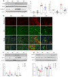
IF improves hepatic autophagy flux and enhances lysosomes in HFD-fed mice. (a) Western blot analysis and quantification of LC3B and p62 protein in liver lysates. (b) Electron micrographs of hepatocytes. Arrows indicate regions of contact between the lysosomal and LD compartment (Nu, nucleus; LD, lipid droplet; Ly, lysosome; CV, central vein). Scale bar, 1 µm. (c,d) Western blot analysis and quantification of LAMP1 (c) and LAL (d) protein levels in liver lysates. β-Actin was used as a loading control. Significance was determined by two-way ANOVA. * p < 0.05 vs. ND. † p < 0.05 vs. HFD.
Figure 5
Figure 5
IF reduces hepatic inflammation in HFD-fed mice. (a) Proinflammatory cytokines and anti-inflammatory cytokines. (b) Western blot analysis and quantification of galectin-3 protein in liver lysates. (c) Representative galectin-3 and F4/80 staining of liver sections. Nuclei were counterstained with DAPI. Scale bar, 50 µm. (d) Western blot analysis and quantification of HO-1 protein in liver lysates. β-Actin was used as a loading control. Significance was determined by two-way ANOVA. * p < 0.05 vs. ND. † p < 0.05 vs. HFD.
Figure 6
Figure 6
IF reduces hepatic fibrosis in HFD-fed mice. (a,b) Representative images (a) and quantification (b) of Picro-Sirius Red staining in liver sections. Scale bar, 100 µm. (c) Hepatic hydroxyproline concentration. (d,e) Western blot analysis and quantification of lumican (d), LCN2, MMP9, pSTAT3, and total STAT3 (e) proteins in liver lysates. β-Actin was used as a loading control. Significance was determined using a two-way ANOVA. * p < 0.05 vs. ND. † p < 0.05 vs. HFD.

References

    1. Eslam M., Newsome P.N., Sarin S.K., Anstee Q.M., Targher G., Romero-Gomez M., Zelber-Sagi S., Wong V.W.-S., Dufour J.F., Schattenberg J.M., et al. A new definition for metabolic dysfunction-associated fatty liver disease: An international expert consensus statement. J. Hepatol. 2020;73:202–209. doi: 10.1016/j.jhep.2020.03.039. - DOI - PubMed
    1. Mashek D.G. Hepatic lipid droplets: A balancing act between energy storage and metabolic dysfunction in NAFLD. Mol. Metab. 2021;50:101115. doi: 10.1016/j.molmet.2020.101115. - DOI - PMC - PubMed
    1. Brunt E.M., Tiniakos D.G. Histopathology of nonalcoholic fatty liver disease. World J. Gastroenterol. 2010;16:5286–5296. doi: 10.3748/wjg.v16.i42.5286. - DOI - PMC - PubMed
    1. Pierantonelli I., Svegliati-Baroni G. Nonalcoholic Fatty Liver Disease: Basic Pathogenetic Mechanisms in the Progression From NAFLD to NASH. Transplantation. 2019;103:e1–e13. doi: 10.1097/TP.0000000000002480. - DOI - PubMed
    1. Grefhorst A., van de Peppel I.P., Larsen L.E., Jonker J.W., Holleboom A.G. The Role of Lipophagy in the Development and Treatment of Non-Alcoholic Fatty Liver Disease. Front. Endocrinol. 2020;11:601627. doi: 10.3389/fendo.2020.601627. - DOI - PMC - PubMed