[Preprint]. 2023 Nov 3:2023.11.01.565151.
doi: 10.1101/2023.11.01.565151.

Ciliary intrinsic mechanisms regulate dynamic ciliary extracellular vesicle release from sensory neurons

Affiliations

Ciliary intrinsic mechanisms regulate dynamic ciliary extracellular vesicle release from sensory neurons

Juan Wang et al. bioRxiv. .

Update in

Abstract

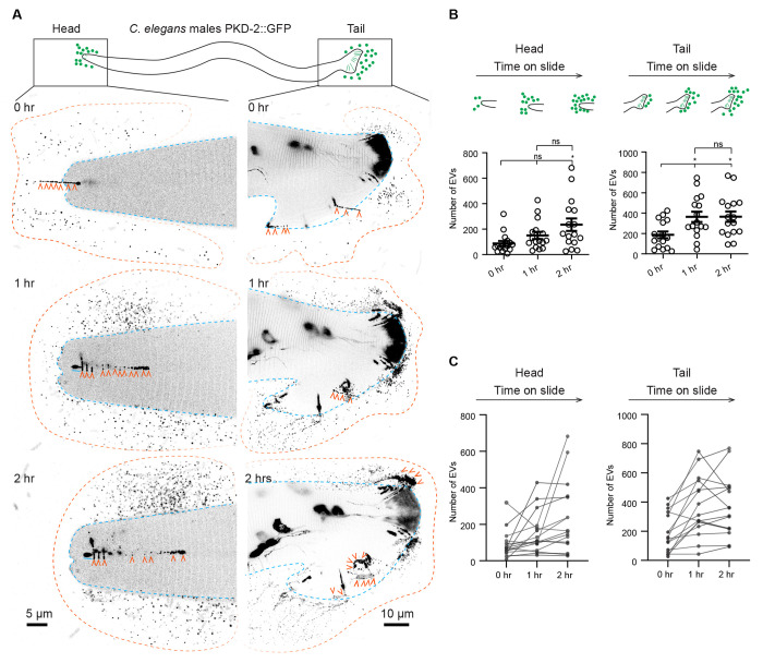
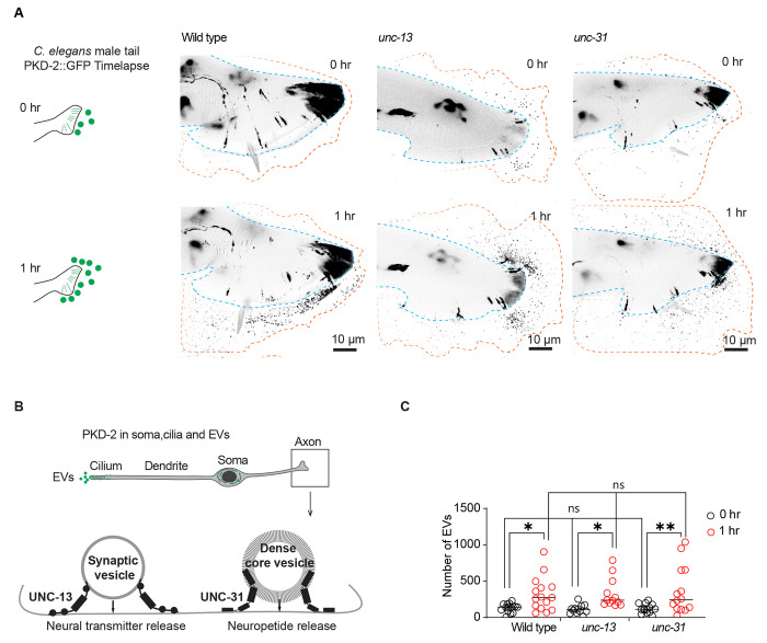
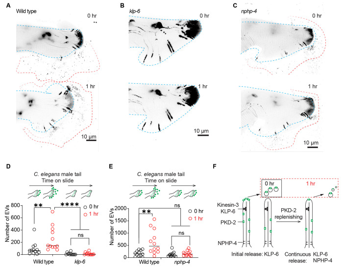
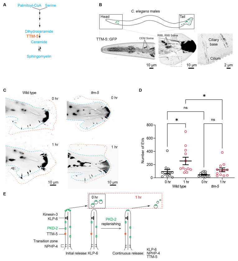
Cilia-derived extracellular vesicles (EVs) contain signaling proteins and act in intercellular communication. Polycystin-2 (PKD-2), a transient receptor potential channel, is a conserved ciliary EVs cargo. Caenorhabditis elegans serves as a model for studying ciliary EV biogenesis and function. C. elegans males release EVs in a mechanically-induced manner and deposit PKD-2-labeled EVs onto the hermaphrodite vulva during mating, suggesting an active release process. Here, we study the dynamics of ciliary EV release using time-lapse imaging and find that cilia can sustain the release of PKD-2-labeled EVs for a two-hour duration. Intriguingly, this extended release doesn't require neuronal synaptic transmission. Instead, ciliary intrinsic mechanisms regulate PKD-2 ciliary membrane replenishment and dynamic EV release. The ciliary kinesin-3 motor KLP-6 is necessary for both initial and extended ciliary EV release, while the transition zone protein NPHP-4 is required only for sustained EV release. The dihydroceramide desaturase DEGS1/2 ortholog TTM-5 is highly expressed in the EV-releasing sensory neurons, localizes to cilia, and is required for sustained but not initial ciliary EV release, implicating ceramide in ciliary ectocytosis. The study offers a comprehensive portrait of real-time ciliary EV release, and mechanisms supporting cilia as proficient EV release platforms.

Keywords: C. elegans; DEGS1/2; KLP-6; NPHP-4; PKD-2; ceramide; cilia; extracellular vesicles; kinesin-3; polycystin.


Conflict of interest statement

Declaration of interests. The authors declare no competing interests.

Figures

Figure 1.
Figure 1.. Extended release of PKD-2::GFP-labeled extracellular vesicles (EVs) by C. elegans male sensory neurons.
(A) Representative images capturing the head and tail regions of C. elegans males at 0, 1, and 2 hours following mounting on a glass slide. Blue lines indicate the outline of the male head or tail. Orange lines indicate the outline of the EV clouds released by the head and the tail. Orange arrowheads point to EVs that are released in a string-like pattern. (B) Quantification of EV counts from the head and tail regions at 0, 1, and 2 hours. The scatter plot with lines indicates the mean ± SEM. Each data point represents the total EV count released by an individual C. elegans male, either from its head (left) or tail (right). n = 16 and the data were scored over 4 days. Statistical analysis was performed using one-way ANOVA with Bonferroni correction. ns denotes not significant (p ≥ 0.05), and * denotes p < 0.05. (C) Individual trajectories depicting the EV release pattern from the head and tail regions of C. elegans males. Each trajectory represents EV counts from a single animal. The EV count at new time points includes newly released EVs in addition to previously released EVs that remain visible post-photobleaching. Photobleaching explains the decline in EV counts among certain animals at the 2-hour mark, where newly released EVs are fewer than the photobleached older EVs (photobleached twice). See also Figure S1, Video S1 - S2.
Figure 2.
Figure 2.. Neuronal transmission-independent release of PKD-2::GFP-labeled ciliary EVs.
(A) Representative images showing EV release from the tail at the initial imaging (0 hr) and the second imaging (1 hr) while the animals were mounted on slides, in wild-type, unc-13, and unc-31 males. (B) Schematic diagram depicts the functional roles of UNC-13 and UNC-31 in synaptic vesicle- and dense core vesicle-mediated neuronal transmission in the axon. (C) Quantification of EV release from the tail at the initial imaging (0 hr) and second imaging (1 hr) in wild-type, unc-13, and unc-31 males. The scatter plot with lines indicates the mean ± SEM. Each data point represents the total EV count released by an individual C. elegans male. 12-15 animals were imaged for each genotype. Statistical analysis was performed by two-way ANOVA with Bonferroni correction. ns denotes not significant, * denotes p < 0.05, and ** denotes p < 0.01.
Figure 3.
Figure 3.. Continued PKD-2 ciliary EV release relies on effective replenishment of PKD-2 at the ciliary tip.
(A-C) Representative images of PKD-2::GFP ciliary EV release in male tail of wild-type, klp-6, and nphp-4 males. (D) Quantification of EV counts from wild-type and klp-6 mutant males in the tail regions at 0 and 1 hour. The scatter plot with lines indicates the mean ± SEM. Each data point represents the total EV count released by an individual C. elegans male. (E) Quantification of EV counts from wild-type and nphp-4 mutant males in the tail regions at 0 and 1 hour. The scatter plot with lines indicates the mean ± SEM. Each data point represents the total EV count released by an individual C. elegans male. For D-E, statistical analysis was performed by two-way ANOVA with Bonferroni correction. n = 12-15 animals per genotype. ns denotes not significant, * denotes p < 0.05, and ** denotes p < 0.01., **** p<0.0001. (F) Model of PKD-2 ciliary EV dynamics. NPHP-4 regulates the release of PKD-2::GFP ciliary EVs by replenishing PKD-2::GFP in the ciliary membrane. Meanwhile, KLP-6 controls ciliary PKD-2 EV release by concentrating PKD-2 at the ciliary tip. The sustained release of PKD-2 ciliary EVs over an hour necessitates the replenishment of PKD-2 in both the ciliary membrane and tip. See also Figure S2.
Figure 4.
Figure 4.. TTM-5 facilitates the dynamic release of PKD-2 ciliary EVs.
(A) Schematic cartoon of TTM-5 action site. TTM-5 is the ortholog of DEGS1/2 (delta-4-desaturase, sphingolipid 1/2), an enzyme that converts dihydroceramides to ceramides. The unsaturated fatty acid chain creates a kink in the chain, making the cone shape of mature ceramides to support the high curvature membrane of cilia and EV. (B) A CRISPR reporter of TTM-5::GFP is expressed in male-specific EV-releasing neurons, including four cephalic male (CEM) neurons and 17 neurons in the tail (soma of R8B and R9B are shown, for the full tail images, see Figure S3C). In the tail EV-releasing neurons, TTM-5 primarily localizes to the soma, ciliary base, and cilium and is not detectable in EVs. (C) Representative images of PKD-2::GFP ciliary EV release in male tail sensory neurons at 0 and 1 hour in wild-type and ttm-5 mutant males. Blue lines indicate the outline of the male tail. Orange lines indicate the outline of the EV clouds released by the tail. (D) Quantification of EV counts at 0 and 1 hour of wild-type and ttm-5 mutant males. The scatter plot with lines indicates the mean ± SEM. Each data point represents the total EV count released by an individual C. elegans male. n = 12. Statistical analysis was performed using one-way ANOVA with Bonferroni correction. ns denotes not significant (p ≥ 0.05), and * denotes p < 0.05. See also Figure S3–4.

References

    1. Buzas E.I. (2023). The roles of extracellular vesicles in the immune system. Nat Rev Immunol 23, 236–250. 10.1038/s41577-022-00763-8. - DOI - PMC - PubMed
    1. Dixson A.C., Dawson T.R., Di Vizio D., and Weaver A.M. (2023). Context-specific regulation of extracellular vesicle biogenesis and cargo selection. Nat Rev Mol Cell Biol 24, 454–476. 10.1038/s41580-023-00576-0. - DOI - PMC - PubMed
    1. Ojeda Naharros I., and Nachury M.V. (2022). Shedding of ciliary vesicles at a glance. J Cell Sci 135. 10.1242/jcs.246553. - DOI - PMC - PubMed
    1. Wang J., and Barr M.M. (2018). Cell-cell communication via ciliary extracellular vesicles: clues from model systems. Essays Biochem 62, 205–213. 10.1042/EBC20170085. - DOI - PMC - PubMed
    1. Wood C.R., Huang K., Diener D.R., and Rosenbaum J.L. (2013). The cilium secretes bioactive ectosomes. Curr Biol 23, 906–911. S0960-9822(13)00427-2 [pii] 10.1016/j.cub.2013.04.019. - DOI - PMC - PubMed

Publication types