[Preprint]. 2023 Oct 27:2023.10.24.563743.
doi: 10.1101/2023.10.24.563743.

Multiple steps of prion strain adaptation to a new host

Affiliations

Multiple steps of prion strain adaptation to a new host

Olga Bocharova et al. bioRxiv. .

Update in

Abstract

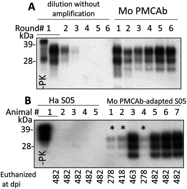
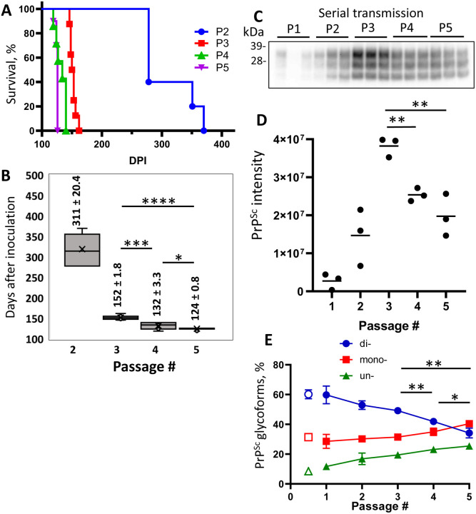
The transmission of prions across species is a critical aspect of their dissemination among mammalian hosts, including humans. This process often necessitates strain adaptation. In this study, we sought to investigate the mechanisms underlying prion adaptation while mitigating biases associated with the history of cross-species transmission of natural prion strains. To achieve this, we utilized the synthetic hamster prion strain S05. Propagation of S05 using mouse PrPC in Protein Misfolding Cyclic Amplification did not immediately overcome the species barrier. This finding underscores the involvement of factors beyond disparities in primary protein structures. Subsequently, we performed five serial passages to stabilize the incubation time to disease in mice. The levels of PrPSc increased with each passage, reaching a maximum at the third passage, and declining thereafter. This suggests that only the initial stage of adaptation is primarily driven by an acceleration in PrPSc replication. During the protracted adaptation to a new host, we observed significant alterations in the glycoform ratio and sialylation status of PrPSc N-glycans. These changes support the notion that qualitative modifications in PrPSc contribute to a more rapid disease progression. Furthermore, consistent with the decline in sialylation, a cue for "eat me" signaling, the newly adapted strain exhibited preferential colocalization with microglia. In contrast to PrPSc dynamics, the intensity of microglia activation continued to increase after the third passage in the new host. In summary, our study elucidates that the adaptation of a prion strain to a new host is a multi-step process driven by several factors.

Keywords: N-linked glycans; cross-species barrier; neurodegenerative diseases; neuroinflammation; prion; prion adaptation; prion diseases; prion strains; reactive astrocytes; reactive microglia; sialylation.


Conflict of interest statement

Conflict of interest. The authors declare that they have no conflicts of interest with the contents of this article.

Figures

Figure 1.
Figure 1.. Adaptation of hamster S05 strain to mouse PrPC.
(A) For adapting the hamster S05 strain to mouse PrPC, sPMCAb was seeded with 103-fold diluted S05 BHs and carried out using mouse normal BH as a substrate. Corresponding serial dilutions of S05 BHs in the absence of amplification are shown as references. Immunodetection with SAF-84. (B) Western blot analysis of PrPSc in mice inoculated IC with 10% of S05 BH or mouse sPMCAb-adapted S05. Products of the tenth sPMCAb round were used for inoculation. Animals # 1, 2 and 4, marked by asterisks, were euthanized for testing the presence of PrPSc at the early stages. Immunodetection with ab3531. In A and B, all samples, with the exception of lanes indicated as -PK were treated with PK.
Figure 2.
Figure 2.. Adaptation of S05 to mice.
Mice were inoculated IC with Mo sPMCAb-adapted S05 (P1) and then serially passaged four times (P2–P5). (A) Kaplan-Meier survival plot for the serial passages (P2 to P5) of Mo sPMCAb-adapted S05 in mice. (B) Box-and-whisker plot of incubation time to terminal disease in mice as a function of serial passage number. The midline of the boxand-whisker plot denotes the median, the x represents the mean, and the ends of the box plot denote the 25th and 75th percentiles (n=5 in 2nd, n=8 in 3d, n=7 in 4th and n=10 in 5th passages). Data presented as means ± SD, *P≤0.05, ***P≤0.001, ****P≤0.0001, two-tailed unpaired t-test. (C) Western blot analysis of PrPSc in five passages of Mo sPMCAb-adapted S05. All samples are 10% BHs, treated with PK and detected using D18 antibody. (D) Quantification of PrPSc amounts in five serial passages of Mo sPMCAb-adapted S05 in mice using densitometry of Western blots. (E) Change in the percentage of di-, mono-, and unglycosylated PrPSc as a function of serial passage (filled symbols), and the percentage of PrPSc glycoforms in the original hamster S05 strain (empty symbols). Mean ± SD (n=3). In D and E, *P≤0.05, **P≤0.01, ***P≤0.001 by two-tailed unpaired t-test.
Figure 3.
Figure 3.. Analysis of PrPSc glycosylation and sialylation ratios.
(A) Western blot analysis of PrPSc in Mo S05 (5th passage), Mo SSLOW and 22L animals. All samples are 10% BHs, treated with PK and detected using D18 antibody (B) The percentage of di-, mono-, and unglycosylated PrPSc. Mean±SEM (n=3 for Mo SSLOW and 22L, and n=5 for Mo S05). (C) An example of a 2D Western blot of Mo S05 PrPSc showing the division of sialoglycoforms into hypo- and hypersialylated isoforms. Western blot was stained with D18 antibody. Arrows indicate the position of di-, mono- and unglycosylated sialoglycoforms. (D) Change in relative populations of hypo- and hypersialylated isoforms in 2nd to 5th passages of Mo S05 and relative population of isoforms in the original S05 strain in hamsters (Ha). Mean ± SD (n=3).
Figure 4.
Figure 4.. Co-localization of PrPSc and microglia, astrocytes, and neurons.
(A) Representative images of co-immunostaining for PrPSc (SAF-84 antibody, red) with astrocytes (anti-GFAP, green), microglia (anti-Iba1, green), or neurons (anti-MAP2, green) in somatosensory (SS) cortex and thalamus in mice from the 5th passage of Mo S05 or age-matched control mice. Arrows point to small PrPSc deposits colocalized with microglia. Scale bars = 50 μm. (B) The percentage of PrPSc signal overlapping with Iba1+ microglia, GFAP+ astrocytes, and MAP2+ neurons in animals from the 5th passage of S05 and age-matched control mice (designated as C). Mean ± SEM (n= 7 fields of view from 3 animals per experimental group; n= 3 fields of view from 3 animals per non-infected control group).
Figure 5.
Figure 5.. Analysis of neuroinflammation during the course of S05 adaptation to mice.
(A) Representative images of somatosensory (SS) cortex and thalamus (TH) in mice from five serial passages of S05, and non-infected control mice (designated as C) co-immunostained with anti-GFAP (green) and anti-Iba1 (red) antibodies. The age of control mice matched the age of mice from the 4th and 5th passages. Scale bar = 50 μm. (B) Quantification of areas covered by GFAP+ or Iba1+ cells in somatosensory cortex (SS, squares) and thalamus (TH, triangles) in mice from five serial passages of S05, and non-infected control mice (C, open squares and triangles). Mean ± SD (n=7–9 fields of view from 3 animals per group).

References

    1. Prusiner S. B. (1997) Prion diseases and the BSE crisis. Science 278, 245–251 - PubMed
    1. Legname G., Baskakov I. V., Nguyen H. O. B., Riesner D., Cohen F. E., DeArmond S. J., and Prusiner S. B. (2004) Synthetic mammalian prions. Science 305, 673–676 - PubMed
    1. Wang F., Wang X., Yuan C. G., and Ma J. (2010) Generating a Prion Bacterially Expressed Recombinant Prion Protein. Science 327, 1132–1135 - PMC - PubMed
    1. Deleault N. R., Harris B. T., Rees J. R., and Supattapone S. (2007) Formation of native prions from minimal components in vitro. Proc.Acad.Natl.Sci.U.S.A. 104, 9741–9746 - PMC - PubMed
    1. Collinge J., and Clarke A. R. (2007) A General Model of Prion Strains and Their Pathogenicity. Science 318, 930–936 - PubMed

Publication types