[Preprint]. 2023 Nov 1:2023.10.28.564535.
doi: 10.1101/2023.10.28.564535.

RNA isoform expression landscape of the human dorsal root ganglion (DRG) generated from long read sequencing

Affiliations

RNA isoform expression landscape of the human dorsal root ganglion (DRG) generated from long read sequencing

Asta Arendt-Tranholm et al. bioRxiv. .

Update in

Abstract

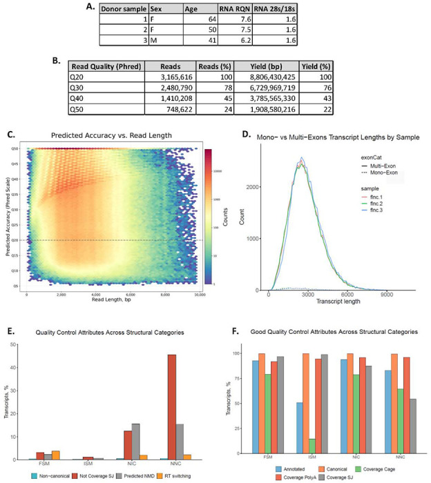
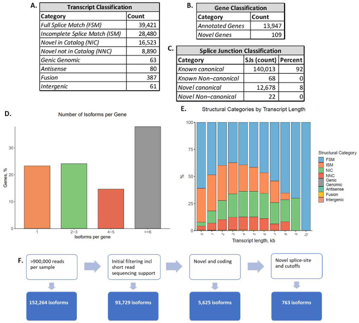
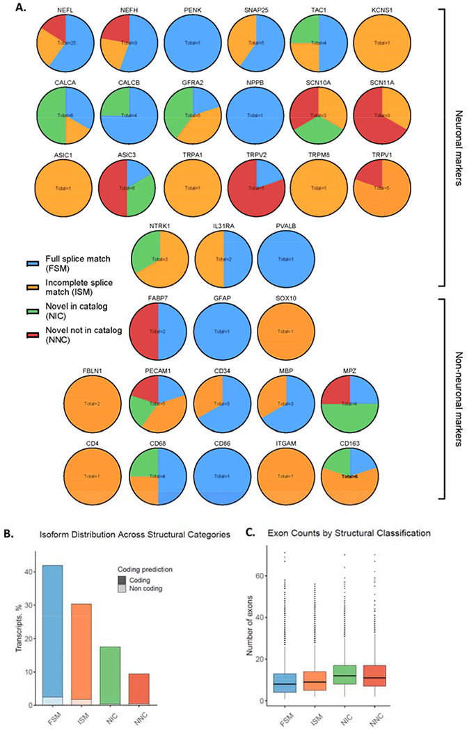
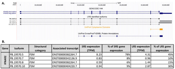
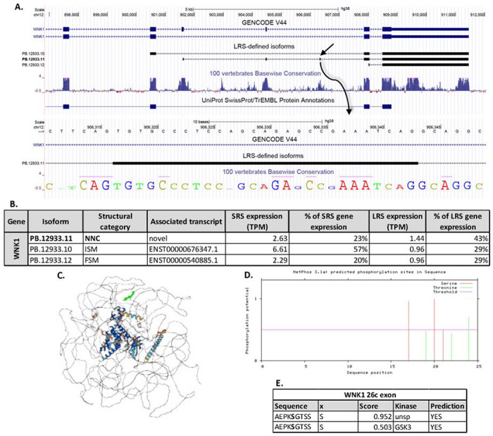
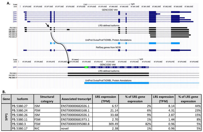
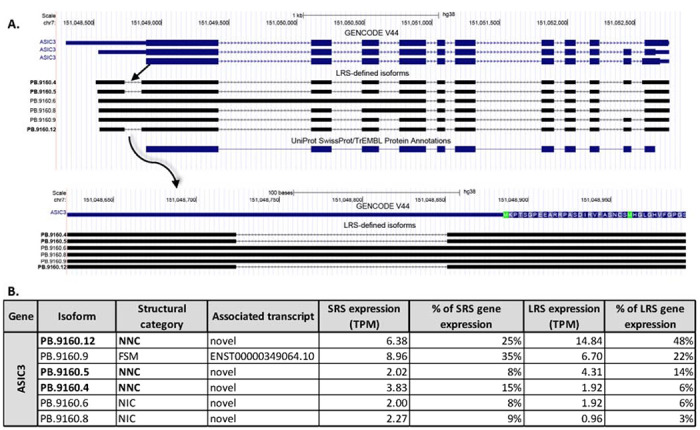
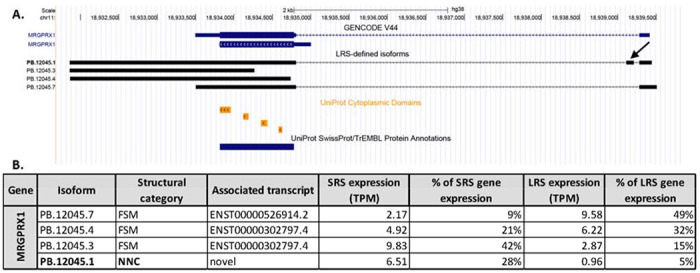
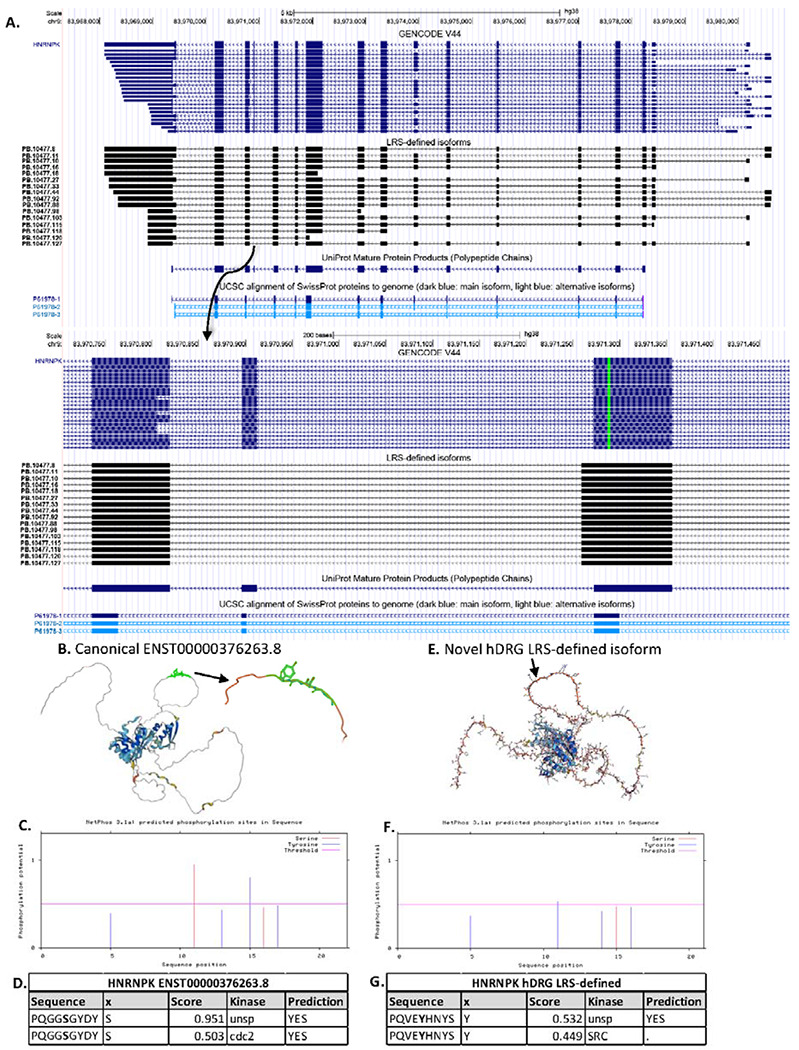
Splicing is a post-transcriptional RNA processing mechanism that enhances genomic complexity by creating multiple isoforms from the same gene. Diversity in splicing in the mammalian nervous system is associated with neuronal development, synaptic function and plasticity, and is also associated with diseases of the nervous system ranging from neurodegeneration to chronic pain. We aimed to characterize the isoforms expressed in the human peripheral nervous system, with the goal of creating a resource to identify novel isoforms of functionally relevant genes associated with somatosensation and nociception. We used long read sequencing (LRS) to document isoform expression in the human dorsal root ganglia (hDRG) from 3 organ donors. Isoforms were validated in silico by confirming expression in hDRG short read sequencing (SRS) data from 3 independent organ donors. 19,547 isoforms of protein-coding genes were detected using LRS and validated with SRS and strict expression cutoffs. We identified 763 isoforms with at least one previously undescribed splice-junction. Previously unannotated isoforms of multiple pain-associated genes, including ASIC3, MRGPRX1 and HNRNPK were identified. In the novel isoforms of ASIC3, a region comprising ~35% of the 5'UTR was excised. In contrast, a novel splice-junction was utilized in isoforms of MRGPRX1 to include an additional exon upstream of the start-codon, consequently adding a region to the 5'UTR. Novel isoforms of HNRNPK were identified which utilized previously unannotated splice-sites to both excise exon 14 and include a sequence in the 5' end of exon 13. The insertion and deletion in the coding region was predicted to excise a serine-phosphorylation site favored by cdc2, and replace it with a tyrosine-phosphorylation site potentially phosphorylated by SRC. We also independently confirm a recently reported DRG-specific splicing event in WNK1 that gives insight into how painless peripheral neuropathy occurs when this gene is mutated. Our findings give a clear overview of mRNA isoform diversity in the hDRG obtained using LRS. Using this work as a foundation, an important next step will be to use LRS on hDRG tissues recovered from people with a history of chronic pain. This should enable identification of new drug targets and a better understanding of chronic pain that may involve aberrant splicing events.

Keywords: WNK1; dorsal root ganglion; hnRNPK; long read sequencing; splicing.


Conflict of interest statement

Conflict of interest: The authors declare no conflicts of interest.

Figures

Figure 1:
Figure 1:. Descriptive characteristics and quality measures of LRS data.
A. Donor demographics (sex and age) and RNA quality (RQN and 28s/18s) of hDRGs used for LRS. B. Table of read quality distribution of 3,165,616 reads show the number of reads and the bp yield grouped by estimated accuracy of bp measured as Phred score (Q20 = 99% to Q50 = 99.999% accuracy). C. Predicted accuracy of the bp call measured as Phred score compared to the length of reads. D. The number and length of mono- and multi-exonic reads for the 3 LRS samples showing comparable expression pattern with low interindividual variability. E. Quality control measures (usage of non-canonical splice-site, splice-junctions without coverage, predicted nonsense-mediated decay (NMD) and RT switching) are generally low for the 4 primary isoform subtypes (Full Splice Match, Incomplete Splice Match, Novel in catalog and Novel not in catalog), apart from usage of novel splice-junctions in novel isoforms as expected. F. Measures of good quality (annotated and canonical coverage, as well as coverage of poly-a motif, CAGE and splice-junction) were high for all 4 primary isoform subtypes, apart from coverage cage for ISM as expected. Figures were generated with SQANTI 3 (Pardo-Palacios, Arzalluz-Luque et al. 2023).
Figure 2:
Figure 2:. Analysis of LRS data.
A. The number of transcripts identified with LRS and validated with SRS for each transcript category. B. The number of genes identified for each gene classification validated with SRS. C. The count and percent of splice-junctions categorized as known canonical, known non-canonical, novel canonical or novel non-canonical. D. The number of isoforms per gene expressed as percentages. E. The proportion of isoform classifications detected for each transcript length. F. Pathway of filtering from in silico validation using SRS to categorization by predicted coding potential, as well as novel splice-sites and expression cutoffs for both LRS and SRS ultimately identifies 763 novel coding isoforms. Figures 2D and 2E were generated with SQANTI 3 (Pardo-Palacios, Arzalluz-Luque et al. 2023).
Figure 3:
Figure 3:. Structural variants of isoforms detected for neuronal and non-neuronal markers in hDRG.
A. Isoforms of 21 neuronal markers and 13 non-neuronal markers detected with LRS and validated with SRS. B. The percent of coding and non-coding isoforms for the 4 primary structural categories. C. The average number of exons for the 4 primary structural categories. Figures 3B and 3C were generated with SQANTI 3 (Pardo-Palacios, Arzalluz-Luque et al. 2023).
Figure 4:
Figure 4:. Isoforms of IFNAR2 detected with long read sequencing.
A. The isoforms of IFNAR2 previously annotated in the GENCODE V44 library and the LRS-identified isoforms are shown. The italicized isoforms are identified but fall below the strict cutoffs imposed for detection. The UniProt protein and cytoplasmic domain annotation are shown. B. A table of the expression level (measured as TPM) for each isoform detected with LRS showing the percentage of total gene expression measured with SRS and LRS for each isoform.
Figure 5:
Figure 5:. Isoforms of WNK1 detected with long read sequencing.
A. The isoforms of WNK1 previously annotated in the GENCODE V44 library and the 3 LRS-identified isoforms of WNK1. The italicized isoforms are identified but fall below the strict cutoffs imposed for detection. The UniProt protein and cytoplasmic domain annotation are shown. A detailed look at the previously unannotated exon 26c is provided. B. A table of the expression level (measured as TPM) for each isoform detected with LRS showing the percentage of total gene expression measured with SRS and LRS for each isoform. C. Protein structure for the novel isoform, PB. 12933.11, of WNK1 predicted with AlphaFold v2.3.2. The previously unannotated region, exon 26c, is highlighted in bold green. D. Predicted phosphorylation sites for serine and threonine are annotated in the novel exon 26c with NetPhos3.1. A cutoff of 0.5 for phosphorylation potential is imposed. E. A table of the predicted serine-phosphorylation sites in the WNK1 26c exon sequence shows a predicted GSK3 phosphorylation site.
Figure 6:
Figure 6:. Isoforms of SPP1 detected with long read sequencing.
A. The isoforms of SPP1 previously annotated in the GENCODE V44 library and the 6 LRS-identified isoforms. The isoform ID in bold indicates a novel isoform. The RefSeq sequence and the UniProt protein annotation are shown. A detailed look at the excised exon is shown, illustrating the localization of the start-codon, corresponding to the UniProt protein annotation. B. A table of the expression level (measured as TPM) for each isoform detected with LRS showing the percentage of total gene expression measured with SRS and LRS for each isoform.
Figure 7:
Figure 7:. Isoforms of ASIC3 detected with long read sequencing.
A. The isoforms of ASIC3 previously annotated in the GENCODE V44 library and the 6 LRS-identified ASIC3 isoforms. The isoform ID in bold indicates a novel isoform. The UniProt protein annotation is additionally shown. A detailed look at the excised region in the 5’ UTR illustrates the localization of the start-codon. B. A table of the expression level (measured as TPM) for each isoform detected with LRS showing the percentage of total gene expression measured with SRS and LRS for each isoform.
Figure 8:
Figure 8:. Isoforms of MRGPRX1 detected with long read sequencing.
A. The isoforms of MRGPRX1 previously annotated in the GENCODE V44 library and the 4 LRS-identified MRGPRX1 isoforms. The isoform ID in bold indicates a novel isoform. An arrow shows the novel exon identified in isoform PB.12045.1. The UniProt cytoplasmic domains and protein annotation is additionally shown. B. A table of the expression level (measured as TPM) for each isoform detected with LRS showing the percentage of total gene expression measured with SRS and LRS for each isoform.
Figure 9:
Figure 9:. Isoforms of HNRNPK detected with long read sequencing.
A. The isoforms of HNRNPK previously annotated in the GENCODE V44 library and the 16 novel LRS-identified isoforms are shown along with the UniProt canonical protein annotation, as well as the UCSC protein isoforms. A detailed view of the deletion of exon 14 and extension of exon 13 is shown. B. The protein structure for the canonical isoform, ENST00000376263.8, of HNRNPK is illustrated with AlphaFold, highlighting the 6aa region which is excised in the previously unannotated isoform. C. Predicted phosphorylation sites for serine and tyrosine in the canonical isoform are annotated with NetPhos3.1. A cutoff of 0.5 for phosphorylation potential is imposed. D. A table of the predicted serine-phosphorylation site in the hnRNP K canonical isoform in the region of the 6aa sequence shows a cdc2-phosphorylation site. E. The protein structure for the novel hDRG HNRNPK isoform predicted with AlphaFold v2.3.2, indicating the 5aa inserted region. F. Predicted phosphorylation sites for serine and tyrosine in the inserted novel region of the hDRG isoform annotated with NetPhos3.1 with a cutoff of 0.5 for phosphorylation potential. G. The predicted tyrosine-phosphorylation site in the inserted 5aa region most likely utilized by SRC.

References

    1. Au P. Y. B., Goedhart C., Ferguson M., Breckpot J., Devriendt K., Wierenga K., Fanning E., Grange D. K., Graham G. E., Galarreta C., Jones M. C., Kini U., Stewart H., Parboosingh J. S., Kline A. D., Innes A. M., & Care for Rare Canada C. (2018). Phenotypic spectrum of Au-Kline syndrome: a report of six new cases and review of the literature. Eur J Hum Genet, 26(9), 1272–1281. 10.1038/s41431-018-0187-2 - DOI - PMC - PubMed
    1. Au P. Y. B., Innes A. M., & Kline A. D. (1993). Au-Kline Syndrome. In Adam M. P., Feldman J., Mirzaa G. M., Pagon R. A., Wallace S. E., Bean L. J. H., Gripp K. W., & Amemiya A. (Eds.), GeneReviews(®). University of Washington, Seattle - PubMed
    1. Copyright © 1993-2023, University of Washington, Seattle. GeneReviews is a registered trademark of the University of Washington, Seattle. All rights reserved.
    1. Barragan-Iglesias P., Franco-Enzastiga U., Jeevakumar V., Shiers S., Wangzhou A., Granados-Soto V., Campbell Z. T., Dussor G., & Price T. J. (2020). Type I Interferons Act Directly on Nociceptors to Produce Pain Sensitization: Implications for Viral Infection-Induced Pain. J Neurosci, 40(18), 3517–3532. 10.1523/JNEUROSCI.3055-19.2020 - DOI - PMC - PubMed
    1. Bhuiyan S. A., Xu M., Yang L., Semizoglou E., Bhatia P., Pantaleo K. I., Tochitsky I., Jain A., Erdogan B., Blair S., Cat V., Mwirigi J. M., Sankaranarayanan I., Tavares-Ferreira D., Green U., McIlvried L. A., Copits B. A., Bertels Z., Del Rosario J. S.,. … Renthal W. (2023). Harmonized cross-species cell atlases of trigeminal and dorsal root ganglia. bioRxiv. 10.1101/2023.07.04.547740 - DOI - PMC - PubMed

Publication types