[Preprint]. 2023 Nov 2:2023.10.31.564805.
doi: 10.1101/2023.10.31.564805.

Decreased fucosylation impacts epithelial integrity and increases risk for COPD

Affiliations

Decreased fucosylation impacts epithelial integrity and increases risk for COPD

Carter Swaby et al. bioRxiv. .

Abstract

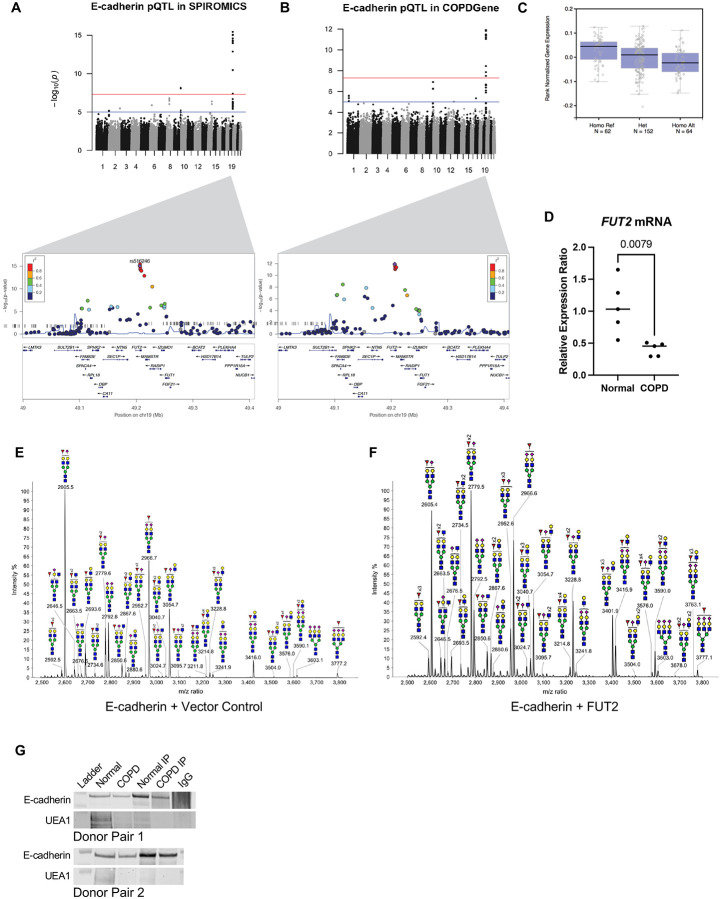
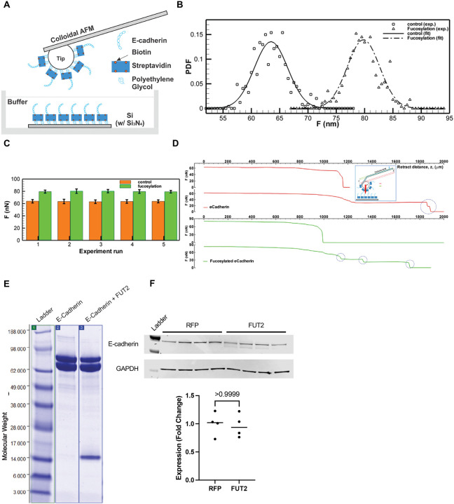
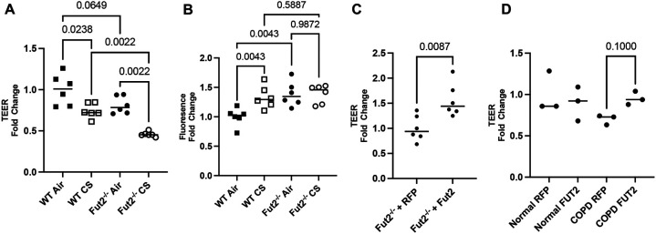
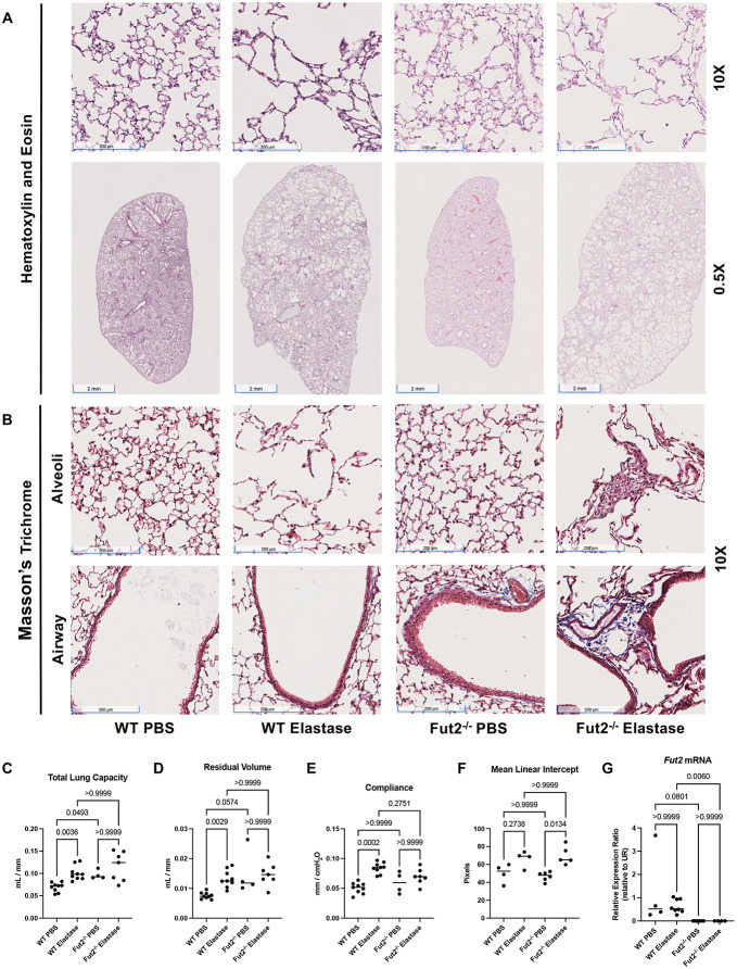
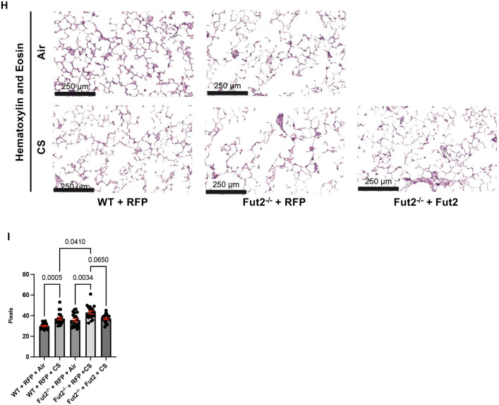
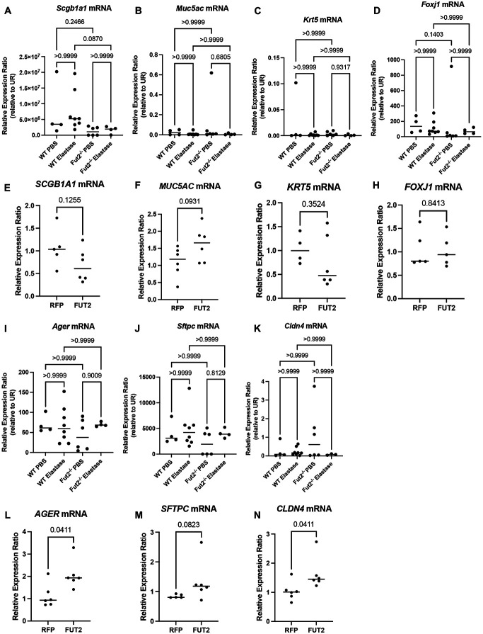
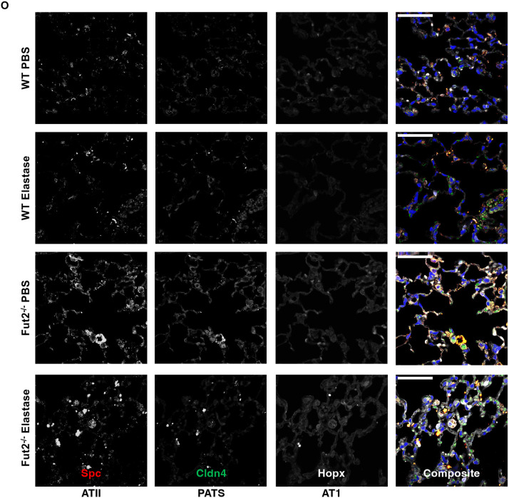
COPD causes significant morbidity and mortality worldwide. Epithelial damage is fundamental to disease pathogenesis, although the mechanisms driving disease remain undefined. Published evidence from a COPD cohort (SPIROMICS) and confirmed in a second cohort (COPDgene) demonstrate a polymorphism in Fucosyltransferese-2 (FUT2) is a trans-pQTL for E-cadherin, which is critical in COPD pathogenesis. We found by MALDI-TOF analysis that FUT2 increased terminal fucosylation of E-cadherin. Using atomic force microscopy, we found that FUT2-dependent fucosylation enhanced E-cadherin-E-cadherin bond strength, mediating the improvement in monolayer integrity. Tracheal epithelial cells from Fut2-/- mice have reduced epithelial integrity, which is recovered with reconstitution of Fut2. Overexpression of FUT2 in COPD derived epithelia rescues barrier function. Fut2-/- mice show increased susceptibility in an elastase model of disease developing both emphysema and fibrosis. We propose this is due to the role of FUT2 in proliferation and cell differentiation. Overexpression of FUT2 significantly increased proliferation. Loss of Fut2 results in accumulation of Spc+ cells suggesting a failure of alveolar type 2 cells to undergo transdifferentiation to alveolar type 1. Using a combination of population data, genetically manipulated mouse models, and patient-derived cells, we present a novel mechanism by which post-translational modifications modulate tissue pathology and serve as a proof of concept for the development of a disease-modifying target in COPD.


Figures

Figure 1:
Figure 1:
Fucosyltransferase-2 fucosylated E-cadherin. A. Protein quantitative trait loci (pQTL) analysis of E-cadherin variants in the SPIROMICS database. B. Protein quantitative trait loci (pQTL) analysis of E-cadherin variants in the COPDGene database. C. Measured expression quantitative trait loci (eQTL) transcript levels of FUT-2 by rs516246 genotype in lung tissue from Genotype-Tissue Expression (GTEx). D. FUT2 mRNA transcript is significantly decreased in COPD derived bronchial epithelia (p = 0.0079). E. Partial MALDI-TOF mass spectra (m/z 2500-3800) of E-cadherin N-glycans. F. Partial MALDI-TOF mass spectra (m/z 2500-3800) of E-cadherin co-expressed with FUT2 N-glycans, demonstrating an increase in fucosylation. G. An immunoprecipitation of E-cadherin indicates less UEA-1+ E-cadherin in bronchial epithelial cells derived from COPD patients.
Figure 2:
Figure 2:
FUT2-fucosylated E-cadherin has higher bond strength than non-FUT2-fucosylated E-cadherin. A. Schematic of the experimental setup for AFM based nano-indentation measurements of e-cadherin with or without fucosylation. B. Probability Density Function (PDF) of maximum adhesion forces between e-cadherin filaments. Square: non-fucosylated e-cadherin. Delta: fucosylated e-cadherin. Lines: normal distribution fits for non-fucosylated (solid: F¯=63.47nN, σ = 2.8585 nN) and fucosylated e-cadherin (dashed: F¯=79.801nN, σ = 2.9432 nN) C. Bar graph showing mean maximum adhesion force (nN) of non-fucosylated (dark) and fucosylated (light) e-cadherin. Error bar: one standard deviation. Total five pairs of experimental runs are presented. (Each run contains ~100 measurements, Supplementary Table 1) D. Sample adhesion curve showing the maximum binding force and break-offs (cliffs, blue circle). Single (Row 1) and two (Row 2) break-offs of the bonded non-fucosylated e-cadherin filaments, while Row 3 & 4 show maximum & partial break-offs of the bonded fucosylated e-cadherin filaments. Inset: schematics of adhesion measurement using AFM. E. Immunoblot depicting the presence of N-terminal fragment of E-cadherin when co-expressed with FUT2. F. E-cadherin expression does not change with FUT2 overexpression.
Figure 3:
Figure 3:
FUT2 is necessary to maintain integrity of the airway epithelium through its regulation of E-cadherin. A. Fut2−/− mTEC have lower TEER than WT mTEC, which is worsened by CS-induced injury (n = 6). B. Fut2−/− mTEC have higher permeability than WT mTEC, but it is not further worsened by CS exposure (n = 6). C. Reconstitution of Fut2 in Fut2−/− mTEC recovers TEER (n = 6). D. Overexpression of FUT2 improves TEER in COPD derived epithelia (n = 3).
Figure 4:
Figure 4:
Fut2−/− mice develop fibrosis and emphysema with elastase administration. A. Hematoxylin and eosin staining of WT and Fut2−/− mice administered PBS or elastase intratracheally (n = 3). B. Masson’s Trichrome staining indicates increased fibrosis of the alveoli and airways in Fut2−/− mice (n = 3). C. Fut2−/− mice have increased total lung capacity and D. residual volume (n = 4 to 9). E. Compliance does not significantly change in Fut2−/− mice (n = 4 to 9). F. Elastase induces alveolar destruction, measured by mean linear intercept (n = 4 to 6). G. Fut2−/− mice have undetectable Fut2 transcript in the lung (n = 4-8). H. Hematoxylin and eosin staining of of WT and Fut2−/− PCLS exposed to air or CS. I. Fut2−/− PCLS show increased susceptibility to CS as measured by mean linear intercept, which is rescued by reconstitution of Fut2.
Figure 4:
Figure 4:
Fut2−/− mice develop fibrosis and emphysema with elastase administration. A. Hematoxylin and eosin staining of WT and Fut2−/− mice administered PBS or elastase intratracheally (n = 3). B. Masson’s Trichrome staining indicates increased fibrosis of the alveoli and airways in Fut2−/− mice (n = 3). C. Fut2−/− mice have increased total lung capacity and D. residual volume (n = 4 to 9). E. Compliance does not significantly change in Fut2−/− mice (n = 4 to 9). F. Elastase induces alveolar destruction, measured by mean linear intercept (n = 4 to 6). G. Fut2−/− mice have undetectable Fut2 transcript in the lung (n = 4-8). H. Hematoxylin and eosin staining of of WT and Fut2−/− PCLS exposed to air or CS. I. Fut2−/− PCLS show increased susceptibility to CS as measured by mean linear intercept, which is rescued by reconstitution of Fut2.
Figure 5:
Figure 5:
FUT2 is required for sufficient proliferation of the lung epithelium. A. Overexpression of FUT2 decreases doubling time of A549s (n = 8 to 12). B and C. Overexpression of FUT2 increases the number of KI67+ cells (n = 12); scale bar (upper left) = 250 microns. D and E. Knockdown of CDH1 decreases the number of KI67+ cells, overexpression of FUT2 in CDH1 knockdown A549s has a limited effect (n = 6); scale bar (upper left) = 125 microns.
Figure 6:
Figure 6:
FUT2 is required for regeneration of the alveolar and airway epithelia. A. Scgb1a1 transcripts are decreased in Fut2−/− mice instilled with elastase when compared to WT. B and C. Muc5ac and Krt5 transcripts are unchanged with loss of Fut2 or elastase. D. Foxj1 expression trends to be lower with loss of Fut2. E. SCGB1A1 expression slightly decreases with FUT2 overexpression. F. MUC5AC expression slightly increases with FUT2 overexpression. G and H. KRT5 and FOXJ1 expression are relatively unchanged with FUT2 overexpression. I, J, and K. There is no change in Ager, Sfptc, or Cldn4 expression with loss of Fut2 or elastase. L, M, and N. AGER, SFTPC, and CLDN4 expression increase with FUT2 overexpression. O. Fut2−/− mice have increased Spc+ cells in both PBS and elastase. Elastase induces increased Cldn4 expression (n = 2); scale bar (upper left) = 50 microns.
Figure 6:
Figure 6:
FUT2 is required for regeneration of the alveolar and airway epithelia. A. Scgb1a1 transcripts are decreased in Fut2−/− mice instilled with elastase when compared to WT. B and C. Muc5ac and Krt5 transcripts are unchanged with loss of Fut2 or elastase. D. Foxj1 expression trends to be lower with loss of Fut2. E. SCGB1A1 expression slightly decreases with FUT2 overexpression. F. MUC5AC expression slightly increases with FUT2 overexpression. G and H. KRT5 and FOXJ1 expression are relatively unchanged with FUT2 overexpression. I, J, and K. There is no change in Ager, Sfptc, or Cldn4 expression with loss of Fut2 or elastase. L, M, and N. AGER, SFTPC, and CLDN4 expression increase with FUT2 overexpression. O. Fut2−/− mice have increased Spc+ cells in both PBS and elastase. Elastase induces increased Cldn4 expression (n = 2); scale bar (upper left) = 50 microns.

References

    1. Halpin DMG, Vogelmeier CF, Agusti A. Lung Health for All: Chronic Obstructive Lung Disease and World Lung Day 2022. Am J Respir Crit Care Med 2022;206:669–671. - PMC - PubMed
    1. The Lancet. UK COPD treatment: failing to progress. Lancet 2018;391:1550. - PubMed
    1. O’Reilly S. Chronic Obstructive Pulmonary Disease. Am J Lifestyle Med 2017;11:296–302. - PMC - PubMed
    1. Adeloye D, Song P, Zhu Y, Campbell H, Sheikh A, Rudan I. Global, regional, and national prevalence of, and risk factors for, chronic obstructive pulmonary disease (COPD) in 2019: a systematic review and modelling analysis. Lancet Respir Med 2022;10:447–458. - PMC - PubMed
    1. Yuksel H, Ocalan M, Yilmaz O. E-Cadherin: An Important Functional Molecule at Respiratory Barrier Between Defence and Dysfunction. Front Physiol 2021;12:1–14. - PMC - PubMed

Publication types