[Preprint]. 2024 Jun 12:2023.10.23.563672.
doi: 10.1101/2023.10.23.563672.

Structure of the human heparan-α-glucosaminide N-acetyltransferase (HGSNAT)

Affiliations

Structure of the human heparan-α-glucosaminide N-acetyltransferase (HGSNAT)

Vikas Navratna et al. bioRxiv. .

Update in

Abstract

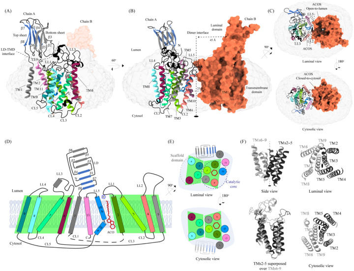
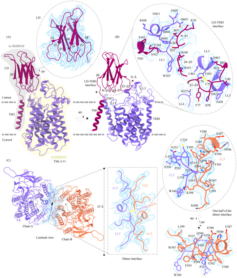
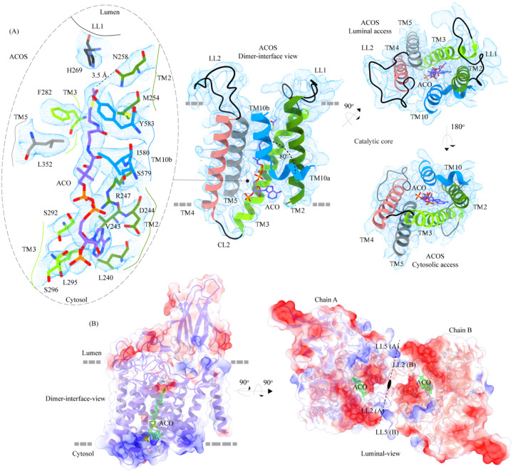
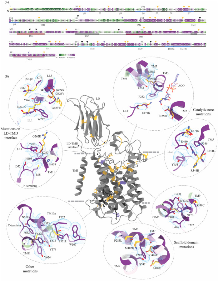
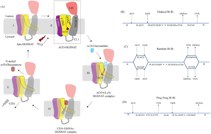
Degradation of heparan sulfate (HS), a glycosaminoglycan (GAG) comprised of repeating units of N-acetylglucosamine and glucuronic acid, begins in the cytosol and is completed in the lysosomes. Acetylation of the terminal non-reducing amino group of a-D-glucosamine of HS is essential for its complete breakdown into monosaccharides and free sulfate. Heparan-a-glucosaminide N-acetyltransferase (HGSNAT), a resident of the lysosomal membrane, catalyzes this essential acetylation reaction by accepting and transferring the acetyl group from cytosolic acetyl-CoA to terminal a-D-glucosamine of HS in the lysosomal lumen. Mutation-induced dysfunction in HGSNAT causes abnormal accumulation of HS within the lysosomes and leads to an autosomal recessive neurodegenerative lysosomal storage disorder called mucopolysaccharidosis IIIC (MPS IIIC). There are no approved drugs or treatment strategies to cure or manage the symptoms of, MPS IIIC. Here, we use cryo-electron microscopy (cryo-EM) to determine a high-resolution structure of the HGSNAT-acetyl-CoA complex, the first step in HGSNAT catalyzed acetyltransferase reaction. In addition, we map the known MPS IIIC mutations onto the structure and elucidate the molecular basis for mutation-induced HGSNAT dysfunction.


Conflict of interest statement

Conflict of interest The authors declare no conflict of interest.

Figures

Figure 1:
Figure 1:. Structure of HGSNAT
Panels (A) and (B) show two different orientations of HGSNAT dimer that highlight (dashed lines) the LD-TMD interface and dimer interface respectively. Micelle is displayed in gray. Chain A is displayed as a cartoon and chain B as orange surface. All the luminal loops (LLs), cytosolic loops (CLs), and the loops that connect β-sheets are shown in black. The top and bottom sheets in the luminal domain (LD) are colored blue and gray, respectively. The two-fold rotation axis is displayed as a dashed line with an ellipsoid. (C) Luminal (top) and cytosolic (bottom) views of the protein. The surface representation of chain B suggests that the acetyl-CoA binding site (ACOS) is more accessible from the luminal side (top) than the cytosolic side (bottom). (D) 2D topology of HGSNAT and YeiB family. The helices and strands in the topology are colored similarly to the 3D structure. TMs 2–5 and 6–9 form two bundles (4+4), highlighted by green parallelograms, that are related to each other by a 2-fold rotation parallel to the plane of the membrane. TMs 1, 10, and 11 do not seem involved in this internal symmetry, with TM10 being bent in the plane of the membrane into two halves TM10a and TM10b. The relative position of bound ACO and active site H269 of LL1 are indicated. (E) Luminal (top) and cytosolic (bottom) views of the protein topology. TMs 2–5 and TM10 enclose ACOS (red hexagon) and are referred to as catalytic core (blue dashed oval). TMs 6–9 will be referred to as scaffold domain (gray dashed oval). (F) 4+4 bundle formed by TMs 2–5 (black) and TMs 6–9 (gray) are related by a 2-fold rotation. The last sub-panel (bottom left) shows a superposition of TMs 2–5 on TMs 6–9.
Figure 2:
Figure 2:. Domain organization, and LD-TMD and dimer interfaces of HGSNAT
(A) HGSNAT is predicted to be proteolyzed into two chains of unequal size – α-HGSNAT (dark magenta cartoon, gray shaded area) and β-HGSNAT (purple cartoon, yellow shaded area). The site for proteolysis remains debated. Based on our structure and prediction of HGSNAT structures from other kingdoms (Fig S4), we have represented α- and β-HGSNAT fragments as shown in panel A. The inset (dashed oval) shows the luminal domain (dark magenta) fit to cryo-EM density (blue; display level 0.21 of the composite map in ChimeraX) (Fig S3). The lysosomal membrane is shown as a dashed gray line. (B) LD-TMD interface is highlighted (dashed line). Inset highlights the residues that interact at the LD-TMD interface, and cryo-EM density for the same (blue; display level 0.25 of the 3.26 Å C2 refined map in ChimeraX). C76-C79 disulfide of β2-β3 turn is shown as yellow sticks, while the residue sidechains are colored the same as their secondary structure elements, with heteroatoms highlighted. (C) Luminal-view of the protein with dimer interface highlighted (dashed line). Inset (dashed rectangle) highlights LL2 and LL5 that line the dimer interface, and the C334-C334 inter-chain disulfide (yellow) between the chains A (purple) and B (orange). The dashed oval inset shows one-half of the dimer interface with LL2 and LL5 of chains A and B, respectively, contributing other hydrophobic interactions that stabilize the dimer interface. The cryo-EM density in panel C is displayed as blue mesh (display level 0.22 of the C2 refine map in ChimeraX).
Figure 3:
Figure 3:. Acetyl-CoA binding site (ACOS)
(A) Catalytic core (chain A) of HGSNAT comprised of TMs 2–5 and TM 10. LLs and CLs are shown in black, and the helices are colored as in Fig 1. Acetyl-CoA (ACO) is colored (purple), the same as chain A in Fig 2 with heteroatoms highlighted. The inset (dashed oval) shows ACOS and highlights the amino acids of HGSNAT that interact with ACO. The amino acids are colored same as the corresponding TMs, with heteroatoms highlighted. Cryo-EM density for ACOS is displayed as blue mesh (display level 0.3 of the 3.26 Å C2 refine map in ChimeraX). ACO could be modeled into the densities at chain A and B ACOSs with a mean correlation coefficient (CC) of 0.77. The nucleoside headgroup of ACO plugs in the cytosolic access of ACOS, and the luminal access seems relatively more accessible. (B) Electrostatic potential and surface charge distribution of HGSNAT, with the surface display colored based on the potential contoured from −10 kT (red) to +10 kT (blue). ACO bound at the ACOS is highlighted in golden yellow. Luminal and cytosolic sides of the protein show a conspicuous polarity. The lysosomal membrane is shown as a dashed gray line in both sub-panels.
Figure 4:
Figure 4:. Molecular basis for MPS IIIC mutation-induced dysfunction
(A) Evolutionary sequence conservation of HGSNAT. Amino acids are color coded according to the conservation scores generated by ConSurf webserver using a Clustal multiple sequence alignment of homologs identified by PSI-BLAST (Ashkenazy et al, 2016). The positions of the mutations - missense (orange), nonsense (black), and polymorphisms (purple) – are indicated on the sequence by triangles. (B) MPS IIIC-causing mutations mapped on the HGSNAT structure. The color coding of the positions is the same as in panel A. Some of the missense mutants are highlighted in the insets (dashed ovals). We grouped them based on their position within the protein – LD-TMD interface, catalytic core, scaffold domain, and other C-terminal mutations. The insets show the 3D environment of the mutant sites on the wild-type HGSNAT color coded as per their evolutionary sequence conservation scores, and the potential disturbance to it caused by the mutation (orange side chains). The coordinates for mutant side chains were generated based on wild-type HGSNAT structure as input in FoldX webserver (Schymkowitz et al, 2005).
Figure 5:
Figure 5:. Proposed mechanism of acetyl transfer by HGSNAT
(A) HGSNAT (I) catalyzes a bisubstrate reaction of transferring acetyl group from cytosolic acetyl-CoA (ACO, red lightning) to terminal non-reducing α-D-Glucosamine (GlcN, blue hexagon) of luminal heparan sulfate (III and IV). After the acetyl group transfer, COA (gray lightning) and acetylated glucosamine (GlcNAc, red hexagon) are believed to be released to cytosol and lumen respectively (V). Depending on the order of binding and release of substrates and products, enzyme-catalyzed bisubstrate reactions could either be sequential reactions (B and C) or ping pong reactions (D). The mechanism of reaction catalyzed by HGSNAT has been a longstanding debate. We believe that the acetyl-CoA bound HGSNAT structure presented in this work (II, dashed box) is in a cofactor primed conformation which could proceed by any of the bisubstrate reaction mechanisms shown in B-D. The function of LD is unclear, and we believe it plays essential role in recognition of substrate and its positioning at the active site.

Similar articles

References

    1. Ashkenazy H, Abadi S, Martz E, Chay O, Mayrose I, Pupko T, Ben-Tal N (2016) ConSurf 2016: an improved methodology to estimate and visualize evolutionary conservation in macromolecules. Nucleic Acids Res 44: W344–350 - PMC - PubMed
    1. Ausseil J, Landry K, Seyrantepe V, Trudel S, Mazur A, Lapointe F, Pshezhetsky AV (2006) An acetylated 120-kDa lysosomal transmembrane protein is absent from mucopolysaccharidosis IIIC fibroblasts: a candidate molecule for MPS IIIC. Mol Genet Metab 87: 22–31 - PubMed
    1. Bame KJ, Rome LH (1985) Acetyl coenzyme A: alpha-glucosaminide N-acetyltransferase. Evidence for a transmembrane acetylation mechanism. J Biol Chem 260: 11293–11299 - PubMed
    1. Bame KJ, Rome LH (1986a) Acetyl-coenzyme A:alpha-glucosaminide N-acetyltransferase. Evidence for an active site histidine residue. J Biol Chem 261: 10127–10132 - PubMed
    1. Bame KJ, Rome LH (1986b) Genetic evidence for transmembrane acetylation by lysosomes. Science 233: 1087–1089 - PubMed

Publication types