[Preprint]. 2023 Oct 23:2023.10.21.562994.
doi: 10.1101/2023.10.21.562994.

Patterns of subregional cerebellar atrophy across epilepsy syndromes: An ENIGMA-Epilepsy study

Rebecca Kerestes  1 Andrew Perry  2 Lucy Vivash  1 Terence J O'Brien  1   3 Marina K M Alvim  4   5 Donatello Arienzo  6 Ítalo K Aventurato  4   5 Alice Ballerini  7 Gabriel F Baltazar  4   5 Núria Bargalló  8   9   10 Benjamin Bender  11 Ricardo Brioschi  4   5 Eva Bürkle  11 Maria Eugenia Caligiuri  12 Fernando Cendes  4   5 Jane de Tisi  13 John S Duncan  13 Jerome P Engel Jr  14 Sonya Foley  15 Francesco Fortunato  16 Antonio Gambardella  12   16 Thea Giacomini  17 Renzo Guerrini  18 Gerard Hall  19 Khalid Hamandi  15   20 Victoria Ives-Deliperi  21 Rafael B João  4   5 Simon S Keller  22   23 Benedict Kleiser  24 Angelo Labate  25   26 Matteo Lenge  18   27 Cassandra Marotta  28 Pascal Martin  24 Mario Mascalchi  29   30 Stefano Meletti  7   31 Conor Owens-Walton  32 Costanza B Parodi  33 Saül Pascual-Diaz  8 David Powell  2 Jun Rao  6 Michael Rebsamen  34 Johannes Reiter  35 Antonella Riva  33 Theodor Rüber  35 Christian Rummel  34 Freda Scheffler  21 Mariasavina Severino  33 Lucas S Silva  4   5 Richard J Staba  14 Dan J Stein  1   2   3   4   5   6   7   8   9   10   11   12   13   14   15   16   17   18   19   20   21   22   23   24   25   26   27   28   29   30   31   32   33   34   35   36   37   38   39   40 Pasquale Striano  17   33 Peter N Taylor  13   19 Sophia I Thomopoulos  32 Paul M Thompson  32 Domenico Tortora  33 Anna Elisabetta Vaudano  7   31 Bernd Weber  36 Roland Wiest  34 Gavin P Winston  13   37   38 Clarissa L Yasuda  4   5 Hong Zheng  32 Carrie R McDonald  6 Sanjay M Sisodiya  13   39 Ian H Harding  1   40 ENIGMA-Epilepsy Working Group
Affiliations

Patterns of subregional cerebellar atrophy across epilepsy syndromes: An ENIGMA-Epilepsy study

Rebecca Kerestes et al. bioRxiv. .

Update in

  • Patterns of subregional cerebellar atrophy across epilepsy syndromes: An ENIGMA-Epilepsy study.
    Kerestes R, Perry A, Vivash L, O'Brien TJ, Alvim MKM, Arienzo D, Aventurato ÍK, Ballerini A, Baltazar GF, Bargalló N, Bender B, Brioschi R, Bürkle E, Caligiuri ME, Cendes F, de Tisi J, Duncan JS, Engel JP Jr, Foley S, Fortunato F, Gambardella A, Giacomini T, Guerrini R, Hall G, Hamandi K, Ives-Deliperi V, João RB, Keller SS, Kleiser B, Labate A, Lenge M, Marotta C, Martin P, Mascalchi M, Meletti S, Owens-Walton C, Parodi CB, Pascual-Diaz S, Powell D, Rao J, Rebsamen M, Reiter J, Riva A, Rüber T, Rummel C, Scheffler F, Severino M, Silva LS, Staba RJ, Stein DJ, Striano P, Taylor PN, Thomopoulos SI, Thompson PM, Tortora D, Vaudano AE, Weber B, Wiest R, Winston GP, Yasuda CL, Zheng H, McDonald CR, Sisodiya SM, Harding IH. Kerestes R, et al. Epilepsia. 2024 Apr;65(4):1072-1091. doi: 10.1111/epi.17881. Epub 2024 Feb 27. Epilepsia. 2024. PMID: 38411286 Free PMC article.

Abstract

Objective: The intricate neuroanatomical structure of the cerebellum is of longstanding interest in epilepsy, but has been poorly characterized within the current cortico-centric models of this disease. We quantified cross-sectional regional cerebellar lobule volumes using structural MRI in 1,602 adults with epilepsy and 1,022 healthy controls across twenty-two sites from the global ENIGMA-Epilepsy working group.

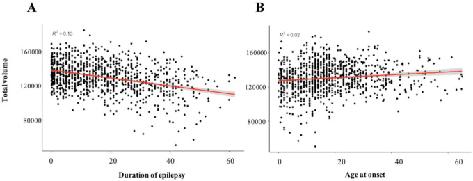
Methods: A state-of-the-art deep learning-based approach was employed that parcellates the cerebellum into 28 neuroanatomical subregions. Linear mixed models compared total and regional cerebellar volume in i) all epilepsies; ii) temporal lobe epilepsy with hippocampal sclerosis (TLE-HS); iii) non-lesional temporal lobe epilepsy (TLE-NL); iv) genetic generalised epilepsy; and (v) extra-temporal focal epilepsy (ETLE). Relationships were examined for cerebellar volume versus age at seizure onset, duration of epilepsy, phenytoin treatment, and cerebral cortical thickness.

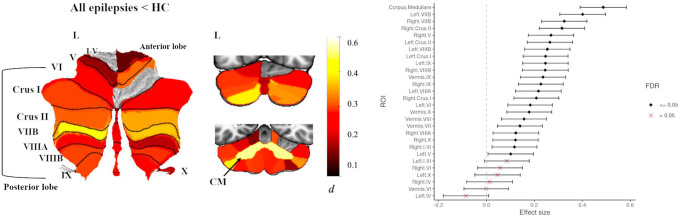
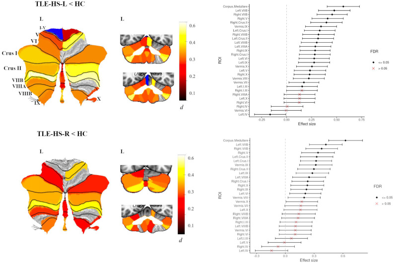
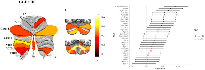
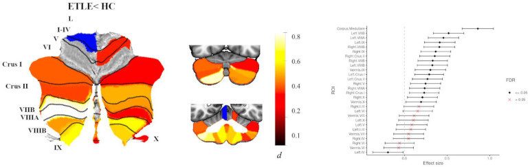
Results: Across all epilepsies, reduced total cerebellar volume was observed (d=0.42). Maximum volume loss was observed in the corpus medullare (dmax=0.49) and posterior lobe grey matter regions, including bilateral lobules VIIB (dmax= 0.47), Crus I/II (dmax= 0.39), VIIIA (dmax=0.45) and VIIIB (dmax=0.40). Earlier age at seizure onset (ηρ2max=0.05) and longer epilepsy duration (ηρ2max=0.06) correlated with reduced volume in these regions. Findings were most pronounced in TLE-HS and ETLE with distinct neuroanatomical profiles observed in the posterior lobe. Phenytoin treatment was associated with reduced posterior lobe volume. Cerebellum volume correlated with cerebral cortical thinning more strongly in the epilepsy cohort than in controls.

Significance: We provide robust evidence of deep cerebellar and posterior lobe subregional grey matter volume loss in patients with chronic epilepsy. Volume loss was maximal for posterior subregions implicated in non-motor functions, relative to motor regions of both the anterior and posterior lobe. Associations between cerebral and cerebellar changes, and variability of neuroanatomical profiles across epilepsy syndromes argue for more precise incorporation of cerebellum subregions into neurobiological models of epilepsy.

Keywords: MRI; anterior lobe; cerebellum; epilepsy; posterior lobe.


Conflict of interest statement

L.Vivash. reports research funding from Biogen Australia, Life Molecular Imaging and Eisai. T.J. O’Brien has received consulting fees from Eisai, UCB, Supernus, Biogen, ES Therapeutics, Epidarex, LivaNova, Kinoxis Therapeutics. He participates on the Data Safety Monitoring Board for ES Therapeutics, Kinoxis Therapeutics. He has served as President (past) for Epilepsy Society of Australia, and is the current chair for Australian Epilepsy Clinical Trials Network (AECTN) and the American Epilepsy Society (Translational Research Committee). B. Bender is the cofounder of AIRAmed GmbH, a company that offers brain segmentation. P. Martin. has received honorary as an advisory board member from Biogen unrelated to the submitted work. P. Striano received speaker fees and advisory boards for Biomarin, Zogenyx, GW Pharmaceuticals; research funding by ENECTA BV, GW Pharmaceuticals, Kolfarma srl., Eisai. P.M. Thompson received a research grant from Biogen, Inc., and was a paid consultant for Kairos Venture Capital, Inc., USA, for projects unrelated to this work. C.L. Yasuda has received personal payments from Torrent, Zodiac and UCB. S.M Sisodiya has received research grants from UCB Pharma and Jazz Pharmaceuticals, speakers fees from UCB, Eisai and Zogenix; honoraria or other fees from Eisai, Jazz Pharma, Stoke Therapeutics, UCB and Zogenix. (payments to institution) The remaining authors have no conflicts of interest.

Figures

Fig 1.
Fig 1.
Atlas-based effect size (Cohen’s d) maps, MNI-based coronal slices (top: y= −72; bottom: y= −54) and forest plots (Cohen’s d +/− 95% confidence interval) of the significant between-group differences for all epilepsies vs. healthy controls (HC). Note: positive effect sizes reflect epilepsy patients < HC. Regions significant at P < 0.05 FDR corrected are depicted in colour (see Table S4 for full tabulation).
Fig 2.
Fig 2.
Scatter-plots showing the association between a) duration of illness and b) age at seizure onset, and total cerebellar volume in epilepsy patients, P < 0.001.
Fig 3.
Fig 3.
Atlas-based effect size (Cohen’s d) maps, MNI-based coronal slices (top: y= −72; bottom: y= −54) and forest plots (Cohen’s d +/− 95% confidence interval) of the significant between-group differences for TLE-HS-L (top panel) and TLE-HS-R (lower panel) vs. healthy controls (HC). Positive effect sizes reflect TLE-HS-L and TLE-HS-R < HC respectively. Regions significant at P < 0.05 FDR corrected are depicted in colour (red-yellow for epilepsy < HC; blue for epilepsy > HC); see Table S5 for full tabulation).
Fig 4.
Fig 4.
Atlas-based effect size (Cohen’s d) maps, MNI-based coronal slices (top: y= −72; bottom: y= −54) and forest plots (Cohen’s d +/− 95% confidence interval) of the significant between-group differences for GGE vs. healthy controls (HC). Note: positive effect sizes reflect epilepsy < HC. Regions significant at P < 0.05 FDR corrected are depicted in colour. See Table S7 for full tabulation.
Fig 5.
Fig 5.
Atlas-based effect size (Cohen’s d) maps, MNI-based coronal slices (top: y= −72; bottom: y= −54) and forest plots (Cohen’s d +/− 95% confidence interval) of the significant between-group differences for ETLE vs. healthy controls (HC). Note: positive effect sizes reflect epilepsy patients < HC. Regions significant at P < 0.05 FDR corrected are depicted in colour (red-yellow for ETLE < HC; blue for ETLE > HC); see Table S8 for full tabulation.

References

    1. Thijs RD, Surges R, O’Brien TJ, Sander JW. Epilepsy in adults. Lancet. Feb 16 2019;393(10172):689–701. doi:10.1016/s0140-6736(18)32596-0 - DOI - PubMed
    1. Téllez-Zenteno JF, Hernández-Ronquillo L. A review of the epidemiology of temporal lobe epilepsy. Epilepsy Res Treat. 2012;2012:630853. doi:10.1155/2012/630853 - DOI - PMC - PubMed
    1. Hatton SN, Huynh KH, Bonilha L, et al. White matter abnormalities across different epilepsy syndromes in adults: an ENIGMA-Epilepsy study. Brain. Aug 1 2020;143(8):2454–2473. doi:10.1093/brain/awaa200 - DOI - PMC - PubMed
    1. Whelan CD, Altmann A, Botía JA, et al. Structural brain abnormalities in the common epilepsies assessed in a worldwide ENIGMA study. Brain. Feb 1 2018;141(2):391–408. doi:10.1093/brain/awx341 - DOI - PMC - PubMed
    1. Kandel A, Buzsáki G. Cerebellar neuronal activity correlates with spike and wave EEG patterns in the rat. Epilepsy Res. Sep 1993;16(1):1–9. doi:10.1016/0920-1211(93)90033-4 - DOI - PubMed

Publication types