[Preprint]. 2024 Jul 5:2023.10.27.23297507.
doi: 10.1101/2023.10.27.23297507.

CXCL12 drives natural variation in coronary artery anatomy across diverse populations

Affiliations

CXCL12 drives natural variation in coronary artery anatomy across diverse populations

Pamela E Rios Coronado et al. medRxiv. .

Update in

  • CXCL12 drives natural variation in coronary artery anatomy across diverse populations.
    Rios Coronado PE, Zhou J, Fan X, Zanetti D, Naftaly JA, Prabala P, Martínez Jaimes AM, Farah EN, Kundu S, Deshpande SS, Evergreen I, Kho PF, Ma Q, Hilliard AT, Abramowitz S, Pyarajan S, Dochtermann D; Million Veteran Program; Damrauer SM, Chang KM, Levin MG, Winn VD, Paşca AM, Plomondon ME, Waldo SW, Tsao PS, Kundaje A, Chi NC, Clarke SL, Red-Horse K, Assimes TL. Rios Coronado PE, et al. Cell. 2025 Apr 3;188(7):1784-1806.e22. doi: 10.1016/j.cell.2025.02.005. Epub 2025 Mar 5. Cell. 2025. PMID: 40049164

Abstract

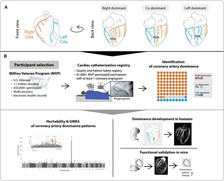
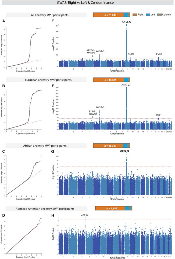
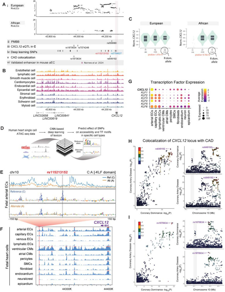
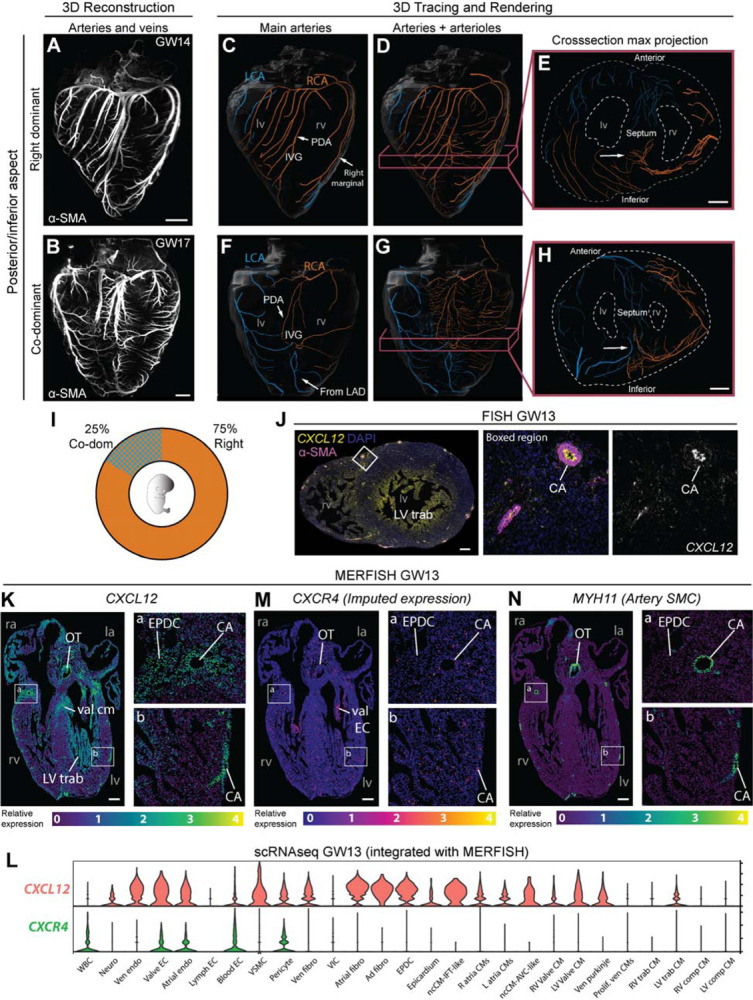
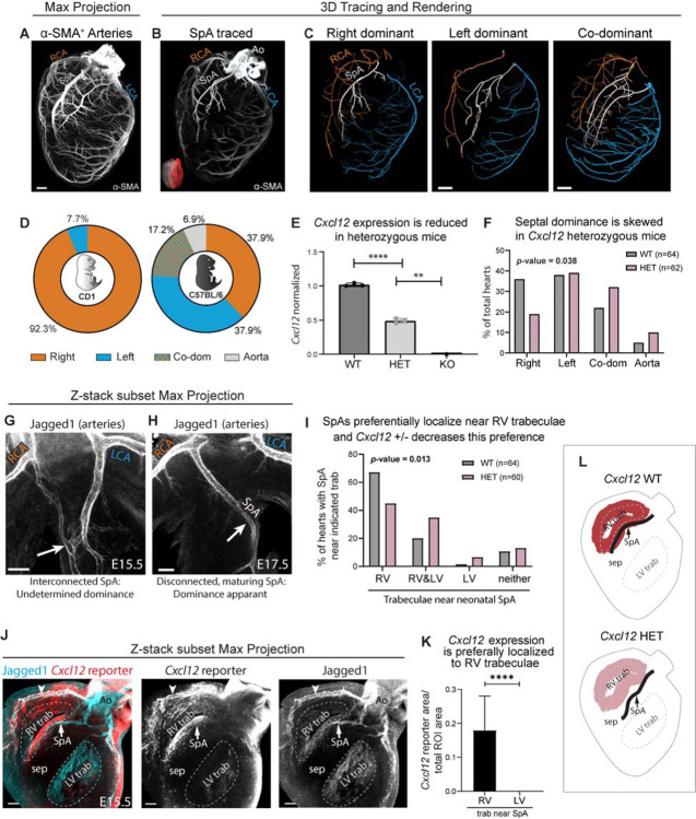
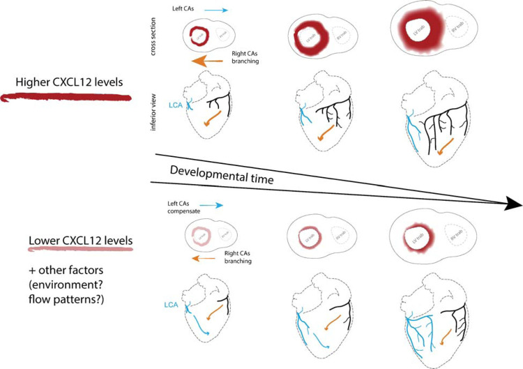
To efficiently distribute blood flow to cardiac muscle, the coronary artery tree must follow a specific branching pattern over the heart. How this pattern arises in humans is unknown due to the limitations of studying human heart development. Here, we leveraged a natural variation of coronary artery anatomy, known as coronary dominance, in genetic association studies to identify the first known driver of human coronary developmental patterning. Coronary dominance refers to whether the right, left, or both coronary arteries branch over the posterior left ventricle, but whether this variability is heritable and how it would be genetically regulated was completely unknown. By conducting the first large-scale, multi-ancestry genome-wide association study (GWAS) of coronary dominance in 61,043 participants of the VA Million Veteran Program, we observed moderate heritability (27.7%) with ten loci reaching genome wide significance. An exceptionally strong association mapped DNA variants to a non-coding region near the chemokine CXCL12 in both European and African ancestries, which overlapped with variants associated with coronary artery disease. Genomic analyses predicted these variants to impact CXCL12 levels, and imaging revealed dominance to develop during fetal life coincident with CXCL12 expression. Reducing Cxcl12 in mice to model the human genetics altered septal artery dominance patterns and caused coronary branches to develop away from Cxcl12 expression domains. Cxcl12 heterozygosity did not compromise overall artery coverage as seen with full deletion, but instead changed artery patterning, reminiscent of the human scenario. Together, our data support CXCL12 as a critical determinant of human coronary artery growth and patterning and lay a foundation for the utilization of developmental pathways to guide future precision 'medical revascularization' therapeutics.


Conflict of interest statement

Competing interests: A.K. is on the scientific advisory board of SerImmune, TensorBio and OpenTargets, and a consultant with Arcadia Science and Inari Agriculture. A.K. was a scientific co-founder of RavelBio, a paid consultant with Illumina, was on the SAB of PatchBio and owns shares in DeepGenomics, Immunai, Freenome, and Illumina. All other authors declare they have no competing interests.

Figures

Figure 1.
Figure 1.. Study design to identify and understand the genetic determinants of coronary artery dominance.
(A) Variation in the posterior descending artery (PDA) determines dominance in the human heart. (B) A multi-ancestry genome-wide association study (GWAS) was performed using genotype data from participants in MVP whose coronary arteries were imaged via angiogram. Analyses were conducted in all participants combined and in cohorts stratified by genetically inferred ancestry. Whole-organ immunolabeling of fetal hearts investigated when dominance is established, and mouse studies validated the most significant genetic association. CAs, coronary arteries.
Figure 2.
Figure 2.. Genome-wide association study (GWAS) of right versus left/co-dominance in the Million Veteran Program (MVP).
GWAS was conducted in all participants independent of ancestry (All ancestry) as well as in each of the three largest genetically inferred ancestry subgroups: European, African, and Admixed American. (A-D) Quantile-quantile plots (Q-Q) show the observed versus expected P-values from association for all genetic variants. Red line indicates null hypothesis. (E-H) Manhattan plots for the corresponding Q-Q plots with P-values of genetic variants. Gene(s) mapped to regions reaching genome wide significance (GWS) are labeled. Red line indicates GWS threshold (P-value<5×10⁻⁸).
Figure 3.
Figure 3.. Dominance variants localize to putative CXCL12 regulatory regions.
(A) SNPs reaching genome wide significance in the indicated MVP populations aligned with variants from additional analyses: Penn Medicine Biobank (PMBB) replication study, Genotype-Tissue Expression (GTEx) eQTLs, deep learning-based predictions, and coronary artery disease (CAD) colocalization. An in vivo validated Cxcl12 enhancer is also shown. (B) Location of accessible chromatin in human fetal heart cell types in relation to dominance SNPs in A. (C) Violin plots of two lead SNP eQTLs revealed that a higher prevalence of right dominance is associated with increased CXCL12 in GTEx data. Left is rs1870634; right is rs7074248. Testis expression levels by genotype are shown, but directionality is the same in arterial and coronary tissues (see Supplementary Figure 5A, B). (D) Schematic of convolutional neural network (CNN) deep learning models used to predict the effect of alternate alleles. (E) Predicted per-base accessibility for reference and alternate alleles of rs115213152 suggest a disrupted KLF motif. (F) Sequencing tracks of chromatin accessibility near CXCL12. Red highlights rs115213152 region where open chromatin coincides with CXCL12 promoter accessibility in select cardiac cell types, including coronary artery cells. (G) ScRNAseq from fetal hearts revealed co-expression of CXCL12 and KLFs, supporting a potential role for these transcription factors in the activity predicted in E. (H and I) LocusCompare ouput displaying colocalization plot (left), and LocusZoom plots for each trait (right). The SNP with the highest H4 posterior probability using COLOC is labelled in purple with additional variants in linkage disequilibrium with labelled SNP colored according to R2 value. Top panel (H) is unmodified analysis and bottom panel (I) shows result when the first colocalization signal at rs649192 (44650000–44850000) is masked.
Figure 4.
Figure 4.. Coronary dominance is apparent during fetal development when CXCL12 is expressed.
(A and B) Whole-organ immunolabeling of coronary artery smooth muscle (α-SMA) in hearts from the indicated gestational weeks (GW). (C-H) Representative artery tracings highlighted branches of the main arteries (C, F) or main and lower order arteries (D, E, G, H) that originated from the right (RCA, orange) or left (LCA, blue) coronary ostia. (C-E) Right dominance was indicated when the artery in the interventricular groove (IVG), i.e., the posterior descending artery (PDA), originated from the RCA (C and D) and when the inferior septum was occupied by right branches (arrow in E). (F-H) Co-dominance was indicated when arteries within the IVG stemmed from both the RCA and LCA (F and G) and the inferior septum was occupied by RCA and LCA branches (arrow in H). (I) Distribution in a cohort of n=8 human fetal hearts. (J) CXCL12 fluorescence in situ hybridization (FISH) and α-SMA immunolabeling on a transverse section through a GW13 heart. Expression was in coronary arteries (CA) and trabecular cardiomyocytes (LV trab). DAPI labeled nuclei. (K-N) Spatial expression plots (K, M, L) and scRNAseq-derived violin plots (L) in GW13 human fetal hearts. MERFISH localized CXCL12 and MYH11 while CXCR4 was imputed by integrating both datasets. ad, adventitial; AVC, atrioventricular; comp, compact; EC, endothelial cell; CM, cardiomyocyte; EPDC, epicardial-derived cells; Endo, endocardial cell; Fibro, fibroblast; IFT, inflow tract; Neuro, neuronal; la, left atrium; Lymph, lymphatic; lv, left ventricle; LV trab, left ventricle trabeculae; ncCM, non-chambered cardiomyocyte; OT, outflow tract; ra, right atrium; RV, right ventricle; trab, trabecular; val, valve; ven, ventricular; VIC, valve interstitial cell; VSMC, vascular smooth muscle; WBC, white blood cell. Scale bars: A, B, E, and J, 500μm; H, 1mm; J, 200μm; K-O, 250μm.
Figure 5.
Figure 5.. Cxcl12 influences septal artery dominance in mice.
(A) Whole-organ immunolabeling of coronary artery smooth muscle (α-SMA) in a postnatal day 6 mouse heart. (B) Septal artery (SpA) tracing overlayed onto a Z-stack subset max projection of internal coronary arteries (red region in insert). (C) Artery tracings in whole-organ images demonstrating various SpA connections. (D) SpA dominance ratios are strain specific. N=13 CD1 neonatal hearts and N=29 for C56BL/6J. (E) QPCR using whole hearts from embryonic day (E) 16.5. WT, Cxcl12+/+; HET, Cxcl12DsRed/+; KO, Cxcl12DsRed/DsRed. Each dot is the average value for genotypes within single litters normalized to WT littermates. Error bars, mean±st dev: **, p=0.0027, p=0.0016, p=0.0011; ****, p<0.0001 by two-sided Student’s t-test. (F) Blinded scoring of septal artery dominance in WT and HET neonatal mice. P-value by two-tailed Chi-square test. (G and H) Whole-organ immunolabeling of WT embryonic hearts. First, an immature artery network is attached to both sides (G) followed by maturation into a SpA with dominance shortly before birth (H). (I) Blinded scoring of SpA location as related to trabeculae (see Supplementary Figure 8I). P-value by two-tailed Chi-square test. (J) Cxcl12 reporter allele shows that SpA develops in regions of high expression. Arrowhead, expression in artery endothelial cells. (K) Reporter allele fluorescence in standardized regions of interest (ROI) along right or left trabeculae. N=14 hearts. Error bars, mean±st dev: ****, p<0.0001 by two-sided Student’s t-test. (L) Schematic of Cxcl12 bias in trabeculae, and HET’s effect on SpA localization. Ao, aorta; LCA, left coronary artery; LV, left ventricle; RCA, right coronary artery; RV, right ventricle; sep, septum; trab, trabecular cardiomyocytes. Scale bars: A, C 400μm; G - I, 100μm.
Figure 6.
Figure 6.. Working model of how CXCL12 genetic variants influence human coronary artery development and result in different dominance configurations.
Genomic data suggested that dominance associated SNPs influence CXCL12 gene expression with right CA dominance being associated with higher CXCL12 levels. Mouse developmental genetics showed that Cxcl12 levels influence SpA dominance and indicated that arteries are attracted to form near expression domains. We propose that arterial endothelial cells are attracted to CXCL12 expression in the epicardium, myocardium, and artery endothelial cells to preferentially extend the right coronary artery on the back of the heart. When CXCL12 levels are lower, extension of right branches is delayed or misplaced, and the left grows to compensate. Colored arrows indicate the extension path(s) that right (orange) and left (blue) CAs will take based on CXCL12 levels.

References

    1. Baroldi G., Scomazzoni G., and United States. Surgeon-General’s, O. (1967). Coronary circulation in the normal and the pathologic heart (Office of the Surgeon General, Dept. of the Army; for sale by the Superintendent of Documents, U.S. Govt. Print. Off.).
    1. Libby P., Bonow R.O., Mann D.L., Tomaselli G.F., Bhattt D.L., and Solomon S. (2021). Braunwald’s heart disease : a textbook of cardiovascular medicine, 12. Edition (Elsevier; ).
    1. Martin S.S., Aday A.W., Almarzooq Z.I., Anderson C.A.M., Arora P., Avery C.L., Baker-Smith C.M., Barone Gibbs B., Beaton A.Z., Boehme A.K., et al. (2024). 2024 Heart Disease and Stroke Statistics: A Report of US and Global Data From the American Heart Association. Circulation 149, e347–e913. 10.1161/CIR.0000000000001209. - DOI - PMC - PubMed
    1. Thiene G., Frescura C., Padalino M., Basso C., and Rizzo S. (2021). Coronary Arteries: Normal Anatomy With Historical Notes and Embryology of Main Stems. Front Cardiovasc Med 8, 649855. 10.3389/fcvm.2021.649855. - DOI - PMC - PubMed
    1. Bruell J.H., Leininger J.T., and Hellerstein H.K. (1970). Inheritance of a cardiac arterial asymmetry in mice. Science (New York, N.Y 167, 199–200. 10.1126/science.167.3915.199. - DOI - PubMed

Publication types