. 2024 Jan 5;52(D1):D442-D455.
doi: 10.1093/nar/gkad1058.

ELM-the Eukaryotic Linear Motif resource-2024 update

Affiliations

ELM-the Eukaryotic Linear Motif resource-2024 update

Manjeet Kumar et al. Nucleic Acids Res. .

Abstract

Short Linear Motifs (SLiMs) are the smallest structural and functional components of modular eukaryotic proteins. They are also the most abundant, especially when considering post-translational modifications. As well as being found throughout the cell as part of regulatory processes, SLiMs are extensively mimicked by intracellular pathogens. At the heart of the Eukaryotic Linear Motif (ELM) Resource is a representative (not comprehensive) database. The ELM entries are created by a growing community of skilled annotators and provide an introduction to linear motif functionality for biomedical researchers. The 2024 ELM update includes 346 novel motif instances in areas ranging from innate immunity to both protein and RNA degradation systems. In total, 39 classes of newly annotated motifs have been added, and another 17 existing entries have been updated in the database. The 2024 ELM release now includes 356 motif classes incorporating 4283 individual motif instances manually curated from 4274 scientific publications and including >700 links to experimentally determined 3D structures. In a recent development, the InterPro protein module resource now also includes ELM data. ELM is available at: http://elm.eu.org.


Figures

Graphical Abstract
Graphical Abstract
Figure 1.
Figure 1.
Data growth trends within the ELM resource since the last update paper (37). (A) This depicts the cumulative count of both motif classes and instances inserted into ELM in the last two decades. (B) The pie chart illustrates the allocation of new instances among both new and updated ELM classes (lighter and darker shades in the outer circle, respectively) belonging to 5 different functional categories. Note that the pie chart focuses only on the classes where new instances were added in this update. Visualisations were done using ggplot2 in RStudio (http://www.rstudio.com/).
Figure 2.
Figure 2.
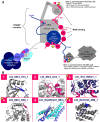
SLiMs in nuclear RNA processing and decay. (A) Schematic representation of the motif-mediated interactions maintaining the inter-subunit interactions of the TRAMP complex and facilitating the recruitment of the complex and the associated exosome to different types of degradable RNA. Motif-mediated interactions are highlighted with numbered pink boxes, with the ones newly added or extensively revised since the last release marked by a star. Where the given motif is employed by several yeast factors belonging to different protein families, the factors and their respective bound RNA types are listed. Abbreviations: CID, CTD-interacting domain, Znk, zinc knuckle, ncRNA - non-coding RNA, AIM, arch-interacting motif, NIM, Nrd1-interacting motif. (B) Structural snapshots of the numbered motif-mediated interactions. Colours for the domains and/or motifs depicted in each box match those of the corresponding proteins in the schematic figure of panel A. Within each box, all protein domains are depicted in cartoon representation. In most cases (boxes 1, 4, 5 and 6) cartoon representation was also applied to the SLiMs for simplicity. In the case of more extensive binding interfaces (boxes 2 and 3), where the motif residues would be difficult to guess, residues belonging to the SLiMs are shown in stick representation. The structural snapshots do not always show the interaction of the exact same proteins as depicted in panel A, in some cases, an X-ray structure was only available for the same type of motif from another protein. (1) PDB: 4U4C (Mtr4 with a Trf4-Air2 fusion protein, Trf4 motif depicted); (2) PDB:4U4C (Mtr4 with a Trf4-Air2 fusion protein, Air2 motif depicted); (3) PDB:3NYB (Trf4 with Air2 regions including the 4th and 5th zink knuckles and the motif in the linker connecting those; motif residues in stick representation); (4) PDB:5OOQ (Mtr4 KOW domain with the AIM motif from Nop53); (5) PDB:4WFD (the PMC2NT domain of Rrp6 forms a heterodimer with cofactor Rrp47 and the resulting dimer interface binds the motif within the N-terminal tail of Mtr4); (6) PDB: 6O3W (CID domain of Nrd1 bound by a Sen1 NIM motif).
Figure 3.
Figure 3.
The diverse functions of C-terminal degrons. These motifs (red) can be part of native, endogenous protein C-termini (A), or be at an internal location and exposed by proteolysis (B). Degron SLiMs may also originate from erroneously translated mRNA sequences (C) or be present in virus-encoded proteins (D). These degrons are subsequently recognized by specific ubiquitin ligases (E3) and the proteins are polyubiquitylated, targeting them into the proteasome.
Figure 4.
Figure 4.
Protein interaction network centred on the key anti-pathogen and inflammatory kinase TBK1 generated by the STRING resource (122). The network shows how TBK1 plays a core role in bringing together the TRAF and IRF3 signalling systems. Edges with SLiMs annotated in ELM are shown by thick red lines. TNF (extracellular) interactions with cytosolic proteins are indirect (dotted lines). STRING settings for building the interaction network were as follows: only experimentally identified interactors of TBK1 and the 1st shell of ‘no more than 20 interactors’ in max number of interactors.

References

    1. Pawson T., Nash P.. Assembly of cell regulatory systems through protein interaction domains. Science. 2003; 300:445–452. - PubMed
    1. Gibson T.J. Cell regulation: determined to signal discrete cooperation. Trends Biochem. Sci. 2009; 34:471–482. - PubMed
    1. Wright P.E., Dyson H.J.. Intrinsically disordered proteins in cellular signalling and regulation. Nat. Rev. Mol. Cell Biol. 2015; 16:18–29. - PMC - PubMed
    1. Quaglia F., Mészáros B., Salladini E., Hatos A., Pancsa R., Chemes L.B., Pajkos M., Lazar T., Peña-Díaz S., Santos J.et al. .. DisProt in 2022: improved quality and accessibility of protein intrinsic disorder annotation. Nucleic Acids Res. 2022; 50:D480–D487. - PMC - PubMed
    1. Bah A., Forman-Kay J.D.. Modulation of intrinsically disordered protein function by post-translational modifications. J. Biol. Chem. 2016; 291:6696–6705. - PMC - PubMed