. 2023 Dec 29;14(1):jkad260.
doi: 10.1093/g3journal/jkad260.

Humanization reveals pervasive incompatibility of yeast and human kinetochore components

Affiliations

Humanization reveals pervasive incompatibility of yeast and human kinetochore components

Guðjón Ólafsson et al. G3 (Bethesda). .

Abstract

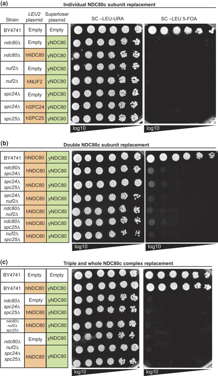
Kinetochores assemble on centromeres to drive chromosome segregation in eukaryotic cells. Humans and budding yeast share most of the structural subunits of the kinetochore, whereas protein sequences have diverged considerably. The conserved centromeric histone H3 variant, CenH3 (CENP-A in humans and Cse4 in budding yeast), marks the site for kinetochore assembly in most species. A previous effort to complement Cse4 in yeast with human CENP-A was unsuccessful; however, co-complementation with the human core nucleosome was not attempted. Previously, our lab successfully humanized the core nucleosome in yeast; however, this severely affected cellular growth. We hypothesized that yeast Cse4 is incompatible with humanized nucleosomes and that the kinetochore represented a limiting factor for efficient histone humanization. Thus, we argued that including the human CENP-A or a Cse4-CENP-A chimera might improve histone humanization and facilitate kinetochore function in humanized yeast. The opposite was true: CENP-A expression reduced histone humanization efficiency, was toxic to yeast, and disrupted cell cycle progression and kinetochore function in wild-type (WT) cells. Suppressors of CENP-A toxicity included gene deletions of subunits of 3 conserved chromatin remodeling complexes, highlighting their role in CenH3 chromatin positioning. Finally, we attempted to complement the subunits of the NDC80 kinetochore complex, individually and in combination, without success, in contrast to a previous study indicating complementation by the human NDC80/HEC1 gene. Our results suggest that limited protein sequence similarity between yeast and human components in this very complex structure leads to failure of complementation.

Keywords: CENP-A; centromere; histone variant; humanization; kinetochore; yeast.


Conflict of interest statement

Conflicts of interest J.D.B. is a founder and director of CDI Labs, Inc.; a founder of and consultant to Neochromosome, Inc.; and a founder, SAB member of, and consultant to ReOpen Diagnostics, LLC, and serves or served on the scientific advisory board of the following: LogoMix Inc.; Modern Meadow Inc.; ROME Therapeutics, Inc.; Sample6 Inc.; Sangamo, Inc.; Tessera Therapeutics Inc., and the Wyss Institute. G.O. and M.A.B.H. declare no competing interests.

Figures

Fig. 1.
Fig. 1.
Humanization of the Cse4 nucleosome with the CENP-A nucleosome in yeast. a) Illustration of the centromere and kinetochore in budding yeast and human cells. The kinetochore cartoon highlights the similarities and differences between yeast and human kinetochores and their subcomplexes. The cartoon is not to scale and is not meant to signify an actual kinetochore structure. The kinetochore cartoon is based on a previous model of the kinetochore architecture (Biggins 2013) and is adapted from Klemm et al. (2020) and Ólafsson and Thorpe (2020). b) Overview of the Superloser dual-plasmid shuffle assay (see Supplementary Fig. 1a for schematics of the Superloser and CEN nucleosome humanization plasmids and methods for details). c) Example images of agar plates from the histone humanization assay of yHsΔ dad1E50D CSE4+ shuffle strain showing histone-humanized colonies after 20 days of growth at 30°C on SC–Trp 5-FOA media. For histone humanization experiments, we use a positive control: a plasmid containing hHs and yeast CSE4 (instead of CENP-A) and 2 negative controls: 1 containing hHs only and the other an empty plasmid to control for recombination. As an additional control, we also performed our experiments using the dad1E50D histone shuffle strain containing endogenous CSE4 (CSE4+). Note that the few large colonies that typically appear within 3 days represent plasmid recombinants or spontaneous ura3 mutants that have gained 5-FOA resistance (see main text for details). d) Schematics of the various Cse4–CENP-A chimeras are shown and are in line with the data in panel e). e) The histone humanization rate data are summarized in a table format (see bar graph of the same data in Supplementary Fig. 1d and the raw data in Supplementary Table 3). Insert (top) displays aa sequence alignment of the loop 1 of Cse4 and CENP-A, and the nonconserved three amino acid sequence region is highlighted. Insert (bottom) shows the alignment of the region of yH2B and hH2B. The nonconserved arginine residue that is important for interaction with the CBF3 complex is highlighted (see main text for details).
Fig. 2.
Fig. 2.
Overexpression of human CENP-A is lethal in WT yeast and perturbs mitotic progression. a) A spot assay showing that CENP-A overexpression is lethal in WT cells in contrast to controls. Log-phase cultures of WT strain (BY4741) with the indicated plasmids were diluted to the same OD600 level, and 10-fold serial dilutions were prepared and spotted onto media containing dextrose (expression off) and galactose (expression on). b) Same as panel a), but the indicated plasmids were transformed into a stable histone-humanized dad1E50D strain (yDT180). c) Cell cycle analysis by flow cytometry of WT cells containing the indicated plasmids. Asynchronous log-phase cultures were grown in media containing galactose for 6 h before cell fixing and DNA staining with SYTOX Green and DNA content quantification (see Materials and methods for details). The experiment was performed in duplicate with identical results. d) Cell cycle analysis by fluorescence microscopy of WT cells containing an empty vector (n = 190), pGAL1-CENP-AW86R mutant (n = 89), and pGAL1-CENP-A (n = 154) plasmids and GFP-tagged Ndc80, an outer kinetochore subunit of the KMN network. Asynchronous log-phase cultures were grown in media containing galactose for 5 h before imaging. Mother-bud cell morphology and kinetochore foci status were used as proxy for cell cycle phases. For example, large-budded cells with 2 kinetochore foci in close proximity were categorized as metaphase cells (see Materials and methods for details). Fisher’s exact statistical test; P-values *P < 0.05; ***P < 0.0005. Error bars indicate 95% binomial confidence intervals (CI).
Fig. 3.
Fig. 3.
CENP-A overexpression disrupts the inner and outer kinetochore in yeast. a) The kinetochore foci of the Ndc80-GFP cells in Fig. 2d were analyzed using fluorescence microscopy, and abnormal declustered Ndc80-GFP foci were quantified. Fisher’s exact statistical test; P-values ****P < 10−5. Error bars indicate 95% binomial CI. Representative micrographs are shown on the right. Scale bars indicate 5 µm. b) The Ndc80-GFP signal in panel a) was also quantified by measuring the GFP intensity along the mitotic spindle. A 5-μm line with 30 points (illustrated by a yellow line in the inset on the top right) was used to include background signal and to cover the spread GFP signal in cells overexpressing CENP-A (40 randomly selected mitotic spindles were measured for each condition). The shadowed area indicates standard deviation. CENP-A-overexpressing cells have a more dispersed arrangement of Ndc80-GFP, as indicated by the flatter profile. Representative micrographs are shown on the right. Bottom right insert highlights the declustered Ndc80-GFP phenotype. c) Kinetochore foci analysis of WT cells containing the CCAN subunit Ctf19-GFP and the empty vector (n = 71), pGAL1-CENP-AW86R (n = 67), pGAL1-Chimera C (n = 89), and pGAL1-CENP-A (n = 45) plasmids. Asynchronous log-phase cell cultures were grown in media containing 2% galactose for 5 h before imaging. Cells containing no or diffused Ctf19-GFP signal were quantified. Fisher’s exact statistical test; P-values ****P < 10−5. Error bars indicate 95% binomial CI. Representative micrographs are shown on the right. Scale bars indicate 5 µm. d) Violin plot showing fluorescence intensities of Ctf19-GFP kinetochore foci in cells containing empty vector (n = 107), pGAL1-CENP-AW86R (n = 112), pGAL1-CSE4 (n = 110), pGAL1-CENP-A (n = 40), pGAL1-chimera A (n = 129), pGAL1-chimera B (n = 113), pGAL1-chimera C (n = 144), and pGAL1-chimera D (n = 76) plasmids. Culture conditions are the same as those in panel c). Statistical significance was evaluated using unpaired 2-tailed Student's t-test; P-value *P < 0.05; **P < 0.005; ****P < 10−5. Cells containing no detectable or very diffused Ctf19-GFP signals were excluded from the analysis.
Fig. 4.
Fig. 4.
Identification of genetic interactions of CENP-A and chimera overexpression using genome-wide screening of nonessential gene deletions. a) Overview of the genetic screens. We used SPA to introduce the indicated plasmids into the whole nonessential gene knockout collection (see main text and Materials and methods for details). b) Example cropped images of arrayed colonies showing suppression of the pGAL1-CENP-A growth phenotype by deletions of INO80/SWR1 and RSC complex subunits. The images were captured after 5 days of growth at 30°C on synthetic media containing 5-FOA. c) GO enrichment analysis of CENP-A overexpression suppressors. d) Protein–protein interaction (PPI) network of CENP-A overexpression suppressors. The network was generated using the STRING database (Szklarczyk et al. 2023) and consists of a subset of the deleted genes identified as suppressors (see Supplementary Table 5 for the completed list). * indicates that these genes were not directly detected, but these 2 dubious open reading frames, YLR358C and YLR322W/VPS65, partially overlap with the essential RSC subunits RSC2 and SFH1, respectively. Edge intensity that connects nodes indicates the confidence of the PPI (based on the STRING database): dark gray, high; light gray, low. Node color indicates different protein complexes. The size of the nodes or length of the edges do not reflect any quantifiable scores. e) GO enrichment analysis of negative genetic interactors of chimera C overexpression. f) PPI network of the negative interactors of chimera C overexpression. The PPI network shows a subset of negative interactors (see Supplementary Table 5 for the completed list). Edge intensity that connects nodes indicates the confidence of the PPI: dark gray, high; light gray, low. Node color indicates different protein complexes. The size of the nodes or length of the edges do not reflect any quantifiable scores.
Fig. 5.
Fig. 5.
Complementation of the yeast NDC80c subunits by the human counterparts. a) Example complementation spot assay of individual replacements of yeast NDC80c subunits by episomal human orthologs. Complementation experiments were also performed by plating various amounts of cells (see Supplementary Table 6). b) Example complementation spot assay of double replacements of yeast NDC80c subunits by episomal human orthologs. c) Example complementation spot assay of triple and complete replacements of yeast NDC80c subunits by episomal human orthologs.

Similar articles

Cited by

References

    1. Abdullah M, Greco BM, Laurent JM, Garge RK, Boutz DR, Vandeloo M, Marcotte EM, Kachroo AH. 2023. Rapid, scalable, combinatorial genome engineering by marker-less enrichment and recombination of genetically engineered loci in yeast. Cell Rep Methods. 3(5):100464. doi:10.1016/J.CRMETH.2023.100464. - DOI - PMC - PubMed
    1. Agmon N, Temple J, Tang Z, Schraink T, Baron M, Chen J, Mita P, Martin JA, Tu BP, Yanai I. et al. et al. 2019. Phylogenetic debugging of a complete human biosynthetic pathway transplanted into yeast. Nucleic Acids Res. 48(1):486–499. doi:10.1093/nar/gkz1098. - DOI - PMC - PubMed
    1. Altemose N, Logsdon GA, Bzikadze AV, Sidhwani P, Langley SA, Caldas GV, Hoyt SJ, Uralsky L, Ryabov FD, Shew CJ, et al. . 2022. Complete genomic and epigenetic maps of human centromeres. Science. 376(6588):eabl4178. doi:10.1126/SCIENCE.ABL4178. - DOI - PMC - PubMed
    1. Athwal RK, Walkiewicz MP, Baek S, Fu S, Bui M, Camps J, Ried T, Sung M-H, Dalal Y. 2015. CENP-A nucleosomes localize to transcription factor hotspots and subtelomeric sites in human cancer cells. Epigenetics Chromatin. 8:2. doi:10.1186/1756-8935-8-2. - DOI - PMC - PubMed
    1. Au WC, Dawson AR, Rawson DW, Taylor SB, Baker RE, Basrai MA. 2013. A novel role of the N terminus of budding yeast histone H3 variant cse4 in ubiquitin-mediated proteolysis. Genetics 194(2):513–518. doi:10.1534/genetics.113.149898. - DOI - PMC - PubMed

Publication types

MeSH terms

LinkOut - more resources