. 2024 Feb;59(2):95-108.
doi: 10.1007/s00535-023-02055-x. Epub 2023 Nov 14.

Marked intestinal trans-differentiation by autoimmune gastritis along with ectopic pancreatic and pulmonary trans-differentiation

Affiliations

Marked intestinal trans-differentiation by autoimmune gastritis along with ectopic pancreatic and pulmonary trans-differentiation

Chihiro Takeuchi et al. J Gastroenterol. 2024 Feb.

Abstract

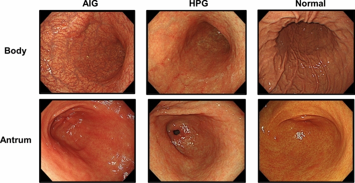
Background: Autoimmune gastritis (AIG) is a prevalent chronic inflammatory disease with oncogenic potential that causes destruction of parietal cells and severe mucosal atrophy. We aimed to explore the distinctive gene expression profiles, activated signaling pathways, and their underlying mechanisms.

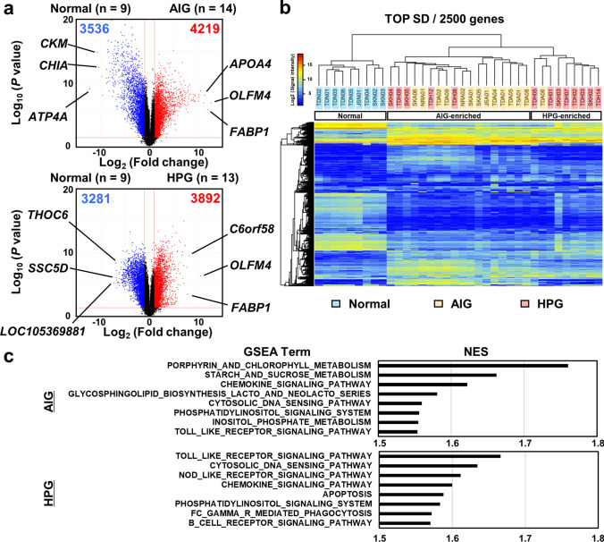
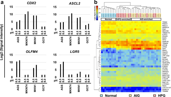
Methods: A comprehensive gene expression analysis was conducted using biopsy specimens from AIG, Helicobacter pylori-associated gastritis (HPG), and non-inflammatory normal stomachs. Gastric cancer cell lines were cultured under acidic (pH 6.5) conditions to evaluate changes in gene expression.

Results: Gastric mucosa with AIG had a unique gene expression profile compared with that with HPG and normal mucosa, such as extensively low expression of ATP4A and high expression of GAST and PAPPA2, which are involved in neuroendocrine tumorigenesis. Additionally, the mucosa with AIG and HPG showed the downregulation of stomach-specific genes and upregulation of small intestine-specific genes; however, intestinal trans-differentiation was much more prominent in AIG samples, likely in a CDX-dependent manner. Furthermore, AIG induced ectopic expression of pancreatic digestion-related genes, PNLIP, CEL, CTRB1, and CTRC; and a master regulator gene of the lung, NKX2-1/TTF1 with alveolar fluid secretion-related genes, SFTPB and SFTPC. Mechanistically, acidic conditions led to the downregulation of master regulator and stemness control genes of small intestine, suggesting that increased environmental pH may cause abnormal intestinal differentiation in the stomach.

Conclusions: AIG induces diverse trans-differentiation in the gastric mucosa, characterized by the transactivation of genes specific to the small intestine, pancreas, and lung. Increased environmental pH owing to AIG may cause abnormal differentiation of the gastric mucosa.

Keywords: Autoimmune gastritis; Diverse trans-differentiation; Increased pH; Intestinal differentiation; Molecular epidemiology.


Conflict of interest statement

YT has received an endowed chair from AI Medical Service Inc. The other authors declare no conflict of interest for this article.

Figures

Fig. 1
Fig. 1
Typical endoscopic views of the gastric body and antrum in cases of autoimmune gastritis (AIG), H. pylori-associated gastritis (HPG), and non-inflammatory normal mucosa (normal)
Fig. 2
Fig. 2
Results of the comprehensive analysis of gene expression among the AIG (n = 14), HPG (n = 13), and normal samples (n = 9). a Volcano plot analysis using the fold changes of gene expression levels between the normal and AIG samples, and the normal and HPG samples. The number of gene transcripts with twofold change and a small P-value (-log10 (P-values) > 1.301) was higher in the AIG samples than in the HPG samples. b Unsupervised hierarchical cluster analysis using gene expression levels of the total 36 samples. Using the 2500 gene transcripts with the highest standard deviation (TOP SD), the AIG and HPG samples were clearly separated from the normal samples and were further separated into the AIG-enriched cluster and the HPG-enriched cluster. c Pathway enrichment analyses conducted via GSEA using the upregulated genes in the AIG and HPG samples. The top eight activated gene sets in the AIG and HPG samples are shown. NES—normalized enrichment score
Fig. 3
Fig. 3
Marked intestinal differentiation in gastric mucosa with AIG. a Tissue enrichment analysis using top 50 upregulated genes in gastric mucosa with AIG and HPG. AIG showed higher enrichment of genes specific to the duodenum and the small intestine compared with HPG. b Unsupervised hierarchical cluster analysis using gene expression levels of small intestine-specific and stomach-specific genes among the AIG (n = 14), HPG (n = 13), and normal samples (n = 9). The AIG samples were clearly separated from the normal samples using small intestine and stomach-specific genes, while a fraction of the HPG samples were grouped with the normal samples. c Immunostaining of MUC2 and MUC5AC using gastric mucosa with AIG. Scale bar: 100 μm. d Unsupervised hierarchical cluster analysis using gene expression levels of CDX signature genes, along with CDX2/1 expression (cutoff signal intensity = 25). The AIG and HPG samples were clearly separated from the normal samples and were further separated into the AIG-enriched cluster and the HPG-enriched cluster. Moreover, all the AIG samples expressed CDX2/1, while the CDX-negative HPG samples showed a unique cluster
Fig. 4
Fig. 4
Trans-differentiation into the pancreas and lung in gastric mucosa with AIG. a Protein–protein interaction (PPI) network analysis using AIG-specific genes. The observed networks (Network A and Network B) showed the enrichment of gene sets related to digestion and the alveolar lamellar body. FDR—false discovery rate. b Unsupervised hierarchical cluster analysis using gene expression levels of pancreas-specific and lung-specific genes among the AIG (n = 14), HPG (n = 13), and normal samples (n = 9). A fraction of the AIG samples was clearly separated from the other samples. c Immunostaining of AIG-specific genes related to abnormal differentiation of the pancreas (PNLIP and BCL10) and lung (NKX2-1/TTF1, SFTPB, and SFTPC) using gastric mucosa with AIG. Scale bar: 100 μm
Fig. 5
Fig. 5
Abnormal intestinal differentiation by environmental acidic conditions. a Gene expression changes in gastric cancer cell lines (AGS, MKN74, MKN1, and GCIY). Acidic conditions downregulated the expression levels of a master regulator gene of the small intestine, CDX2, and stem marker genes, LGR5, ASCL2, and OLFM4. b Unsupervised hierarchical cluster analysis using gene expression levels of pancreas-specific and lung-specific genes among the AIG (n = 14), HPG (n = 13), and normal samples (n = 9). The AIG and HPG samples were clearly separated from the normal samples and were further separated into the AIG-enriched cluster and the HPG-enriched cluster

References

    1. Strickland RG, Mackay IR. A reappraisal of the nature and significance of chronic atrophic gastritis. Am J Dig Dis. 1973;18:426–440. doi: 10.1007/BF01071995. - DOI - PubMed
    1. Neumann WL, Coss E, Rugge M, et al. Autoimmune atrophic gastritis–pathogenesis, pathology and management. Nat Rev Gastroenterol Hepatol. 2013;10:529–541. doi: 10.1038/nrgastro.2013.101. - DOI - PubMed
    1. Lenti MV, Rugge M, Lahner E, et al. Autoimmune gastritis. Nat Rev Dis Primers. 2020;6:56. doi: 10.1038/s41572-020-0187-8. - DOI - PubMed
    1. Itsuno M, Watanabe H, Iwafuchi M, et al. Multiple carcinoids and endocrine cell micronests in type A gastritis. Their morphology, histogenesis, and natural history. Cancer. 1989;63:881–890. doi: 10.1002/1097-0142(19890301)63:5<881::AID-CNCR2820630515>3.0.CO;2-K. - DOI - PubMed
    1. Müller J, Kirchner T, Müller-Hermelink HK. Gastric endocrine cell hyperplasia and carcinoid tumors in atrophic gastritis type A. Am J Surg Pathol. 1987;11:909–917. doi: 10.1097/00000478-198712000-00001. - DOI - PubMed

Publication types