. 2023 Nov 14;14(1):7363.
doi: 10.1038/s41467-023-43211-4.

Dietary L-Tryptophan consumption determines the number of colonic regulatory T cells and susceptibility to colitis via GPR15

Affiliations

Dietary L-Tryptophan consumption determines the number of colonic regulatory T cells and susceptibility to colitis via GPR15

Nguyen T Van et al. Nat Commun. .

Abstract

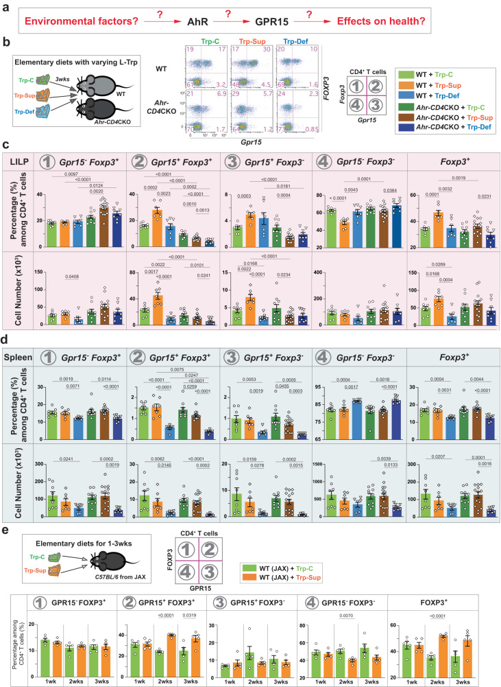
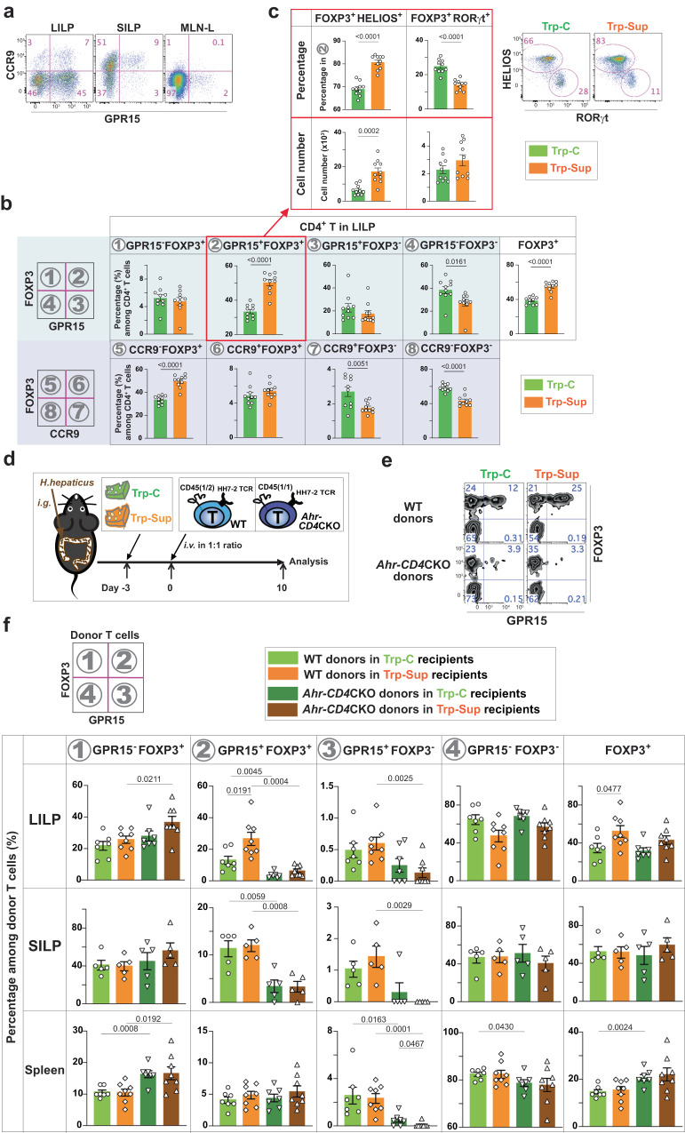
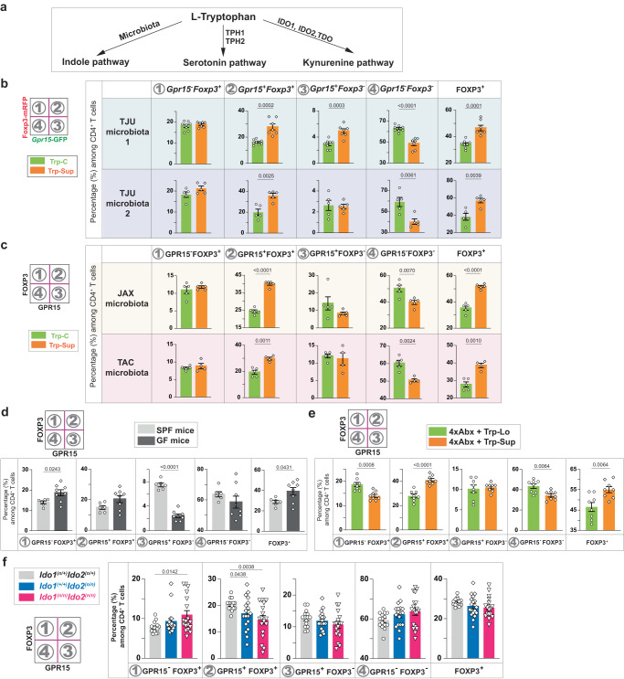
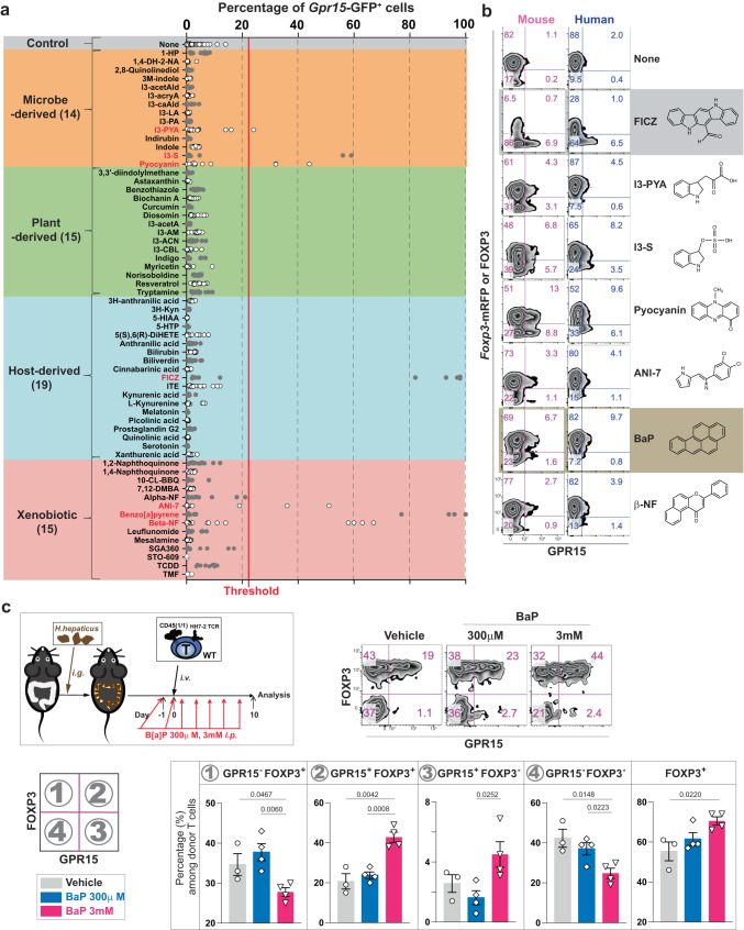
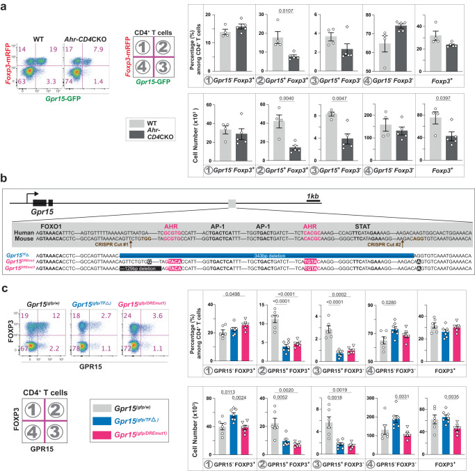
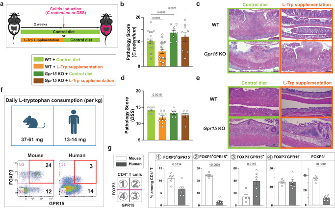
Environmental factors are the major contributor to the onset of immunological disorders such as ulcerative colitis. However, their identities remain unclear. Here, we discover that the amount of consumed L-Tryptophan (L-Trp), a ubiquitous dietary component, determines the transcription level of the colonic T cell homing receptor, GPR15, hence affecting the number of colonic FOXP3+ regulatory T (Treg) cells and local immune homeostasis. Ingested L-Trp is converted by host IDO1/2 enzymes, but not by gut microbiota, to compounds that induce GPR15 transcription preferentially in Treg cells via the aryl hydrocarbon receptor. Consequently, two weeks of dietary L-Trp supplementation nearly double the colonic GPR15+ Treg cells via GPR15-mediated homing and substantially reduce the future risk of colitis. In addition, humans consume 3-4 times less L-Trp per kilogram of body weight and have fewer colonic GPR15+ Treg cells than mice. Thus, we uncover a microbiota-independent mechanism linking dietary L-Trp and colonic Treg cells, that may have therapeutic potential.


Conflict of interest statement

S.V.K. is a co-inventor of a patent (US 9,376,663 B2) related to this work. The remaining authors declare no competing interests.

Figures

Fig. 1
Fig. 1. The amount of ingested dietary L-Trp determines the number of colonic CD4+ T cells via the AhR-GPR15 pathway.
a Major questions addressed in our study. b Three different types of elementary diets (Trp-C (green); Trp-Sup (orange); Trp-Def (blue), Table S1) were fed to mice with TJU microbiota 1 for three weeks. Representative flow cytometry plots for CD4+ T cells in the LILP were shown. c, d Gpr15-GFP and Foxp3-mRFP expression was examined in CD4+ T cells in the LILP (c in the pink shade) and the spleen (d in the light blue shade) from wild-type (WT: Ahr(fl/fl)Gpr15(gfp/+)Foxp3mrfp) and Ahr-CD4CKO mice (Cd4CreAhr(fl/fl)Gpr15(gfp/+)Foxp3mrfp). 8–14-week-old mice were used. Data are presented as mean values ± SEM of the percentage of each population of CD4+ T cells or the cell numbers. The numbers of mice used (n) for WT group were 8 for Trp-C, 7 for Trp-Sup, and 7 for Trp-Def. The mice used for Ahr-CD4CKO group were 10 for Trp-C, 15 for Trp-Sup, 8 for Trp-Def. Representative of three independent experiments. e 8–10-week-old WT mice from the Jackson Laboratory (JAX) were treated with Trp-C or Trp-Sup elementary diets for 1–3 weeks (1wk (n = 5), 2wk (n = 5), 3wk (n = 5)). Data are presented as mean values±SEM of the percentage of each population of CD4+ T cells. Representative of two independent experiments. Each data point represents the result from one mouse, and p values were calculated by two-sided student’s t-test (ce). Source data are provided as a Source Data file (ce).
Fig. 2
Fig. 2. L-Trp supplementation increases GPR15 expression via AhR during CD4+ T cell activation and their migration to the large intestine.
a Representative flow cytometry plot of CD4+ T cells in the LILP, SILP, and mesenteric lymph nodes-draining the proximal and mid-colon (MLN-L) in wild-type (WT) mice from JAX. b CD4+ T cells in the LILP, SILP, and MLN-L of WT mice from JAX with 8–12 weeks of age were analyzed for GPR15 (in the light blue shade) or CCR9 expression (in the light purple shade) in addition to FOXP3 expression after elementary diet treatment. The number of mice used: 10 for Trp-C and 10 for Trp-Sup. c GPR15+ Treg cells in the LILP in b were analyzed for HELIOS and RORγt expression. Trp-C (n = 10), Trp-Sup (n = 10). Combined results of two independent experiments and representative flow cytometry plots (b, c). df WT mice (JAX) were gavaged with H. hepaticus and treated with Trp-C or Trp-Sup elementary diets. After 3 days of elementary diets, recipient mice were transferred intravenously with 1:1 mixture of 104 naive CD4+ T cells from two different mice (WT: HH7-2TgRag1(n/n)Ahr(fl/fl)CD45(1/2); Ahr-CD4CKO: HH7-2Tg Rag1(n/n)CD4creAhr(fl/fl)CD45(1/1)) and analyzed 10 days later. d Schematic plan. e Representative flow cytometry plots of donor cells in the LILP. f The percentages of different populations among total donor T cells are shown. Data are presented as mean values ± SEM. The number of mice used: n = 7 for LILP, n = 5 for SILP, n = 7 for spleen for recipients treated with Trp-C; n = 8 for LILP, n = 5 for SILP, n = 8 for spleen for recipients treated with Trp-Sup. Representative of two independent experiments. 8–12-week-old mice were used (ac, e, f). Data are presented as mean values ± SEM (b, c, f). Each data point represents the result from one mouse, and p values were calculated by two-sided student’s t-test (b, c, f). Source data are provided as a Source Data file (b, c, f).
Fig. 3
Fig. 3. The default response to L-Trp is a selective increase of GPR15+ Treg cells via host IDO1/2 enzymes in a microbiota-independent manner.
a. L-Trp metabolic pathways upon ingestion. b-f. Percentages of each population among CD4+ T cells in the LILP based on Gpr15-GFP and Foxp3-mRFP expression (b) or GPR15 and FOXP3 expression (c–f). b, c Wild-type (WT) mice (Gpr15(gfp/+)Foxp3mrfp) with Thomas Jefferson University (TJU) microbiota (two different microbiota: TJU1 or TJU2) (b), or WT mice with Jackson laboratory microbiota (JAX) or Taconic Biosciences microbiota (TAC) (c) were treated with Trp-C or Trp-Sup elementary diets for 2 wks. The number of mice used (n): 8 for TJU1 (Trp-C), 7 for TJU1 (Trp-Sup), 5 each for TJU2 (Trp-C and Trp-Sup), 5 each for JAX (Trp-C and Trp-Sup), 5 for TAC (Trp-C), 4 for TAC (Trp-Sup). d Specific pathogen-free (SPF) mice (TAC, n = 6) and germ-free mice (n = 8) were compared at a steady state. e WT mice with JAX microbiota were treated with four different antibiotics (4xAbx) in drinking water for four weeks. Elementary diets (Trp-Lo (n = 8) or Trp-Sup (n = 8)) were provided for two weeks in the presence of 4xAbx. Representatives of two independent experiments (b, c, e). f Ido1/2(n/+), Ido2(n/n), and Ido1/2(n/n) mice were analyzed at a steady state. Combined results of five independent experiments. The number of mice used: 15 for Ido1/2(n/+), 18 for Ido2(n/n), 18 for Ido1/2(n/n). 8–14-week-old mice were used (b-f). Data are presented as mean values ± SEM (bf). Each data point represents the result from one mouse, and p values were calculated by two-sided student’s t-test (bf). Source data are provided as a Source Data file (bf).
Fig. 4
Fig. 4. The identity of AhR ligand determines the T cell types expressing GPR15.
a Chemical compounds with known AhR ligand activity (Table S2) were categorized based on their origin and tested on CD4+ naive T cells from Gpr15(gfp/+)Foxp3mrfp mice. The red vertical line is set as a threshold for further analysis. Seven compounds (labeled red) induced a GFP signal above the threshold. b Compounds shown to induce GPR15 expression in Fig. 4a were tested with mouse and human CD4+ T cells for GPR15 and FOXP3 protein expression. Representative flow cytometry plots of at least three independent experiments. c Wild-type mice (JAX) were gavaged and injected intraperitoneally with 300 μl of either PBS (n = 3) or 300 μM (n = 4) or 3 mM (n = 4) of BaP 7 times. GPR15 and FOXP3 expression of T cells with HH7-2 TCRs were analyzed on day 10. The percentage of each population in CD4+ donor T cells in the LILP is shown. Representative of two independent experiments. Data are presented as mean values±SEM (c). Each data point represents the result from one mouse, and p values were calculated by two-sided student’s t-test (c). 8-14-week-old mice were used (ac). Source data are provided as a Source Data file (a, c).
Fig. 5
Fig. 5. AhR directly controls the transcriptional activation of Gpr15 in CD4+ T cells via dioxin-responsive elements (DRE).
a Gpr15 and Foxp3 mRNA expression in CD4+ T cells in the LILP was determined by the reporter expression in the presence (WT: Ahr(fl/fl)Gpr15(gfp/+)Foxp3mrfp, n = 4) or the absence (Ahr-CD4CKO: CD4CreAhr(fl/fl)Gpr15(gfp/+)Foxp3mrfp, n = 5) of AhR. Flow cytometry plots and the percentage/cell number of each population among CD4+ T cells in the LILP are shown. Representatives of at least two independent experiments. b The DNA sequence of the conserved region in the 3′-distal to Gpr15 locus. Our knock-in strategy is shown: Gpr15TFΔ allele has a deletion of 343 bps containing two AhR binding sites, two AP-1 binding sites, and a STAT binding site. Also, we intended to mutate two AhR binding sites to make a Gpr15DREmut allele but ended up with a Gpr15DREmut1 allele having an additional 129 bp deletion of the non-conserved sequence. c Representative flow cytometry plots and the percentage/cell number of two independent experiments are shown for CD4+ T cells in the LILP. The number of mice used: 6 for WT, 7 for Gpr15TFΔ, and 6 for a Gpr15DREmut1. 8–14-week-old mice were used (a, c). Data are presented as mean values±SEM (a, c). Each data point represents the result from one mouse, and p values were calculated by two-sided student’s t-test (a, c). Source data are provided as a Source Data file (a, c).
Fig. 6
Fig. 6. L-Trp supplementation reduces the future risk of developing colitis through GPR15.
ae Wild-type (WT) mice or Gpr15(gfp/gfp) (Gpr15 KO) mice with JAX microbiota were fed with control diet or L-Trp supplemented diet for two weeks. Both mice were switched to control diet and gavaged with C. rodentium or treated with DSS. Mice were analyzed at day 10 (for C. rodentium) or day 7 after colitis induction (for DSS). b Pathology score of the large intestine with C. rodentium-induced colitis. Number of mice used: 18 for WT with Control diet, 20 for WT with L-Trp supplementation, 9 for Gpr15 KO with Control diet, and 9 for Gpr15 KO with L-Trp supplementation. Combined results of four independent experiments. c Representative images of large-intestine tissues (C. rodentium-induced colitis), stained with Hematoxylin and Eosin (H/E). The bars represent 100 μm. d Pathology score of the large intestine with DSS-induced colitis. Number of mice used: 9 for WT with Control diet, 10 for WT with L-Trp supplementation, 7 for Gpr15 KO with Control diet, and 7 for Gpr15 KO with L-Trp supplementation. Combined results of two independent experiments. e Representative images of large-intestine tissues (DSS-induced colitis). The bars represent 100 μm. f, g Estimated daily L-Tryptophan consumption between mice and humans after normalization and the expression of GPR15 and FOXP3 among CD4+ T cells in the large intestine are shown (n = 5 for mice, n = 6 for humans). 8–14-week-old mice were used (bg). Data are presented as mean values±SEM (b, d, g). Each data point represents the result from one mouse or one human, and p values were calculated by two-sided student’s t-test (b, d, g). Source data are provided as a Source Data file (b, d, f, g).

References

    1. Cleynen, I. et al. Inherited determinants of Crohn’s disease and ulcerative colitis phenotypes: a genetic association study. Lancet10.1016/s0140-6736(15)00465-1 (2015). - PMC - PubMed
    1. Alatab S, et al. The global, regional, and national burden of inflammatory bowel disease in 195 countries and territories, 1990–2017: a systematic analysis for the Global Burden of Disease Study 2017. Lancet Gastroenterol. Hepatol. 2019;5:17–30. doi: 10.1016/S2468-1253(19)30333-4. - DOI - PMC - PubMed
    1. Kobayashi T, et al. Ulcerative colitis. Nat. Rev. Dis. Prim. 2020;6:74. doi: 10.1038/s41572-020-0205-x. - DOI - PubMed
    1. Ellinghaus D, et al. Analysis of five chronic inflammatory diseases identifies 27 new associations and highlights disease-specific patterns at shared loci. Nat. Genet. 2016;48:510–518. doi: 10.1038/ng.3528. - DOI - PMC - PubMed
    1. Liu JZ, et al. Association analyses identify 38 susceptibility loci for inflammatory bowel disease and highlight shared genetic risk across populations. Nat. Genet. 2015;47:979–986. doi: 10.1038/ng.3359. - DOI - PMC - PubMed

Publication types