

Cerebellar Volume and Disease Staging in Parkinson's Disease: An ENIGMA-PD Study
- PMID: 37964373
- PMCID: PMC10754393
- DOI: 10.1002/mds.29611
Cerebellar Volume and Disease Staging in Parkinson's Disease: An ENIGMA-PD Study
Abstract
Background: Increasing evidence points to a pathophysiological role for the cerebellum in Parkinson's disease (PD). However, regional cerebellar changes associated with motor and non-motor functioning remain to be elucidated.
Objective: To quantify cross-sectional regional cerebellar lobule volumes using three dimensional T1-weighted anatomical brain magnetic resonance imaging from the global ENIGMA-PD working group.
Methods: Cerebellar parcellation was performed using a deep learning-based approach from 2487 people with PD and 1212 age and sex-matched controls across 22 sites. Linear mixed effects models compared total and regional cerebellar volume in people with PD at each Hoehn and Yahr (HY) disease stage, to an age- and sex- matched control group. Associations with motor symptom severity and Montreal Cognitive Assessment scores were investigated.
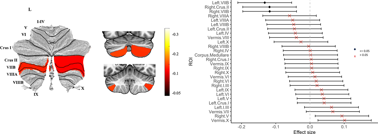
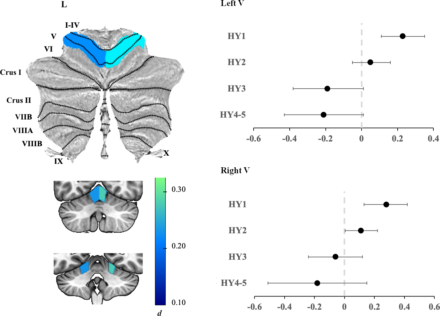
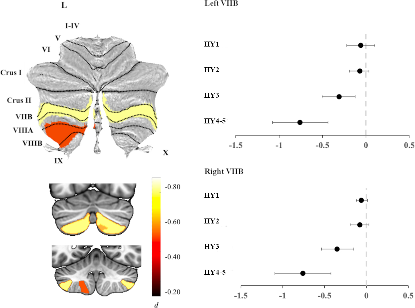
Results: Overall, people with PD had a regionally smaller posterior lobe (dmax = -0.15). HY stage-specific analyses revealed a larger anterior lobule V bilaterally (dmax = 0.28) in people with PD in HY stage 1 compared to controls. In contrast, smaller bilateral lobule VII volume in the posterior lobe was observed in HY stages 3, 4, and 5 (dmax = -0.76), which was incrementally lower with higher disease stage. Within PD, cognitively impaired individuals had lower total cerebellar volume compared to cognitively normal individuals (d = -0.17).
Conclusions: We provide evidence of a dissociation between anterior "motor" lobe and posterior "non-motor" lobe cerebellar regions in PD. Whereas less severe stages of the disease are associated with larger motor lobe regions, more severe stages of the disease are marked by smaller non-motor regions. © 2023 The Authors. Movement Disorders published by Wiley Periodicals LLC on behalf of International Parkinson and Movement Disorder Society.
Keywords: MRI; Parkinson's disease; cerebellum; disease staging.
© 2023 The Authors. Movement Disorders published by Wiley Periodicals LLC on behalf of International Parkinson and Movement Disorder Society.
Conflict of interest statement
Relevant conflicts of interest/financial disclosures
Figures
References
Publication types
MeSH terms
Grants and funding
- T32 AG000258/AG/NIA NIH HHS/United States
- U54EB020403/NH/NIH HHS/United States
- RC4 NS073008/NH/NIH HHS/United States
- NS053488 (P50)/NS/NINDS NIH HHS/United States
- R01 AG066152/AG/NIA NIH HHS/United States
- R01 AG080734/AG/NIA NIH HHS/United States
- R01 NS109260/NS/NINDS NIH HHS/United States
- R01AG080734/NH/NIH HHS/United States
- R01MH116147/NH/NIH HHS/United States
- P30AG072979/NH/NIH HHS/United States
- P30 AG072979/AG/NIA NIH HHS/United States
- NS115114/NS/NINDS NIH HHS/United States
- R01 AG058854/AG/NIA NIH HHS/United States
- P41 EB015922/EB/NIBIB NIH HHS/United States
- R01AG058854/NH/NIH HHS/United States
- U01NS107027/NH/NIH HHS/United States
- R01 AG076832/AG/NIA NIH HHS/United States
- P01AG066597/NH/NIH HHS/United States
- U54 NS092091/NS/NINDS NIH HHS/United States
- NS075097/NS/NINDS NIH HHS/United States
- R01AG066152/NH/NIH HHS/United States
- R01 MH116147/MH/NIMH NIH HHS/United States
- P50 NS053488/NS/NINDS NIH HHS/United States
- R01MH117601/NH/NIH HHS/United States
- U19AG062418/NH/NIH HHS/United States
- R01 AG070885/NH/NIH HHS/United States
- U01 NS107027/NS/NINDS NIH HHS/United States
- U54 EB020403/EB/NIBIB NIH HHS/United States
- R01 MH117601/MH/NIMH NIH HHS/United States
- P01 AG066597/AG/NIA NIH HHS/United States
- 2019NF4100087335/NH/NIH HHS/United States
- R01AG059874/NH/NIH HHS/United States
- R01NS107513/NH/NIH HHS/United States
- R01 NS107513/NS/NINDS NIH HHS/United States
- AG062418 (U19)/NS/NINDS NIH HHS/United States
- P50 NS062684/NS/NINDS NIH HHS/United States
- R56 AG058854/AG/NIA NIH HHS/United States
- RC4 NS073008/NS/NINDS NIH HHS/United States
- 180365/SNSF_/Swiss National Science Foundation/Switzerland
- P50 NS062684/NH/NIH HHS/United States
- K23 NS075097/NS/NINDS NIH HHS/United States
- U54NS092091/NH/NIH HHS/United States
- R01NS109260/NH/NIH HHS/United States
- U19 AG062418/AG/NIA NIH HHS/United States
- R01 AG070885/AG/NIA NIH HHS/United States
- 204593 ("ScanOMetrics")/SNSF_/Swiss National Science Foundation/Switzerland
- R01 NS115114/NS/NINDS NIH HHS/United States
- R01 AG059874/AG/NIA NIH HHS/United States
LinkOut - more resources
Full Text Sources
Medical
Miscellaneous
