. 2023 Oct 27:32:488-501.
doi: 10.1016/j.bioactmat.2023.09.019. eCollection 2024 Feb.

Profibrogenic macrophage-targeted delivery of mitochondrial protector via exosome formula for alleviating pulmonary fibrosis

Affiliations

Profibrogenic macrophage-targeted delivery of mitochondrial protector via exosome formula for alleviating pulmonary fibrosis

Wei Zhang et al. Bioact Mater. .

Abstract

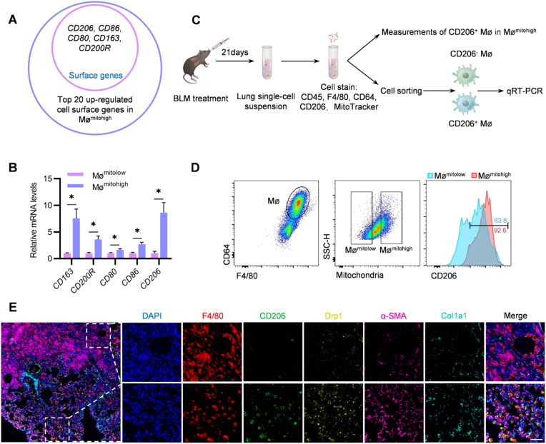
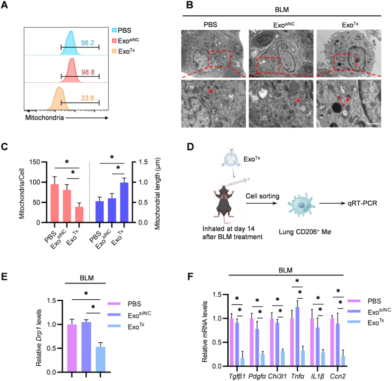
Pulmonary fibrosis (PF) is a devastating lung disease with limited treatment options. During this pathological process, the profibrogenic macrophage subpopulation plays a crucial role, making the characterization of this subpopulation fundamentally important. The present study revealed a positive correlation between pulmonary macrophages with higher mitochondrial mass (Mømitohigh) and fibrosis. Among the Mømitohigh subpopulation of CD206+ M2, characterized by higher expression of dynamin 1-like (Drp1), as determined by flow cytometry and RNA-seq analysis, a therapeutic intervention was developed using an exosome-based formula composed of pathfinder and therapeutics. A pathfinder exosome called "exosomeMMP19 (ExoMMP19)", was constructed to display matrix metalloproteinase-19 (MMP19) on the surface to locally break down the excessive extracellular matrix (ECM) in the fibrotic lung. A therapeutic exosome called "exosome therapeutics (ExoTx)", was engineered to display D-mannose on the surface while encapsulating siDrp1 inside. Prior delivery of ExoMMP19 degraded excessive ECM and thus paved the way for ExoTx to be delivered into Mømitohigh, where ExoTx inhibited mitochondrial fission and alleviated PF. This study has not only identified Mømitohigh as profibrotic macrophages but it has also provided a potent strategy to reverse PF via a combination of formulated exosomes.

Keywords: Drp1; Exosomes; Macrophages; Mitochondrial fission; Pulmonary fibrosis.


Conflict of interest statement

The authors declare that they have no known competing financial interests or personal relationships that could have appeared to influence the work reported in this paper.

Figures

Image 1
Graphical abstract
Scheme 1
Scheme 1
Schematic illustration of the study. ExoMMP19, the pathfinder exosome, and ExoTx, the therapeutic exosome, were engineered. As prior delivery of ExoMMP19 degraded excessive ECM, the way was paved for ExoTx delivery into Mømitohigh, the novel profibrogenic macrophage subtype we identified. The formulated exosome therapy inhibited mitochondrial fission and alleviated pulmonary fibrosis efficiently.
Fig. 1
Fig. 1
Drp1 was highly expressed in Mømitohigh of mice with PF. (A) Schematic illustration of the process for RNA-Seq analysis. (B) Flow cytometric analysis of lung Mitohigh macrophages after intratracheal administration of PBS/BLM. Representative data from three independent experiments. (C) Volcano plot showing DEG between Mømitolow and Mømitohigh in fibrotic lung tissues. (D) GSEA analysis of Mømitolow and Mømitohigh. NES, normalized enrichment score. P, Nominal P value. (E) Heat map diagram of mitochondrial fission related genes expression in Mømitolow and Mømitohigh. n = 3. (F) qRT-PCR analysis of mitochondrial fission related genes in Mømitolow and Mømitohigh in fibrotic lung tissues. Data are expressed as mean ± SEM of three independent experiments. *p < 0.05 by two-tailed unpaired Student's t-test.
Fig. 2
Fig. 2
Interference with Drp1 reversed mitochondrial dysfunction and downregulates profibrotic genes in vitro. (A) Schematic illustration of the experiment. BMDMs were transfected with PBS/siNC/siDrp1. (B) qRT-PCR analysis of relative expression of Drp1 in BMDMs transfected with PBS/siNC/siDrp1. Data are expressed as mean ± SEM of three independent experiments. *p < 0.05 by One-way ANOVA. (C) Western blot analysis of Drp1 expression in BMDMs transfected with PBS/siNC/siDrp1. GAPDH served as the loading control. Representative data from three independent experiments. (D) Flow cytometric analysis of mitochondrial mass in BMDMs transfected with PBS/siNC/siDrp1. Representative data from three independent experiments. (E) Flow cytometric analysis of mtROS in BMDMs transfected with PBS/siNC/siDrp1. Representative data from three independent experiments. (F) Flow cytometric analysis of Δψm in BMDMs transfected with PBS/siNC/siDrp1. Representative data from three independent experiments. (G) Representative TEM images of mitochondrial morphology in BMDMs transfected with PBS/siNC/siDrp1. Scale bars: 1 μm. (H) Calculated mitochondria per cell and mitochondrial length. Data are expressed as mean ± SEM of three independent experiments in which 30 cells were analyzed. *p < 0.05 by One-way ANOVA. (I) qRT-PCR analysis of relative expression of Tgfβ1, Pdgfα, Chi3l1, Tnfα, IL1β and Ccn2 in BMDMs transfected with PBS/siNC/siDrp1. Data are expressed as mean ± SEM of three independent experiments. *p < 0.05 by One-way ANOVA.
Fig. 3
Fig. 3
Drp1 was highly expressed in CD206+ macrophages. (A) Venn diagram of highly-expressed cell surface genes of Mømitohigh compared to Mømitolow. (B) qRT-PCR analysis of macrophage markers in Mømitolow and Mømitohigh in fibrotic lung tissues. Data are expressed as mean ± SEM of three independent experiments. *p < 0.05 by two-tailed unpaired Student's t-test. (C) Schematic illustration of the process for lung flow cytometric analysis and cell sorting. (D) Flow cytometric analysis of mice lung CD206+ macrophages in Mømitohigh after BLM-induced PF. Representative data from three independent experiments. (E) Representative multiplexed immunofluorescence images of Drp1 in macrophages of fibrotic lung tissues. Scale bar: 50 μm. n = 3.
Fig. 4
Fig. 4
ExoTarget can target CD206+ macrophages in vivo. (A) Schematic illustration of the experimental procedure. (B) Biodistribution of DiR-labeled ExoNone/ExoTraptavidin/ExoTarget among organs after the intratracheal administration. n = 5. (C) Schematic illustration of the experimental procedure. (D) Representative fluorescence images of the DiI-labeled ExoNone/ExoTraptavidin/ExoTarget in lung tissues. Scale bar:10 μm. n = 5. (E) The percentage of triple-positive cells in DiI-positive cells. n = 5. *p < 0.05 by One-way ANOVA. (F) Flow cytometric analysis of DiO-positive CD206+ macrophages in fibrotic lung tissues of mice treated with ExoNone/ExoTraptavidin/ExoTarget. Representative data from five independent experiments.
Fig. 5
Fig. 5
ExoTx decreased mitochondrial mass and downregulated the profibrotic genes in vivo. (A) Flow cytometric analysis of mitochondrial mass in CD206+ macrophages in fibrotic lung tissues of mice treated with PBS/ExosiNC/ExoTx. Representative data from five independent experiments. (B) Representative TEM images of mitochondrial morphology in lung macrophages treated with PBS/ExosiNC/ExoTx. Scale bars: 1 μm. (C) Calculated mitochondria per cell and mitochondrial length. Data are expressed as mean ± SEM of five independent experiments in which 50 cells were analyzed. *p < 0.05 by One-way ANOVA. (D) Schematic illustration of the process for lung cells sorting after exosomes treated. (E) qRT-PCR analysis of Drp1 in lung CD206+ macrophages of BLM-induced mice treated with PBS/ExosiNC/ExoTx. Data are expressed as mean ± SEM of five independent experiments. *p < 0.05 by One-way ANOVA. (F) qRT-PCR analysis of Tgfβ1, Pdgfα, Chi3l1, Tnfα, IL1β and Ccn2 in lung CD206+ macrophages of BLM-induced mice treated with PBS/ExosiNC/ExoTx. Data are expressed as mean ± SEM of five independent experiments. *p < 0.05 by One-way ANOVA.
Fig. 6
Fig. 6
ExoMMP19degraded excessive ECM and improved the targeting efficacy of ExoTarget. (A) Schematic illustration of the experimental procedure. (B) Western blot analysis of fibronectin, Col1a1 and α-SMA of L929 cells. GAPDH served as the loading control. Representative data from three independent experiments. (C) Schematic illustration of the experimental procedure. (D) Biodistribution of DiR-labeled ExoNone/ExoEmpty vector/ExoMMP19 among organs after the intratracheal administration. n = 5. (E) Schematic illustration of the experimental procedure. (F) Representative fluorescence images of the DiI-labeled ExoNone/ExoEmpty vector/ExoMMP19 in lung tissues. Scale bar:20 μm. n = 5. (G) Col1a1 relative MFI was analyzed. MFI, mean fluorescence intensity. n = 5. *p < 0.05 by One-way ANOVA. (H) Schematic illustration of the experimental procedure. (I) Representative fluorescence images of the DiI-labeled ExoTarget in lung tissues. Scale bar:10 μm. n = 5. (J) The percentage of triple-positive cells in DiI-positive cells. n = 5. *p < 0.05 by two-tailed unpaired Student's t-test. (K) Flow cytometric analysis of DiO-positive CD206+ macrophages in fibrotic lung tissues of mice treated with ExoTarget + ExoNone or ExoTarget + ExoMMP19. Representative data from five independent experiments.
Fig. 7
Fig. 7
Therapeutic efficacy of ExoTx combined with ExoMMP19in vivo. (A) Schematic illustration of the experimental procedure and exosomes treatment time course. (B) Survival curve of the mice after treatments. n = 20. *p < 0.05. (C) Representative H&E, Masson's trichrome, Sirius red staining images and immunohistochemical staining of α-SMA of lung tissues in different groups. Scale bar: 200 μm. n = 5. (D) qRT-PCR analysis of Fn1, Col1a1 and Acta2 of lung tissues with different treatments. *p < 0.05. Data are expressed as mean ± SEM of five independent experiments. *p < 0.05 by One-way ANOVA. (E) Western blot analysis of fibronectin, Col1a1 and α-SMA of lung tissues with different treatments. GAPDH served as the loading control. Representative data from five independent experiments.

References

    1. Selman M., Pardo A. Idiopathic pulmonary fibrosis: an epithelial/fibroblastic cross-talk disorder. Respir. Res. 2002;3:3. - PMC - PubMed
    1. Ley B., Collard H.R., King T.E., Jr. Clinical course and prediction of survival in idiopathic pulmonary fibrosis. Am. J. Respir. Crit. Care Med. 2011;183(4):431–440. - PubMed
    1. Roger A.J., Munoz-Gomez S.A., Kamikawa R. The origin and diversification of mitochondria. Curr. Biol. 2017;27(21):R1177–R1192. - PubMed
    1. Wang Y., Li N., Zhang X., Horng T. Mitochondrial metabolism regulates macrophage biology. J. Biol. Chem. 2021;297(1) - PMC - PubMed
    1. Xiong Q., Tian X., Xu C., Ma B., Li W., Xia Y., Liu W., Sun B., Ru Q., Shu X. Mediation of PM2.5-induced cytotoxicity: the role of P2X7 receptor in NR8383 cells. Int. J. Environ. Health Res. 2023:1–13. - PubMed

LinkOut - more resources