. 2024 Mar;244(3):448-457.
doi: 10.1111/joa.13976. Epub 2023 Nov 15.

Axillary arch (of Langer): A large-scale dissection and simulation study based on unembalmed cadavers of body donors

Affiliations

Axillary arch (of Langer): A large-scale dissection and simulation study based on unembalmed cadavers of body donors

Jeremias T Weninger et al. J Anat. 2024 Mar.

Abstract

Connective or muscular tissue crossing the axilla is named axillary arch (of Langer). It is known to complicate axillary surgery and to compress nerves and vessels transiting from the axilla to the arm. Our study aims at systematically researching the frequency, insertions, tissue composition and dimension of axillary arches in a large cohort of individuals with regard to gender and bilaterality. In addition, it aims at evaluating the ability of axillary arches to cause compression of the axillary neurovascular bundle. Four hundred axillae from 200 unembalmed and previously unharmed cadavers were investigated by careful anatomical dissection. Identified axillary arches were examined for tissue composition and insertion. Length, width and thickness were measured. The relation of the axillary arch and the neurovascular axillary bundle was recorded after passive arm movements. Twenty-seven axillae of 18 cadavers featured axillary arches. Macroscopically, 15 solely comprised muscular tissue, six connective tissue and six both. Their average length was 79.56 mm, width 7.44 mm and thickness 2.30 mm. One to three distinct insertions were observed. After passive abduction and external rotation of the arm, 17 arches (63%) touched the neurovascular axillary bundle. According to our results, 9% of the Central European population feature an axillary arch. Approximately 50% of it bilaterally. A total of 40.74% of the arches have a thickness of 3 mm or more and 63% bear the potential of touching or compressing the neuromuscular axillary bundle upon arm movement.

Keywords: axilla; axillopectoral muscle; chondroepitrochlearis muscle; thoracic outlet syndrome; variation.


Conflict of interest statement

The authors declare that they have no known competing financial interests or personal relationships that could have appeared to influence the work reported in this article.

Figures
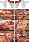
FIGURE 1
FIGURE 1
Frequent insertions (arrowheads) of axillary arches (asterisk). Double headed arrows indicate proximal (P) and distal (D). (a–d) Scheme demonstrating the area exposed by microdissection on the right (a, b) and left body side (c, d). (e–j) Cadaver specimens. Inlays show displayed specimens in lower magnification. The boxed area in the inlay is the field of view of the panel. (e, f) Single insertion of the axillary arch (asterisk) at deep aspect of the pectoralis major muscle tendon (PM). The muscle is cut from its origins and flipped anterolaterally. Note that in F also the axillary arch is cut and flipped medially to expose the neurovascular bundle (NB) transiting the axilla. (g) Insertion of axillary arch in the fascia covering the superficial aspect of the coracobrachialis muscle and the short head of the biceps brachii muscle (BCM). (h) Insertion of axillary arch in the deep fascia and tendon of the pectoralis major, the connective tissue sheath covering the deep surface of the pectoralis minor muscle, before reaching the coracoid process (PMI) and fascia covering the superficial aspect of the coracobrachialis muscle and the short head of the biceps brachii muscle. (i) Insertion at greater tubercle of the humerus, independently from the tendon of the pectoralis major muscle. (j) Insertion at coracoid process and fascia covering the superficial aspect of the coracobrachialis muscle and the short head of the biceps brachii muscle; UL, upper limb; TH, thorax; LD, latissimus dorsi muscle; TM, triceps brachii muscle. Scalebars 2 cm.
FIGURE 2
FIGURE 2
Unusual axillary arches (white asterisk) in an 82‐year‐old female cadaver. Note the insertion at the medial epicondyle of the humerus (ME) via a chondroepitrochlearis muscle tendon (yellow asterisk). Double headed arrow indicates proximal (P) and distal (D). (a) Dissected right upper arm. (b, c) Detail of insertion at the medial epicondyle of the humerus (b) and pectoralis major muscle (PM) (c). (d) Dissected left upper arm. (e, f) Details of insertion at the medial epicondyle of the humerus (e) and the fascia covering the superficial aspect of the coracobrachialis muscle and the short head of the biceps brachii muscle (BCM). LD, latissimus dorsi muscle; NB, neurovascular bundle; TM, triceps brachii muscle; DM, deltoid muscle; UN, ulnar nerve; MN, median nerve; GU, groove for ulnar nerve. Scalebars 2 cm.
FIGURE 3
FIGURE 3
Characterization of axillary arches. (a) Insertions of axillary arches (n = 27) according to the principally reached locations and in respect to combined insertions. (b) Tissue composition in relation to dimensions. (c) Dimensions and macroscopic tissue composition in relation to the ability to touch the axillary neurovascular bundle (nb) at maximal abduction and external rotation of the arm. Brown dots, axillary arches macroscopically solely composed of muscular tissue; blue dots, axillary arches macroscopically solely composed of connective tissue; green dots, axillary arches macroscopically composed of muscular and connective tissue. pm, pectoralis major muscle; bcm, fascia covering the superficial aspect of the coracobrachialis muscle and the short head of the biceps brachii muscle; cp, coracoid process; pmi, pectoralis minor muscle; gt, greater tubercle of the humerus; me, medial epicondyle of the humerus.
FIGURE 4
FIGURE 4
Macroscopical tissue composition of axillary arches (asterisk). Inlay demonstrates laterality of specimen. Double headed arrow indicates proximal (P) and distal (D). (a) Axillary arch macroscopically solely composed of muscular tissue. (b) Axillary arch macroscopically composed of both, muscular and connective tissue. (c) Axillary arch macroscopically solely composed of fibrous tissue. PM, pectoralis major muscle; BCM, fascia covering the superficial aspect of the coracobrachialis muscle and the short head of the biceps brachii muscle; NB, neurovascular bundle; LD, latissimus dorsi muscle; PMI, pectoralis minor muscle. Scalebars 2 cm.

Comment in

References

    1. Ando, J. , Kitamura, T. , Kuroki, Y. & Igarashi, S. (2010) Preoperative diagnosis of the axillary arch with multidetector row computed tomography and the axillary arch in association with anatomical problems of sentinel lymph node biopsy. Breast Cancer, 17, 3–8. - PubMed
    1. Besana‐Ciani, I. & Greenall, M.J. (2005) Langer's axillary arch: anatomy, embryological features and surgical implications. The Surgeon, 3, 325–327. - PubMed
    1. Birmingham, A. (1889) Homology and innervation of the achselbogen and pectoralis quartus, and the nature of the lateral cutaneous nerve of the thorax. Journal of Anatomy and Physiology, 23, 206–223. - PMC - PubMed
    1. Boontje, A.H. (1979) Axillary vein entrapment. British Journal of Surgery, 66, 331–332. - PubMed
    1. Chiba, S. , Suzuki, T. & Kasai, T. (1983) A rare anomaly of the pectoralis major? The chondroepitrochlearis. Okajimas Folia Anatomica Japonica, 60, 175–185. - PubMed