. 2024 Mar;244(3):438-447.
doi: 10.1111/joa.13977. Epub 2023 Nov 15.

Is thoracolumbar fascia shear-wave modulus affected by active and passive knee flexion?

Affiliations

Is thoracolumbar fascia shear-wave modulus affected by active and passive knee flexion?

Eleftherios Kellis et al. J Anat. 2024 Mar.

Abstract

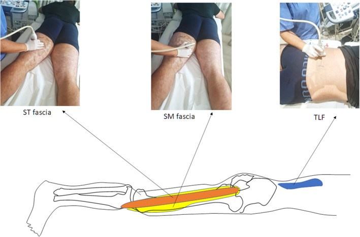
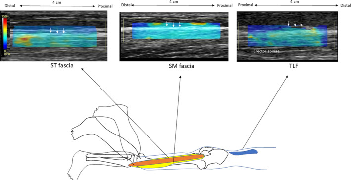
The purpose of this study was to examine the effect of passive and active knee flexion efforts on the stiffness of the thoracolumbar (TLF), semitendinosus (STF), and semimembranosus fascia (SMF). Fourteen young healthy males participated in this study. Using ultrasound shear-wave elastography, fascia elastic modulus was measured at rest (passive condition) and during submaximal isometric knee flexion efforts (active condition) with the hip at neutral position and the knee flexed at 0°, 45°, and 90°. Analysis of variance designs indicated that when the knee was passively extended from 90° to 0°, shear modulus of the TLF, SMF, and STF increased significantly (p < 0.05). Similarly, active knee flexion contractions caused a significant increase in TLF, SMF, and STF shear modulus (p < 0.001). Compared to hamstring fascia, the TLF showed greater thickness but a lower shear modulus (p < 0.05) while STF modulus was greater compared that to SMF during active contraction (p < 0.05). These results indicate that exercising the hamstring muscles can remotely influence the stiffness of the fascia which surrounds the lumbar area.

Keywords: elastography; in vivo; myofascial path; semimembranosus; semitendinosus; spine; stiffness.


Conflict of interest statement

The authors declare no conflict of interest.

Figures

FIGURE 1
FIGURE 1
Experimental setup. The participant assumed the prone position and the knee was passively moved at three knee joint positions. The ultrasound probe was placed 2.5 cm away from the L3 spinous process at the L3–L4 level to visualize the thoracolumbar fascia. The corresponding position for the semitendinosus fascia was in the middle of the muscle belly, approximately at 40% of the distance between the medial condyle and the ischial tuberosity. The SM fascia was identified more medially than ST fascia. SM, semimembranosus fascia; ST, semitendinosus fascia; TLF, thoracolumbar fascia.
FIGURE 2
FIGURE 2
Example elastograms from the semitendinosus, the semimembranosus, and thoracolumbar fasciae. First, elastographic data were obtained using a rectangular color‐coded box to isolate the measurement area. Then, circles of smaller diameter were drawn along each fascia (positions are indicated with vertical arrows) and the software provided the shear modulus. The color scale was extracted from the software and is enlarged so that the measurement scale is easily visible. SM, semimembranosus fascia; ST, semitendinosus fascia; TLF, thoracolumbar fascia.
FIGURE 3
FIGURE 3
Mean group shear modulus values of the thoracolumbar (TLF), semimembranosus (SMF) and semitendinosus (STF) fascia at rest and contraction. Bars indicate average values from all angular positions. Error bars indicate standard deviation. #Significantly different compared to active condition, p < 0.05. *Significantly different compared to STF and SMF, p < 0.05. ^Significantly different compared to SMF, p < 0.05.
FIGURE 4
FIGURE 4
Mean group transverse thickness values of the thoracolumbar (TLF), semimembranosus (SMF) and semitendinosus (STF) fascia which were recorded while the participant relaxed in the prone position with the knee in full extension (error bars indicate standard deviation). *Significantly different compared to STF and SMF, p < 0.05.

References

    1. Arab, A.M. , Soleimanifar, M. & Nourbakhsh, M.R. (2019) Relationship between hip extensor strength and back extensor length in patients with low back pain: a cross‐sectional study. Journal of Manipulative and Physiological Therapeutics, 42(2), 125–131. Available from: 10.1016/j.jmpt.2019.03.004 - DOI - PubMed
    1. Benjamin, M. (2009) The fascia of the limbs and back—a review. Journal of Anatomy, 214(1), 1–18. Available from: 10.1111/j.1469-7580.2008.01011.x - DOI - PMC - PubMed
    1. Berrigan, W.A. , Wickstrom, J. , Farrell, M. & Alter, K. (2020) Hip position influences shear wave elastography measurements of the hamstring muscles in healthy subjects. Journal of Biomechanics, 109, 109930. Available from: 10.1016/j.jbiomech.2020.109930 - DOI - PMC - PubMed
    1. Bierry, G. , Simeone, F.J. , Borg‐Stein, J.P. , Clavert, P. & Palmer, W.E. (2014) Sacrotuberous ligament: relationship to normal, torn, and retracted hamstring tendons on MR images. Radiology, 271(1), 162–171. Available from: 10.1148/radiol.13130702 - DOI - PubMed
    1. Blain, M. , Bedretdinova, D. , Bellin, M.F. , Rocher, L. , Gagey, O. , Soubeyrand, M. et al. (2019) Influence of thoracolumbar fascia stretching on lumbar back muscle stiffness: a supersonic shear wave elastography approach. Clinical Anatomy, 32, 73–80. Available from: 10.1002/ca.23266 - DOI - PubMed

Publication types