. 2023 Nov 15;133(22):e169655.
doi: 10.1172/JCI169655.

Antitumor activity of AZD0754, a dnTGFβRII-armored, STEAP2-targeted CAR-T cell therapy, in prostate cancer

Affiliations

Antitumor activity of AZD0754, a dnTGFβRII-armored, STEAP2-targeted CAR-T cell therapy, in prostate cancer

Peter Zanvit et al. J Clin Invest. .

Abstract

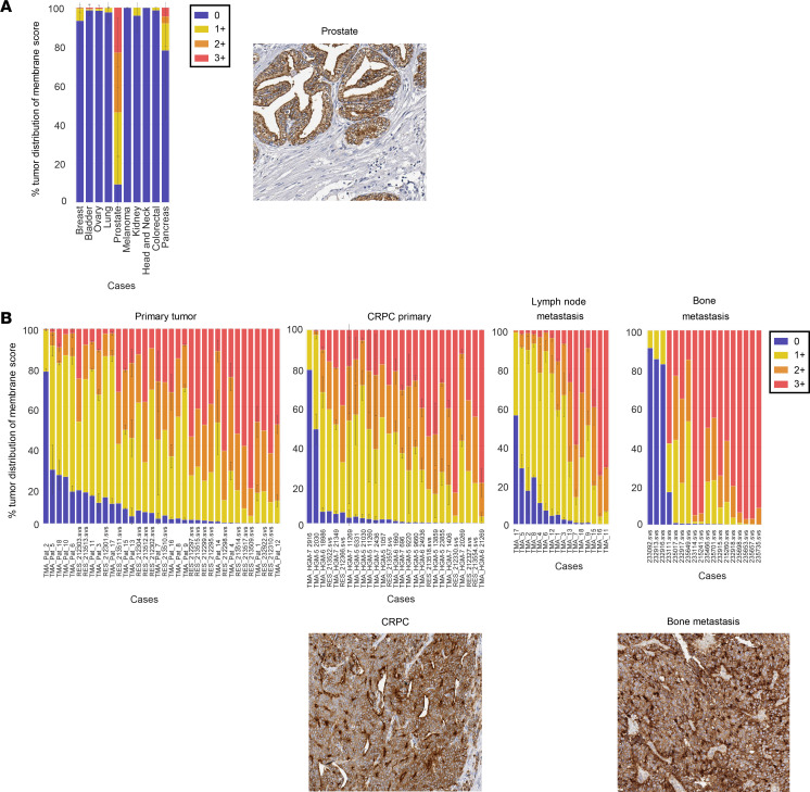
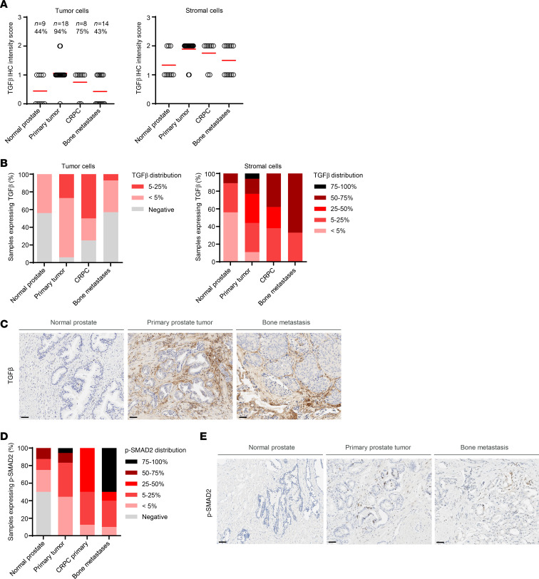
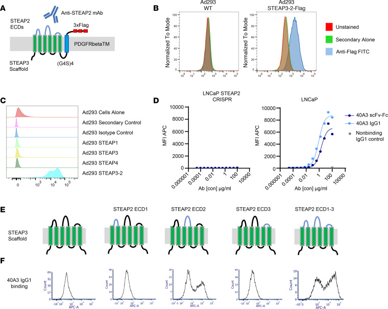
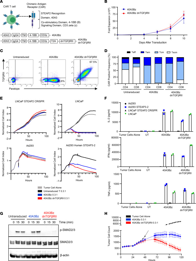
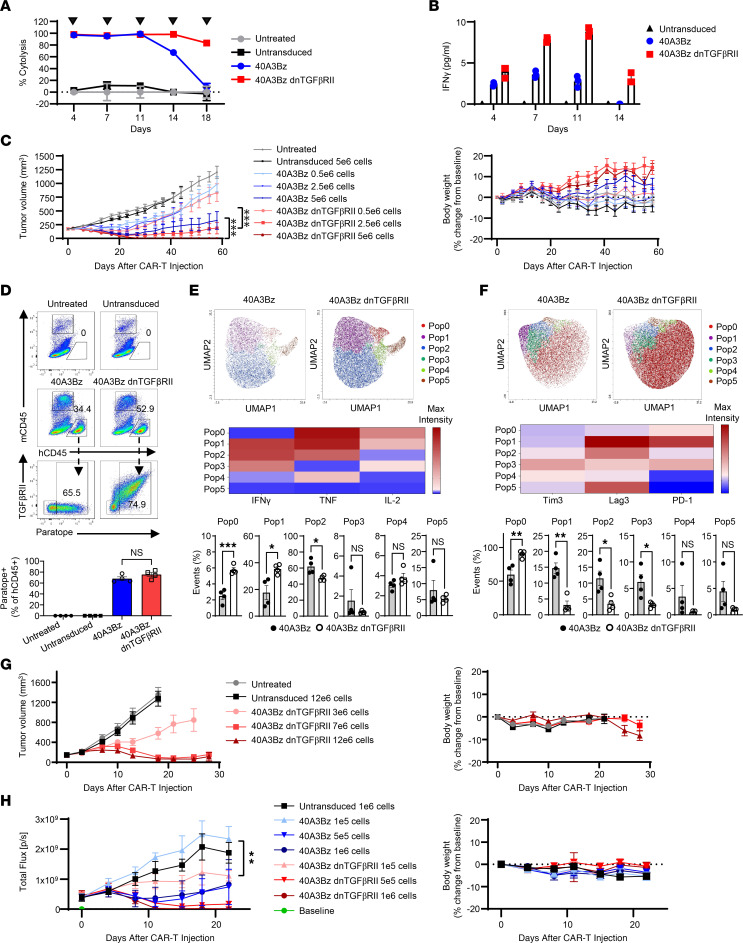
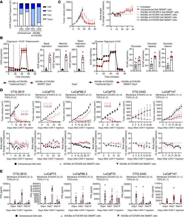
Prostate cancer is generally considered an immunologically "cold" tumor type that is insensitive to immunotherapy. Targeting surface antigens on tumors through cellular therapy can induce a potent antitumor immune response to "heat up" the tumor microenvironment. However, many antigens expressed on prostate tumor cells are also found on normal tissues, potentially causing on-target, off-tumor toxicities and a suboptimal therapeutic index. Our studies revealed that six-transmembrane epithelial antigen of prostate-2 (STEAP2) was a prevalent prostate cancer antigen that displayed high, homogeneous cell surface expression across all stages of disease with limited distal normal tissue expression, making it ideal for therapeutic targeting. A multifaceted lead generation approach enabled development of an armored STEAP2 chimeric antigen receptor T cell (CAR-T) therapeutic candidate, AZD0754. This CAR-T product was armored with a dominant-negative TGF-β type II receptor, bolstering its activity in the TGF-β-rich immunosuppressive environment of prostate cancer. AZD0754 demonstrated potent and specific cytotoxicity against antigen-expressing cells in vitro despite TGF-β-rich conditions. Further, AZD0754 enforced robust, dose-dependent in vivo efficacy in STEAP2-expressing cancer cell line-derived and patient-derived xenograft mouse models, and exhibited encouraging preclinical safety. Together, these data underscore the therapeutic tractability of STEAP2 in prostate cancer as well as build confidence in the specificity, potency, and tolerability of this potentially first-in-class CAR-T therapy.

Keywords: Oncology; Prostate cancer.


Figures

Figure 1
Figure 1. Prevalence of STEAP2, a prostate tumor–associated antigen, throughout disease progression.
(A) IHC studies of a broad TMA demonstrated membrane STEAP2 expression exclusively in prostate cancer. An example of STEAP2 IHC staining in normal prostate tissue is shown. (B) As in A, TMAs containing primary prostate cancer, CRPC, and prostate lymph node metastases, as well as decalcified full-face sections of prostate cancer bone metastases, were evaluated by IHC for STEAP2 membrane expression. Representative images of STEAP2 in CRPC and bone metastases are shown under the IHC graphs. All IHC images shown are at ×20 original magnification.
Figure 2
Figure 2. Prevalence of TGF-β throughout prostate cancer progression.
(A) The same prostate cancer TMA samples shown in Figure 1 were profiled for expression of TGF-β by IHC. The TGF-β staining intensity in tumor and stromal cells was quantified. (B) Distribution of TGF-β staining in tumor and stromal cells. (C) Representative images of TGF-β staining in normal prostate sample, primary prostate tumor, and prostate cancer bone metastatic sample, highlighting the contribution of tumor and stromal cell TGF-β in the TME. (D) IHC for p-SMAD2 was performed on the samples shown in A, and the distribution of expression in each prostate cancer disease subset was quantified. (E) Representative images of p-SMAD2 staining quantified in D. All images include a 50 μm scale bar at bottom left.
Figure 3
Figure 3. Generation of an anti-STEAP2 antibody that demonstrates specificity in model systems.
(A) Chimera containing the STEAP3-GS-PDGFRβTM-FLAG amino acid sequence backbone with the sequence of the STEAP2 extracellular loops grafted on to establish cell surface localization. (B) FACS analysis demonstrating robust and stable expression of the STEAP3-2–FLAG chimera and, by extrapolation, the STEAP3-2 (non-tagged) used for antibody generation, at the cell surface of Ad293 cells. (C) 40A3 scFv-Fc was tested for binding to Ad293 cells expressing human STEAP family members (STEAP1, 2, 3, and 4). (D) Multiple scFv-Fcs and full-length IgG1 antibodies were screened for binding to antigen-positive (Ad293 STEAP3-2, Ad293 STEAP3-2 murine, and LNCaP) cells and antigen-negative (Ad293 and LNCaP STEAP2 CRISPR) cell lines. Representative FACS titration graphs show binding curves for the 40A3 scFv-Fc, 40A3 IgG1, and nonbinding IgG1 as a negative control in the LNCaP STEAP2 CRISPR and LNCaP cell lines. Anti–human Fc secondary antibodies conjugated with Alexa Fluor 647 were used for detection of scFv-Fc or IgG1 binding to cells by flow cytometry. (E) Models of STEAP3-STEAP2 ECD chimeras used to assess 40A3 IgG domain recognition. (F) FACS analysis showing preferential binding of 40A3 IgG to ECD2 of STEAP2 on Ad293 cells expressing STEAP3-2 chimeras. All FACS results are representative of n > 3 experimental replicates.
Figure 4
Figure 4. In vitro cytolytic activity of 40A3Bz dnTGFβRII CAR-Ts.
(A) Components of the STEAP2 unarmored and armored lentivirus constructs used in generation of the CAR-Ts. (B) Viable cell expansion of CAR-Ts was assessed for 10 days after lentivirus transduction in n = 3 donors. Data represent mean ± SEM. (C) The 40A3Bz cells and 40A3Bz dnTGFβRII STEAP2 CAR-Ts were evaluated by flow cytometry at day 9 after transduction to assess CAR positivity and cell surface expression of dnTGFβRII, compared with untransduced T cells from the same donor. (D) CAR-Ts from C were stained for phenotypic surface markers including CD45RO and CD62L and analyzed by flow cytometry. Naive (CD45ROCD62L+), central memory (CD45RO+CD62L+), effector memory (CD45RO+CD62L), and effector (CD45ROCD62L) T cells were used. (E) CAR-Ts from C were cocultured with antigen-positive (Ad293 STEAP3-2 and LNCaP) and antigen-negative (Ad293 and LNCaP STEAP2 CRISPR) cell lines. Killing of target cells was measured over 100 hours with the xCELLigence impedance assay. Data are an average of duplicate. (F) Supernatants from the same coculture experiments were collected 24 hours after addition of CAR-Ts, and cytokines (IFN-γ, TNF-α, and IL-2) were measured by Meso Scale Discovery (MSD) electrochemiluminescence assay. Data are an average of duplicate. (G) STEAP2 CAR-Ts were subjected to FACS for CAR positivity and starved overnight before stimulation with recombinant human TGF-β treatment. Cell lysates were generated during the indicated time course, and Western blotting was performed to evaluate levels of p-SMAD2/3, total SMAD2/3, and β-actin in each sample. (H) STEAP2 CAR-Ts were cocultured with antigen-positive cells (C4-2 cells stably expressing mKate2 red fluorescent protein) at a 0.3:1 ratio in the presence of 30 ng/mL recombinant TGF-β. C4-2 cell viability was monitored over 120 hours with the Incucyte live cell analysis system (Sartorius) in triplicate. Data represent mean ± SEM. Data in BF and H are representative of the results obtained in 3 or more independent experiments with CAR-Ts prepared from 3 healthy donors, and G was performed twice with 2 donors.
Figure 5
Figure 5. dnTGFβRII CAR-T armoring enhances activity and persistence in vitro and in vivo.
(A) Serial killing was measured following coculture of LNCaP and CART-T cells at an E/T ratio of 0.3:1, as indicated by arrowheads. (B) Cytokines were profiled 24 hours after each coculture in A. (C) The 40A3Bz and 40A3Bz CAR-Ts were dosed at 3 concentrations (0.5 × 10⁶, 2.5 × 10⁶, and 5 × 10⁶ CAR-positive cells) by tail vein injection in NSG mice implanted with C4-2 cells overexpressing TGF-β (n = 10). Tumor volumes and body weights were measured. (D) For C, a cohort of animals from each group were sacrificed to examine CAR-T pharmacodynamics and phenotype in dissociated tumors by FACS (n = 4). Representative plots revealed the percentage of tumor-infiltrated human CD45+ (hCD45+) cells in mice dosed with 5 × 10⁶ cells at day 14. Bottom bar graph shows CAR-positive, tumor-infiltrated 40A3Bz cells (paratope+ TGFβRII hCD45+) and 40A3Bz dnTGFβRII CAR-Ts (paratope+ TGFβRII+ hCD45+). (E) Paratope-positive cells from D were analyzed for the percentage of IFN-γ+, TNF+, and IL-2+ cells in mice at day 14. (F) As in E, cells were analyzed for expression of Tim3+, Lag3+, and PD-1+ cells. UMAP plots in E and F show populations identified by a FlowSOM algorithm and further defined by heatmap. (G) Similarly to C, mice bearing 22RV1 cells overexpressing TGF-β, and 40A3Bz dnTGFβRII CAR-Ts were dosed at 3 concentrations (3 × 10⁶, 7 × 10⁶, and 12 × 106 CAR-positive cells) (n = 10). (H) C4-2 luciferase-expressing cells were implanted in the intratibial space of NSG mice, and luciferase signal was monitored. Randomization occurred when the tumor flux reached 4.04 × 10⁸ photons/second (p/s), and CAR-Ts from C were dosed at 3 concentrations (0.1 × 10⁶, 0.5 × 10⁶, and 1 × 10⁶ CAR-positive cells). (n = 5). Data shown in AC and G are representative of 3 independent experiments using CAR-Ts from 2 donors. H and DF were performed twice with material from 2 donors. All error bars represent mean ± SEM. Statistical significance was determined using 1-sided growth rate comparison nonparametric test in C and H and unpaired 2-tailed Student’s t test in E and F (*P < 0.05, **P < 0.01, ***P < 0.001).
Figure 6
Figure 6. Effect of enhanced CAR-T manufacturing on antitumor activity.
(A) 40A3Bz dnTGFβRII CAR-Ts were manufactured according to the SMART process, and CAR positivity, activation, and phenotype of the cells were evaluated at expansion day 4 and compared with those of untransduced T cells from the same donor. (B) Bioenergetic profile of SMART (day 4) versus traditional (day 11) manufactured 40A3Bz dnTGFβRII CAR-Ts as determined by Seahorse analysis. OCR, oxygen consumption rate; ECAR, extracellular acidification rate. (C) 40A3Bz dnTGFβRII SMART CAR-Ts were dosed at 4 concentrations (0.3 × 10⁶, 1 × 10⁶, 3 × 10⁶, and 6 × 10⁶ CAR-positive cells) by tail vein injection in NSG MHC class I/II knockout mice implanted with 22Rv1 cells overexpressing TGF-β (n = 10). Tumor volumes and body weights were measured to 50 days after tumor implantation. (D) PDX fragments from frozen stocks of various prostate cancer PDX models were implanted into NSG MHC I/II knockout mice and randomized when tumor volumes for each model ranged from 125 to 250 mm3. Mice were dosed as described in C with 0.5 × 10⁶ or 5 × 10⁶ 40A3Bz dnTGFβRII SMART CAR-Ts and compared with 5 × 10⁶ untransduced SMART controls (n ranged from 4 to 12 depending on the model). The IHC data inset on each model represents the cell surface STEAP2 expression scoring. (E) Serum levels of IFN-γ across all PDX models described in D, determined by MSD (n = 4 or greater). Experiments are representative of 2 different donor CAR-Ts. Data in AC are representative of multiple independent experiments prepared from 3 healthy donors, while D and E were performed once with material from 1 donor. All data represent mean ± SEM.

References

    1. Siegel RL, et al. Cancer statistics, 2022. CA Cancer J Clin. 2022;72(1):7–33. doi: 10.3322/caac.21708. - DOI - PubMed
    1. Siegel RL, et al. Cancer statistics, 2020. CA Cancer J Clin. 2020;70(1):7–30. doi: 10.3322/caac.21590. - DOI - PubMed
    1. Rathi N, et al. Evolving role of immunotherapy in metastatic castration refractory prostate cancer. Drugs. 2021;81(2):191–206. doi: 10.1007/s40265-020-01456-z. - DOI - PMC - PubMed
    1. Vitkin N, et al. The tumor immune contexture of prostate cancer. Front Immunol. 2019;10:603. doi: 10.3389/fimmu.2019.00603. - DOI - PMC - PubMed
    1. Hubert RS, et al. STEAP: a prostate-specific cell-surface antigen highly expressed in human prostate tumors. Proc Natl Acad Sci U S A. 1999;96(25):14523–14528. doi: 10.1073/pnas.96.25.14523. - DOI - PMC - PubMed

Publication types