

LKB1-SIK2 loss drives uveal melanoma proliferation and hypersensitivity to SLC8A1 and ROS inhibition
- PMID: 37966164
- PMCID: PMC10701601
- DOI: 10.15252/emmm.202317719
LKB1-SIK2 loss drives uveal melanoma proliferation and hypersensitivity to SLC8A1 and ROS inhibition
Erratum in
-
Publisher Correction: LKB1-SIK2 loss drives uveal melanoma proliferation and hypersensitivity to SLC8A1 and ROS inhibition.EMBO Mol Med. 2024 Sep;16(9):2262-2267. doi: 10.1038/s44321-024-00114-1. EMBO Mol Med. 2024. PMID: 39164473 Free PMC article.
Abstract
Metastatic uveal melanomas are highly resistant to all existing treatments. To address this critical issue, we performed a kinome-wide CRISPR-Cas9 knockout screen, which revealed the LKB1-SIK2 module in restraining uveal melanoma tumorigenesis. Functionally, LKB1 loss enhances proliferation and survival through SIK2 inhibition and upregulation of the sodium/calcium (Na+ /Ca2+ ) exchanger SLC8A1. This signaling cascade promotes increased levels of intracellular calcium and mitochondrial reactive oxygen species, two hallmarks of cancer. We further demonstrate that combination of an SLC8A1 inhibitor and a mitochondria-targeted antioxidant promotes enhanced cell death efficacy in LKB1- and SIK2-negative uveal melanoma cells compared to control cells. Our study also identified an LKB1-loss gene signature for the survival prognostic of patients with uveal melanoma that may be also predictive of response to the therapy combination. Our data thus identify not only metabolic vulnerabilities but also new prognostic markers, thereby providing a therapeutic strategy for particular subtypes of metastatic uveal melanoma.
Keywords: LKB1; SIK2; SLC8A1; calcium; uveal melanoma.
© 2023 The Authors. Published under the terms of the CC BY 4.0 license.
Conflict of interest statement
The authors declare that they have no conflict of interest.
Figures
- A
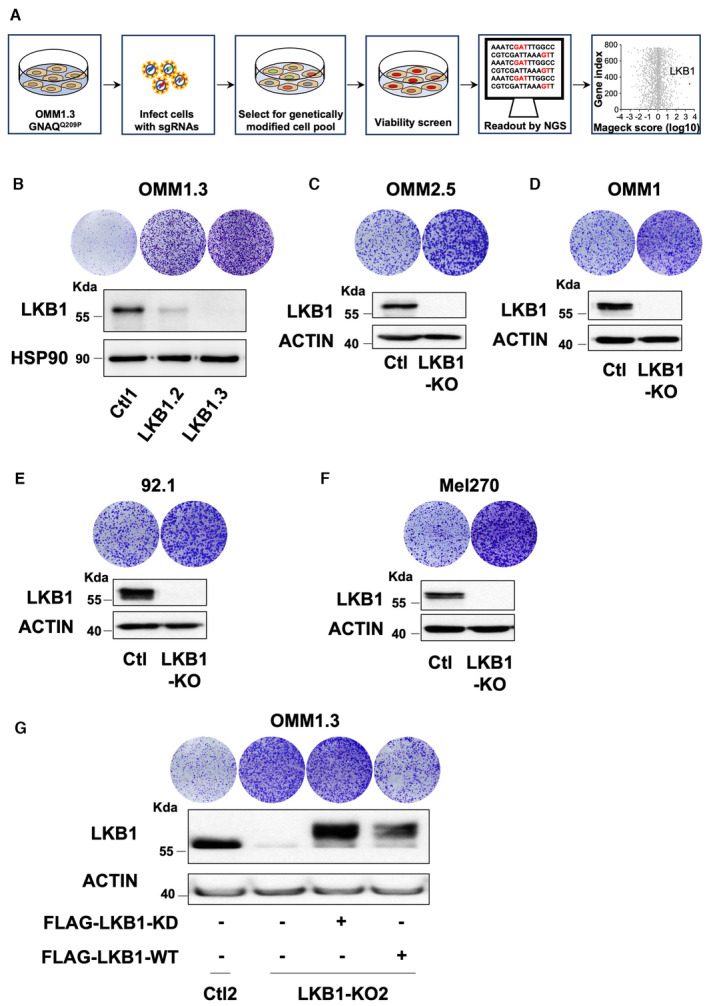
Schematic of the CRISPR‐Cas9 kinome screen with Log10‐transformed MAGeCK robust ranking aggregation (RRA) scores for either depletion (left) or enrichment (right) of sgRNAs in OMM1.3 cells at D35 compared to D0.
- B
(Bottom) Immunoblot of LKB1 in the indicated whole‐cell lysates of OMM1.3 pooled LKB1‐CRISPR cell lines. HSP90 was used as a loading control. (Top) Colony formation assay of OMM1.3 Ctl1 and pooled LKB1‐KD or KO cells grown for 10 days. Representative images of three independent experiments are shown.
- C–F
(Bottom) Immunoblot of LKB1 in the indicated whole‐cell lysates of pooled LKB1‐CRISPR generated in human OMM2.5 and OMM1 metastatic uveal melanoma cells and human 92.1 and Mel270 primary uveal melanoma cells. B‐actin was used as a loading control. (Top) Colony formation assay of Ctl and pooled LKB1‐KO cells grown for 10 days. Representative images of three independent experiments are shown.
- G
OMM1.3 Ctl2 and LKB1‐KO2 cells were noninfected (left) or infected with a vector‐encoding FLAG‐tagged kinase‐dead (FLAG‐LKB1‐KD) or wild‐type (FLAG‐LKB1‐WT) LKB1. Colony formation assay (top) and immunoblot (bottom) of LKB1 for the indicated cell lines are shown. B‐actin was used as a loading control. Representative images of three independent experiments are shown.

Immunoblot of LKB1 in the indicated whole‐cell lysates of OMM1.3 Ctl or LKB1‐KO cells. Three different OMM1.3 Ctl and LKB1‐KO cell lines are shown. B‐actin was used as a loading control.
Proliferation curves of OMM1.3 Ctl and LKB1‐KO cells. Data represent mean ± SD of three biological replicates (unpaired t‐test with Welsch's correction); ****P = 0.000041.
Colony formation assay of OMM1.3 Ctl and LKB1‐KO cells grown for 10 days. Cells were seeded at low density. Representative images of three independent experiments are shown.
OMM1.3 Ctl1 and LKB1‐KO1 cells were noninfected (left) or infected with a vector‐encoding FLAG‐tagged kinase dead (FLAG‐LKB1‐KD) or wild‐type (FLAG‐LKB1‐WT) LKB1. Colony formation assay (top) and immunoblot (bottom) of LKB1 for the indicated cell lines are shown. B‐actin was used as a loading control. Representative images of three independent experiments are shown.
Quantification of tumor volume in nude mice bearing xenograft tumors of LKB1‐WT or LKB1‐KO OMM1.3 cells. Mann–Whitney test was performed for comparison between groups, n = 6. Data are mean ± SEM. **P = 0.0022.

Representative box and whisker plots of LKB1 mRNA level in liver and skin metastasis of human uveal melanomas. The central band denotes the median value, box contains interquartile ranges, while whiskers mark minimum and maximum values. Mann–Whitney test was performed for comparison between groups. All points are represented. NS, not significant, P = 0.8425. Data were obtained from Karlsson et al (2020)).
Immunohistochemical stainings for LKB1 were performed on skin metastasis of human uveal melanomas. Staining intensity is represented as (H) High, (L) Low, and (N) Negative). Scale bars represent 10 μm.
Immunoblot of LKB1 total or phosphorylated on serine 428 and of SIK2 total or phosphorylated on threonine 175 in OMM1.3 cells untreated or treated with HGF at 10 or 25 ng/ml for 10 days. B‐actin was used as a loading control. Representative images of three independent experiments are shown.
Colony formation assay of OMM1.3 treated as in (C) grown for 10 days. Cells were seeded at low density. Representative images of three independent experiments are shown. Data represent mean ± SD of three biological replicates (unpaired t‐test with Welsch's correction); ***P = 0.0004, ****P = 0.000001.

Schematic of the integrated transcriptional profiling of LKB1 loss in OMM1.3 metastatic uveal melanoma cells. Venn diagrams indicating the number of significant differentially expressed genes selected as follow log2(foldchange) ≥ 1 or ≤ −1 and adjusted P‐value ≤ 0.05.
GSEA analysis from LKB1‐KO cells versus OMM1.3 Ctl cells, data queried against the “GOBP” group of gene sets.
Representative box and whisker plots of transient cytosolic Ca2+ changes in two OMM1.3 Ctl and two LKB1‐KO clones. Intracellular calcium concentrations are presented as ratio of the fluorescence signals obtained at 340 and 380 nm (F340/F380). One‐way ANOVA test was performed for comparison between groups. The central band denotes the median value, box contains interquartile ranges, while whiskers mark minimum and maximum values. All technical replicates are shown (n = 53). NS, not significant, P = 0.8571, **P = 0.0035/*P = 0.0399, ***P = 0.0003/**P = 0.0060. All points are represented.
Effect of reducing extracellular calcium with EGTA on Ctl1 and LKB1‐KO1. Indicated cells were seeded at the same density and cultured for 7 days in the presence of DMSO or EGTA (0.25 or 0.5 mM). Relative cell proliferation (percentage). Data represent mean ± SD of three biological replicates (unpaired t‐test with Welsch's correction); **P = 0.0057/***P = 0.0008, ****P = 0.000015/***P = 0.0004.
RT‐qPCR analysis of SLC8A1 mRNA level in OMM1.3 Ctl1 and LKB1‐KO1 noninfected or infected with a vector encoding FLAG‐tagged kinase‐dead (FLAG‐LKB1‐KD) or wild type (FLAG‐LKB1‐WT) LKB1. Data represent mean ± SD of four biological replicates (unpaired t‐test with Welsch's correction); ****P = 0.000047/****P = 0.000024 and ****P = 0.000012.
Disease‐specific survival stratified by SLC8A1 mRNA expression (median) from UM‐TCGA dataset (tumors n = 80). Low SLC8A1 mRNA level in blue and high SLC8A1 mRNA level in red. P‐value, log‐rank test.

- A
Overall survival stratified by SLC8A2 mRNA expression (median) from UM‐TCGA dataset (tumors n = 80); P‐value, log‐rank test.
- B, C
Staining of skin metastasis of human uveal melanomas with human‐positive control probe (PPIB) or with negative control probe (DapB). Scale bars represent 20 μm.
- D
RT–qPCR analysis of SLC8A1 mRNA level in LKB1‐KO2 OMM1.3 cells treated with control siRNA (siCtl) or two different SLC8A1 siRNA (siSLC8A1). Data represent mean ± SD of three biological replicates (unpaired t‐test with Welsch's correction); ***P = 0.0005/**P = 0.0023.
- E
Colony formation assay of LKB1‐KO2 OMM1.3 cells treated as in (D) grown for 10 days. A total of 75,000 cells were seeded. Representative images and crystal violet quantification at OD 561 nm are shown. Data represent mean ± SD of three biological replicates (unpaired t‐test with Welsch's correction); ****P = 0.00001/***P = 0.0002.

Sections from patients were labeled with the RNAscope probe for SLC8A1 (red). The membrane contours were detected by immunofluorescence using CD44 staining. Images were captured by spinning disk confocal microscopy. Scale bars represent 20 μm.
Representative box and whisker plots of Log2 fold change mRNA level of SLC8A1 and LKB1 in liver and skin metastasis of human uveal melanomas (Karlsson et al, 2020). The central band denotes the median value, box contains interquartile ranges, while whiskers mark minimum and maximum values. Mann–Whitney test was performed for comparison between groups. *P = 0.0151; NS, not significant, P = 0.8425. All points are represented.
RT–qPCR analysis of SLC8A1 mRNA level in LKB1‐KO1 OMM1.3 cells treated with control siRNA (siCtl) or two different SLC8A1 siRNA (siSLC8A1). Data represent mean ± SD of three biological replicates (unpaired t‐test with Welsch's correction); **P = 0.0065/**P = 0.0046.
SLC8A1 protein level was studied by immunoblot in membrane‐enriched lysates in the same conditions. HTR2B was used as a loading control. Representative images of three independent experiments are shown.
Colony formation assay of LKB1‐KO1 cells grown for 10 days in the same conditions. A total of 75,000 cells were seeded. Representative images of three independent experiments and crystal violet quantification at OD 561 nm are shown. Data represent mean ± SD of three biological replicates (unpaired t‐test with Welsch's correction); ***P = 0.0008/***P = 0.0004.

Immunoblot of SIK2 in the indicated whole‐cell lysates of OMM1.3 Ctl or SIK2‐KO cells. Three different OMM1.3 Ctl and SIK2‐KO cell lines are shown. B‐actin was used as a loading control.
Colony formation assay of OMM1.3 Ctl and SIK2‐KO cells grown for 10 days. Cells were seeded at low density. Representative images of three independent experiments and crystal violet quantification at OD 561 nm are shown.
LKB1‐KO1 cells were transfected with an empty vector (EV) or vectors encoding either a kinase‐dead (SIK2 K49M) or a constitutively active (SIK2 T175D) form of SIK2. Immunoblot (bottom) of SIK2 for the indicated cell lines and colony formation assay (top) are shown. B‐actin was used as a loading control. Representative images of three independent experiments are shown.
RT–qPCR analysis of SLC8A1 mRNA level in OMM1.3 Ctl1 and SIK2‐KO1 noninfected (left) or infected with an empty vector (EV) or a vector‐encoding SIK2‐WT. Data represent mean ± SD of three biological replicates (unpaired t‐test with Welsch's correction); ****P = 0.000057/**P = 0.0018; NS, not significant, P = 0.2765.
RT–qPCR analysis of SLC8A1 mRNA level in OMM1.3 Ctl1 and LKB1‐KO1 noninfected (left) or infected with an empty vector (EV) or a vector‐encoding SIK2‐WT. Data represent mean ± SD of three biological replicates (unpaired t‐test with Welsch's correction); **P = 0.0036/***P = 0.0005, **P = 0.0040.
RT–qPCR analysis of SLC8A1 mRNA level in OMM1.3 SIK2‐KO1 cells treated with control siRNA (siCtl) or two different SLC8A1 siRNA (siSLC8A1). Data represent mean ± SD of three biological replicates (unpaired t‐test with Welsch's correction); ***P = 0.0008/**P = 0.0013.
Colony formation assay of OMM1.3 SIK2‐KO1 cells grown for 10 days in the same conditions. A total of 75,000 cells were seeded. Representative images of three independent experiments and crystal violet quantification at OD 561 nm are shown. Data represent mean ± SD of three biological replicates (unpaired t‐test with Welsch's correction); ****P = 0.00000085/****P = 0.000002.

- A
(Bottom) Immunoblot of SIK2 in the indicated whole‐cell lysates of OMM1.3‐pooled SIK2‐CRISPR cell lines. HSP90 was used as a loading control. (Top) Colony formation assay of OMM1.3 Ctl1 and pooled SIK2‐KD or KO cells grown for 10 days. Cells were seeded at low density. Representative images of three independent experiments are shown.
- B
Proliferation curves of OMM1.3 Ctl and SIK2‐KO cells. Data represent mean ± SD of three biological replicates (unpaired t‐test with Welsch's correction); ****P = 0.000041.
- C, D
(Bottom) SIK2‐CRISPR‐KO was generated in human OMM2.5 metastatic and human 92.1 primary uveal melanoma cells. Representative western blot assay of SIK2 is shown. B‐actin was used as a loading control. (Top) Colony formation assay of control cells (Ctl) and pooled SIK2‐KO cells grown for 10 days. Cells were seeded at low density. Representative images of three independent experiments are shown. Ctl cells from Fig EV1C and E have been used.
- E, F
OMM1.3 Ctl1, SIK2‐KO1 cells and OMM1.3 Ctl2, SIK2‐KO2 cells were noninfected (left) or infected with an empty vector (EV) or a vector‐encoding SIK2‐WT. Colony formation assay and immunoblot of SIK2 are shown. B‐actin was used as a loading control. Representative images of three independent experiments are shown.

- A
Immunoblot of SIK2, phospho‐SIK2, and LKB1 in the indicated whole‐cell lysates from control, LKB1‐KO, and SIK2‐KO OMM1.3 cells. B‐actin was used as a loading control. Representative images of three independent experiments are shown.
- B
LKB1‐KO2 cells transfected with an empty vector (EV) or vectors encoding either a kinase‐dead (SIK2 K49M) or a constitutively active (SIK2 T175D) form of SIK2. Immunoblot for SIK2 and colony formation assay are shown. B‐actin was used as a loading control. Representative images of three independent experiments are shown.
- C, D
Human OMM2.5 and OMM1 metastatic uveal melanoma cells Ctl or LKB1‐KO cells were noninfected (left) or LKB1‐KO cells were infected with an empty vector (EV) or a vector‐encoding SIK2‐WT. Colony formation assay and immunoblot of SIK2 are shown. B‐actin was used as a loading control. Representative images of three independent experiments are shown.
- E
RT–qPCR analysis of SLC8A1 mRNA level in OMM1.3 Ctl2 and SIK2‐KO2 noninfected (left) or infected with an empty vector (EV) or a vector‐encoding SIK2‐WT. Data represent mean ± SD of three biological replicates (unpaired t‐test with Welsch's correction); **P = 0.0068/***P = 0.004; NS, not significant, P = 0.0844.
- F
RT–qPCR analysis of SLC8A1 mRNA level in OMM1.3 Ctl2 and LKB1‐KO2 noninfected (left) or infected with an empty vector (EV) or a vector‐encoding SIK2‐WT. Data represent mean ± SD of three biological replicates (unpaired t‐test with Welsch's correction) **P = 0.0069/**P = 0076 and *P = 0.0109.

Representative box and whisker plots of Rhod2 fluorescence in control, LKB1‐KO, and SIK2‐KO OMM1.3 cells. The central band denotes the median value, box contains interquartile ranges, while whiskers mark minimum and maximum values. All technical replicates are shown (n = 16); Mann–Whitney test was performed for comparison between groups. ***P = 0.0007 and ****P = 0.00000004. All points are represented.
Representative box and whisker plots of mitochondrial ROS levels in control, LKB1‐KO, and SIK2‐KO OMM1.3 cells measured using a dihydrorhodamine 123 staining. The central band denotes the median value, box contains interquartile ranges, while whiskers mark minimum and maximum values. All biological replicates are shown (n = 5); Mann–Whitney test was performed for comparison between groups. **P = 0.0079. All points are represented.
Analysis of apoptosis in OMM1.3 cells (control, LKB1, and SIK2‐KO) treated with 200 nM MitoQ and/or 5 μM KB‐R7943 for 72 h. Annexin V diagram (left) and quantitation of the percentage of apoptotic cells using the Annexin V assay (right). Data represent mean ± SD of three biological replicates (unpaired t‐test with Welsch's correction); ***P = 0.0004, ***P = 0.0007, ****P = 0.000098, **P = 0.0012, and ***P = 0.0002.

Quantification of tumor volume in nude mice‐bearing xenograft tumors of LKB1‐KO cells treated with MitoQ (5 mg/kg), KB‐R7943 (5 mg/kg), a combination of both, or vehicle three times per week (n = 4 mice, each group). Mann–Whitney test was performed for comparison between groups. Data are mean ± SEM. *P = 0.0286; NS, not significant, P = 0.6857
Representative images of tumors are shown.
Kaplan–Meier analysis of the LKB1‐KO signature in UM‐TCGA dataset (tumors n = 80).
Time‐dependent receiver operating characteristic (ROC) curves show the sensitivity and specificity of our LKB1 signature compared to the gene expression profiling signature (GEP) (Onken et al, 2004) for predicting the patient disease‐specific survival (UM‐TCGA cohort).

- A
Body weights of mice during treatment are shown as the mean ± SEM (n = 4 mice, each group).
- B
Tumor weights of the indicated xenografts at the endpoint (12 days) are shown as the mean ± SD (n = 4 mice, each group). Mann–Whitney test was performed for comparison between groups. *P = 0.0286; NS, not significant, P = 0.1143.
- C
Representative box and whiskers plots of the LKB1 loss signature score based on LKB1 mRNA expression level (low and high) from the UM‐TCGA dataset. Mann–Whitney test was performed for comparison between groups. *P = 0.0487. All points are represented.
- D
Time‐dependent receiver operating characteristic (ROC) curves show the sensitivity and specificity of our LKB1 signature compared to the gene expression profiling signature (GEP) (Onken et al, 2004), for predicting the progression‐free survival (Laurent et al, ; Data ref: Laurent et al, 2011b).
References
-
- Benjamini Y, Hochberg Y (1995) Controlling the false discovery rate: a practical and powerful approach to multiple testing. J R Stat Soc Series B Stat Methodol 57: 289–300
Publication types
MeSH terms
Substances
Associated data
- Actions
Grants and funding
LinkOut - more resources
Full Text Sources
Medical
Molecular Biology Databases
Research Materials
Miscellaneous
